平成 29 年度博士学位論文
Studies on geranylgeranoic acid-induced cell death: with special attention to pyroptosis in human hepatoma cells
ゲラニルゲラノイン酸誘導性細胞死に関する研究
–
特にヒト肝癌細胞におけるパイロトーシスについて–
D3215001
薮田末美
Contents
Chapter 0: Foundation research ... 1
Abstract ... 2
0-1. Introduction ... 3
0-1-1. Telomeres ... 3
0-1-2. Nutritional perturbation of telomere length shortening ... 3
0-1-3. β-Carotene metabolism-related genes ... 4
0-2. Results ... 6
0-2-1. Characteristics of subjects ... 6
0-2-2. Circulating β-carotene levels in each genotype group ... 7
0-2-3. Buccal RTLs and FFQ ... 8
0-2-4. Measurement of transcribed telomere repeats ... 10
0-2-5. Retinoids downregulated TERRA ... 12
0-3. Discussion ... 13
Chapter I: General Introduction ... 16
I-1. Diterpenoid acids or acyclic retinoids ... 17
I-2. Geranylgeranoic acid (GGA) ... 18
I-3. GGA-induced cell death ... 19
I-3-1. GGA-induced mitochondrial impairment ... 21
I-3-2. GGA-induced nuclear translocation of the mutant p53 ... 21
I-3-3. GGA-induced incomplete autophagic response ... 24
II-1-2. Canonical inflammasomes: NLR and ALR inflammasomes ... 42
II-1-3. Mechanisms of NLRP3 inflammasome activation ... 44
II-1-4. NLRP3 inflammasome priming: first hit ... 47
II-1-5. NLRP3 inflammasome activation: second hit ... 48
II-1-6. Expression of NLRP3 through NF-κB ... 49
II-1-7. Downstream signals of TLRs ... 50
II-1-8. Pyroptosis interacts with UPR ... 55
II-1-9. Gasdermin D is emerging as executors of pyroptosis ... 57
II-1-10. GSDMD-N forms functional pores ... 59
II-2. Results ... 60
II-2-1. Activation of caspase-1 activity after GGA treatment ... 60
II-2-2. GGA induced priming of inflammasome activation in HuH-7 cells ... 62
II-2-3. NF-κB relocates from the cytoplasm to the nucleus after GGA treatment ... 64
II-2-4. Translocation for plasma membrane of GSDMD after GGA treatment ... 66
II-2-5. Morphological alterations and membrane damages after GGA treatment ... 68
II-2-6. GGA-induced cell death via TLR4 signaling ... 70
II-2-7. GGA-induced UPR upregulates TLR2 expression ... 76
II-2-8. ATRA did not induce pyroptotic cell death ... 80
II-3. Discussion ... 82
Chapter III: Pyroptotic cell death with GGA through noncanonical inflammasome ... 87
Abstract ... 88
III-1. Introduction ... 89
III-1-1. Non-canonical inflammasome pathway ... 89
III-1-2. Priming of the non-canonical inflammasome ... 90
III-1-3. Non-canonical pathway interacts with UPR ... 91
III-2. Results ... 93
III-2-2. UPR induces the activation of caspase-4 ... 95
III-2-3. Increase of intracellular calcium concentration via UPR in HuH-7 cells ... 97
III-3. Discussion ... 102
Chapter IV: Epigenetic aspect of cancer chemoprevention with GGA ... 105
Abstract ... 106
IV-1. Introduction ... 107
IV-1-1. Epigenetic regulatory mechanisms ... 107
IV-1-2. Histone modification ... 107
IV-1-3. Lysine-specific demethylase 1A (LSD1/KDM1A) ... 109
IV-1-4. Another lysine-specific demethylase for histone H3 (KDM5A) ... 112
IV-1-5. Aim of the present study in this chapter ... 112
IV-2. Results ... 114
IV-2-1. Cytoplasmic translocation of nuclear KDM1A induced by GGA or TCP ... 114
IV-2-2. Specificity for KDM1A inhibitors and nuclear KDMs ... 116
IV-2-3. Subcellular distribution of KDM1A in M-phase cells ... 118
IV-2-4. Release of nuclear KDM1A by GGA or TCP under cell-free conditions ... 120
IV-3. Discussion ... 122
Chapter V: General Discussion ... 126
V-1. How does GGA induce cell death in HuH-7 cells? ... 127
V-1-1. Previous studies on GGA-induced cell death in HuH-7 cells. ... 127
V-1-2. Anti-tumorigenic effects of GGA other than inflammatory cell death. ... 128
V-2. Inflammation ... 129
V-5-2. Primary prevention with GGA ... 136
Chapter VI: Materials and methods ... 139
VI. Materials and methods ... 140
VI-1. Materials ... 140
VI-1-1. Instruments... 140
VI-1-2. Chemical reagents ... 142
VI-1-3. Cell culture compounds ... 144
VI-1-4. Kits ... 145
VI-1-5. Antibodies ... 145
VI-1-6. Cell line ... 146
VI-1-7. Solutions and Buffers... 147
VI-2. Methods ... 149
VI-2-1. Study population ... 149
VI-2-2. Food frequency questionnaire (FFQ) ... 149
VI-2-3. DNA-preparation ... 150
VI-2-4. RTLs determination ... 151
VI-2-5. Genotyping ... 152
VI-2-6. Measurement of serum β-carotene ... 152
VI-2-7. Cell culture ... 153
VI.2-8. Sodium dodecyl sulfate polyacrylamide gel electrophoresis (SDS-PAGE) and immunoblotting ... 153
VI-2-9. Cell-free analysis ... 155
VI-2-10. Isolation of total cellular RNA ... 155
VI-2-11. Reverse transcription for cDNA synthesis ... 155
VI-2-12. Real-time PCR ... 156
VI-2-13. ROS detection ... 159
VI-2-14. Ca2+ fluorescence imaging ... 159
VI-2-15. Fluorescent immunostaining ... 159
VI-2-16. Caspase-Glo1 assay ... 160
VI-2-17. Cell viability assay ... 160
VI-2-18. LDH assay ... 161
VI-2-19. Statistical analysis ... 161
Notes ... 162
Acknowledgments ... 163
References ... 164
List of abbreviations
9CRA AD ADP AIM ALR AM AMP AP APAF APS ASC ATF ATG ATP ATRA BBC BCL BCO1 BiP BH-3 CIITA CARD CASP CD14 CDK CDKN1A cDNA CoA Cont CoREST CP CRABP CREB Ct CTL
9-cis retinoic acid allele discrimination adenosine diphosphate absent in melanoma AIM-like receptor acetoxymethyl
adenosine monophosphate activator protein
apoptotic protease activating factor ammonium peroxydisulfate
apoptosis-associated speck-like protein containing a caspase recruitment domain activating transcription factor
autophagy-related gene adenosine triphosphate all-trans retinoic acid BCL2 binding component B-cell lymphoma
β-carotene oxygenase 1
binding immunoglobulin protein bcl-2 homology domain 3 class II transcription activator
caspase activation and recruitment domain caspase
cluster of differentiation 14 cyclin-dependent kinase CDK inhibitor 1
complementary DNA coenzyme A
control
corepressor for element 1-silencing transcription factor control peptide
cellular retinoic acid binding protein
cyclic AMP-responsive element-binding protein threshold cycle
c-type lectin
DAMP damage-associated molecular pattern DDIT3/CHOP
DIC ΔΨm D-MEM DMSO DNA DP DRAM dsDNA DSS ECSIT eIF ER ES FA FAD FBS FFQ G1 G2 GBP GGA GGOH GGPP GGPPase GPX GRP
DNA-damage-inducible transcript 3 differential interference contrast
mitochondrial inner membrane potential Dulbecco’s modified Eagle’s medium dimethyl sulfoxide
deoxyribonucleic acid
DNA-binding partner of E2Fs
DNA damage-regulated autophagy modulator double-stranded DNA
disuccinimidyl suberate
evolutionarily conserved signaling intermediate in Toll pathway eukaryotic initiation factor
endoplasmic reticulum embryonic stem farnesoic acid
flavin adenine dinucleotide fetal bovine serum
food frequency questionnaire gap phase 1 in cell cycle gap phase 2 in cell cycle guanylate-binding protein geranylgeranoic acid geranylgeraniol
geranylgeranyl diphosphate geranylgeranyl diphosphatase glutathione peroxidase glucose-regulated protein 78
HSC HUGO
hepatic stellate cell
human genome organization IC50
ICE IFN IgG IL IL-1R IκB IKK IPAF IRAK IRE IRF IRGB ISX JARID JNK kDa KDM LC LDH LBP LPS LRR
LSD1/KDM1A LTA
LTL M MAF MAL MAP MAPK MD MDa MHC MKK
inhibitory concentration at a half maximum IL-1β-converting enzyme
interferon
immunoglobulin G interleukin
IL-1 receptor inhibitor of NF-κB IκB kinase
interstitial pneumonia with autoimmune feature IL-1R-associated kinase
inositol requiring
interferon-regulatory factor
immunity-related GTPase family member B intestine-specific homeobox
Jumonji, At rich interactive domain c-jun N-terminal kinase
kilodalton
lysine (K)-specific demethylase
microtubule-associated protein 1 light chain lactate dehydrogenase
LPS-binding protein lipopolysaccharide leucine-rich repeat
histone lysine demethylase 1 lipoteichoic acid
leukocyte telomere length mitosis phase in cell cycle minor allele frequency MyD88 adaptor-like mitogen-activated protein MAP kinase
myeloid differentiation protein megadalton
major histocompatibility complex MAP kinase kinase
MMqPCR mRNA mTOR
monochrome multiplex qPCR messenger RNA
mammalian target of rapamycin mtROS
MyD NADH NAIP NBD N.D.
NEMO NET NF-κB NIK NLRC NLRP nm NOD NR2E1/TLX OA
OCT OD ORC P2X7 P2X7R PA PAD PAMP PARC
mitochondrial ROS
myeloid differentiation primary-response protein β-nicotinamide adenine dinucleotide, reduced neuronal apoptosis inhibitory protein
nucleotide-binding domain not determined
NF-κB essential modulator neutrophil extracellular trap
nuclear factor of kappa-light polypeptide gene enhancer in B-cells NF-κB-inducing kinase
NLR family CARD domain-containing protein NOD-like receptor containing pyrin domain nanometer
nucleotide-binding oligomerization domain nuclear receptor subfamily 2 group E oleic acid
octamer-binding transcription factor optical density
origin recognition complex purinergic receptor P2X 7 P2X7 receptor
palmitic acid
peptidylarginine deiminase
pathogen-associated molecular pattern
p53-associated parkin-like cytoplasmic protein
POU5F1 PRR PTEN
POU domain, class 5, transcription factor 1 pattern-recognition receptor
phosphatase and tensin homolog PTM
PUMA PYD PYHIN PYRIN PVDF QNZ qPCR RAR RB RIP RIPA RIPK RhoA RNA RNase ROI ROS rRNA RT RTL RT-qPCR RXR S SCD scg SCO2 SD
SDS-PAGE siRNA SLC7A11 SNP SQSTM ssRNA
post-translational modification
p53 upregulated modulator of apoptosis pyrin domain
pyrin and HIN domain family PYD or PADD/DAPIN domain polyvinylidene difluoride quinazolinediamine quantitative PCR retinoic acid receptor retinoblastoma protein receptor-interacting protein radio immunoprecipitation assay receptor interacting protein kinase Ras homolog gene family member A ribonucleic acid
endoribonuclease regions-of-interest reactive oxygen species ribosomal RNA
reverse transcription relative telomere length
reverse transcription and quantitative polymerase chain reaction retinoid X receptor
DNA synthesis phase stearoyl-CoA desaturase single copy gene
synthesis of cytochrome c oxidase 2 standard deviation
sodium dodecyl sulfate-polyacrylamide gel electrophoresis small interfering RNA
solute carrier family 7 member 11 single nucleotide polymorphism sequestosome
single-stranded RNA
T3SS TAB TAK
type III secretion system TAK1-binding protein TGF-β-activated kinase tBID
TCP TE TERRA TERT TG TGF TIGAR TIR TIRAP TLR TM TNF TNFR TP TP1 TRAF TRAM TRF TRIF UPR UV XBP1 xCT
truncated p15 BH3 interacting-domain death agonist trans-2-phenylcyclopropylamine
10 mM Tris-HCl, pH 7.4 and 1 mM EDTA buffer telomeric repeat-containing RNA
telomerase reverse transcriptase thapsigargin
transforming growth factor
TP53-induced glycolysis and apoptosis regulator toll-IL-1 receptor
TIR domain-containing adaptor protein toll-like receptor
tunicamycin
tumor necrosis factor TNF receptor
tumor protein
telomerase-associated protein 1 TNF-receptor-associated factor TRIF-related adaptor molecule terminal restriction fragment
TIR-domain-containing adaptor-inducing interferon unfolded protein response
ultra violet
X-box binding protein 1 cysteine transporter
List of tables and figures
Tables
Table 0-1 Subjects characteristics……….…...6
Table 0-2 Circulating β-carotene levels in each genotype group………8
Table 0-3 Association between buccal cell RTL andβ-carotene intakes……….9
Table II-1 Toll-like receptors and their ligands……….….53
Table VI-1 The nucleotide sequences of each primers used for real time RT-PCR………157
Figures Fig.0-1. ISX binds regulatory SNP site (rs6564851) of the BCO1 gene………..5
Fig.0-2. Alternative method of TERRA measurement……….11
Fig.0-3. GGA time-dependently downregulated TERRA……….………...12
Fig.I-1. Chemical structure of geranylgeranoic acid………...19
Fig.I-2. Diverse function of p53 through multiple target genes………..22
Fig.I-3. The formation of phagolysosomes………...25
Fig.I-4. Canonical unfolded protein responses………28
Fig.I-5. Cell cycle………31
Fig.I-6. Genes related G1/S transition……….32
Fig.I-7. Suppression of growth by the UPR………33
Fig.I-8. Programmed cell death………...36
Fig.I-9. Autophagy, apoptosis, necroptosis, pyroptosis and necrosis pathways………..37
Fig.II-1. Canonical inflammasomes………43
Fig.II-2. Mechanisms of NLRP3 inflammasome activation………45
Fig.II-3. Mammalian TLR signaling pathways………...54
Fig.II-4. Schematic representation of the pyroptosis pathways………...……...58
Fig.II-5. GGA activated caspase-1 in HuH-7 cells………..61
Fig.II-6. GGA induced priming of inflammasome in HuH-7 cells………....…..63
Fig.II-7. NF-κB is relocated from the cytoplasm to the nucleus by GGA………...65
Fig.II-8. Translocate for plasma membrane of GSDMD after GGA treatment……….…..67
Fig.II-9. Morphological alterations and membrane damages after GGA treatment………69
Fig.II-10. GGA-induced cell death via TLR4………..73
Fig.II-11. GGA-induced UPR upregulates TLR2………78
Fig.II-12. ATRA was not induced pyroptotic cell death………..81
Fig.III-1. The role of GSDMD in pyroptosis driven by non-canonical and canonical inflammasomes………...90
Fig.III-2. GGA-induced pyroptosis via non-canonical inflammasome pathway in HuH-7 cells……94
Fig.III-3. UPR induces the activation caspase-4……….96
Fig.III-4. Increase intracellular calcium via UPR in HuH-7 cells……..………...99
Fig.IV-1. Characteristic of histone H3K4 demethylase KDM1A………..110
Fig.IV-2. Cytoplasmic translocation of nuclear KDM1A induced by either GGA or TCP treatment………115 Fig.IV-3. Nuclear localization of KDM1A and KDM5A after 3 h treatment with non-inhibitory
Chapter 0
Foundation research
Suemi Yabuta
Molecular and Cellular Biology, Graduate School of Human Health Science, University of Nagasaki, Nagasaki, Japan
Abstract
Telomere length shortening is modulated not only by aging, but also by both genetic and environmental factors. The aim of this study was to investigate the interactions between antioxidant nutrient metabolism-related gene single nucleotide polymorphisms (the genetic factors) and nutrient intake (the environmental factors) in their effects on telomere length shortening. Data were collected on the relative telomere lengths (RTLs) of buccal cells and the habitual food intake of 70 healthy Japanese adults. All subjects were genotyped for two common single nucleotide polymorphisms: rs6564851 in the β-carotene oxygenase 1 (BCO1) gene and rs362090 in the intestine-specific homeobox (ISX) gene. We subdivided the study population into four groups based on combinations of the rs6564851 and rs362090 genotypes. After this subdivision, we showed a positive effect of daily α- or β-carotene intake on buccal RTL in the ISX rs362090 G-allele carrier + BCO1 rs6564851 GG-genotype group. In contrast, negative association of the buccal RTL with β-carotene intakes was unexpectedly detected in ISX G-carrier + BCO1 T-carrier genotype. We speculated telomere length maintenance might affected by retinoids, so we next showed
0-1. Introduction
0-1-1. Telomeres
It is well established that cellular telomere length reflects the replication number of chromosomal DNA, such that telomere length is the most reliable indicator of mitotic history and acts as a biomarker of cellular aging [1]. Indeed, a systematic review reveals that a decrease of leukocyte telomere length (LTL) with age is out of question [2]. Hence, it may be important to measure the telomere length of non-invasive samples to assess the aging degree in public health service.
Several years ago, Cawthon developed a singleplex quantitative polymerase chain reaction (qPCR) assay for “relative” average telomere lengths [3], which uses far less DNA and requires much less time to perform than the traditional Southern blot method for measuring terminal restriction fragment (TRF) lengths. Furthermore, Cawthon has recently improved his previous method in a monochrome multiplex qPCR (MMqPCR) [4,5].
0-1-2. Nutritional perturbation of telomere length shortening
Telomere length is likely shortened not only by cell proliferation, but also by environmental factors through an effect of oxidative stress and inflammation, which is the major causes of accelerated telomere erosion [6]. Both oxidative stress and inflammation form a pathogenic mechanism of lifestyle-related diseases and it is well known that dietary intervention can modulate oxidative stress and inflammation. In
cells from accelerated telomere erosion. Recently, strong evidence for this speculation has been repeatedly brought by Blackburn’s group that LTL increased with decreasing n-6:n-3 ratios in polyunsaturated fatty acid supplementation for 4 months in human clinical trial [7], comprehensive lifestyle changes significantly increased telomerase activity in peripheral immune cells [8] and an inverse relationship between baseline blood levels of marine omega-3 fatty acids and the rate of telomere shortening over 5 years [9]. Another evidence was very recently appended to demonstrate that telomere shortening in elderly individuals with mild cognitive impairment could be attenuated with n-3 fatty acid supplementation [10].
0-1-3. β-Carotene metabolism-related genes
The metabolism of antioxidant nutrients can be affected by genetic background. For example, several common SNPs of β-carotene oxygenase 1 (BCO1) gene are associated with β-carotene metabolism [11].
Indeed, a common promoter SNP (rs6564851) was associated with higher circulating β-carotene level.
Other common nonsynonymous SNPs (rs12934922; rs7501331) in the BCO1 gene, occurring at frequencies similar to those of the poor converter trait, reduced catalytic activity of BCO1 by down to 69%
intestinal lipid absorption including anti-oxidant lipids, carotenoids and vitamin E [14].
Fig.0-1. ISX binds regulatory SNP site (rs6564851) of the BCO1 gene.
BCO1 is an intestinal enzyme responsible for the cleavage of β-carotene into retinal. Several years ago, the gut-specific homeodomain transcription factor ISX has been identified as a putative repressor of intestinal BCO1 gene expression. ISX has been shown to bind to BCO1 SNP-containing 26-bp DNA fragment; G-allele-containing fragment has more affinity to ISX than T-allele fragment. Yabuta S, Masaki M, and Shidoji Y. Atlas of Science (2016)
https://atlasofscience.org/%CE%B2-carotene-requirement-for-anti-aging-depends-on-genetic-back ground/ [15]
The current study addresses a basic question. Are there any interactions between nutrient intake (environment factor) and common SNPs of the BCO1 and ISX genes (genetic factor) in their effects on telomere length shortening? The approach used in this study is the measurement of buccal cell telomere length with the analysis of food frequency questionnaires (FFQs) and genotyping of 2 common SNPs (rs6564851 of BCO1 and rs362090 of ISX).
0-2. Results
0-2-1. Characteristics of subjects
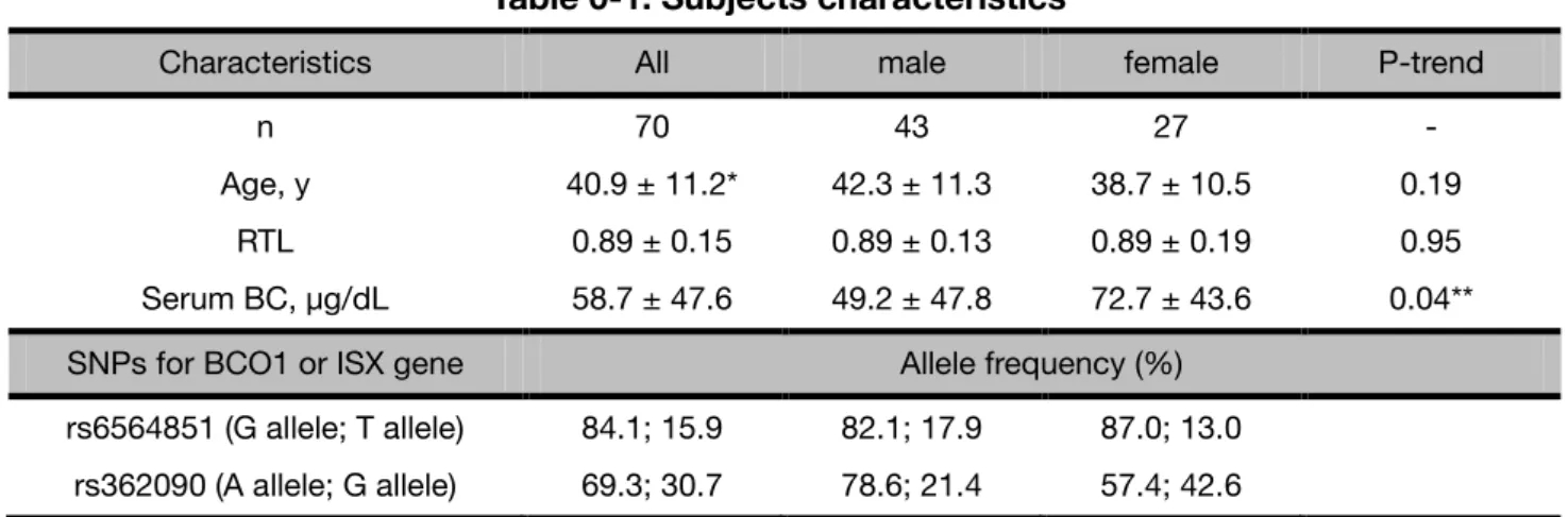
The demographic characteristics, and BCO1 and ISX common SNPs allele frequencies of the participants analyzed in the present study are shown in Table 0-1. There was no difference in the buccal cell relative telomere lengths (RTLs) in between males and females, whereas the average concentration of serum β-carotene was significantly higher in females than that in males.
As for rs6564851, a major allele (frequency, 84.1%) was G in the subjects and no difference of the allele frequency was found in between males and females. A minor allele (30.7%) of rs362090 was G, which frequency was higher in females than males.
Table 0-1. Subjects characteristics
Characteristics All male female P-trend
n 70 43 27 -
Age, y 40.9 ± 11.2* 42.3 ± 11.3 38.7 ± 10.5 0.19
RTL 0.89 ± 0.15 0.89 ± 0.13 0.89 ± 0.19 0.95
Serum BC, µg/dL 58.7 ± 47.6 49.2 ± 47.8 72.7 ± 43.6 0.04**
SNPs for BCO1 or ISX gene Allele frequency (%)
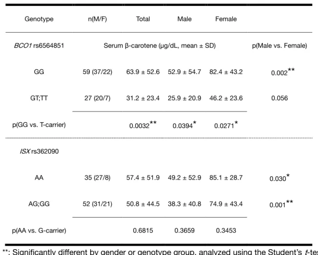
0-2-2. Circulating β-carotene levels in each genotype group
A genome-wide association study revealed that a common SNP rs6564851 near the BCO1 gene affects the circulating levels of carotenoids in three Caucasian population [16], thus we evaluated the effects of SNPs and serum carotenoid levels in Japanese population. Because the two polymorphisms (rs6564851 in BCO1, rs362090 in ISX) tested herein are common SNPs with a minor allele frequency (MAF) of 0.05 or more, two groups of homozygotes carrying major alleles and the other minor-allele carriers were analyzed below for comparison (Table 0-2). The average concentrations of serum β-carotene in the rs6564851 GG homozygotes (the SNP of the BCO1 gene) in total, males and females were significantly higher than those in the T allele carriers (p = 0.003, 0.039 and 0.027, respectively), showing that the GG homozygotes had a 2-fold higher serum β-carotene level compared with that of T allele carriers. In contrast, it was no significant difference in the serum β-carotene concentration between the major homozygotes and the minor allele carriers in the other SNPs rs362090 (the SNP of the ISX gene).
Table 0-2. Circulating β-carotene levels in each genotype group
Genotype n(M/F) Total Male Female
BCO1 rs6564851 Serum β-carotene (µg/dL, mean ± SD) p(Male vs. Female)
GG 59 (37/22) 63.9 ± 52.6 52.9 ± 54.7 82.4 ± 43.2 0.002
**
GT;TT 27 (20/7) 31.2 ± 23.4 25.9 ± 20.9 46.2 ± 23.6 0.056
p(GG vs. T-carrier) 0.0032
**
0.0394*
0.0271*
ISX rs362090
AA 35 (27/8) 57.4 ± 51.9 49.2 ± 52.9 85.1 ± 28.7 0.030
*
AG;GG 52 (31/21) 50.8 ± 44.5 38.3 ± 40.8 74.9 ± 43.4 0.001
**
p(AA vs. G-carrier) 0.6815 0.3659 0.3453
*, **: Significantly different by gender or genotype group, analyzed using the Student’s t-test (*, **: p<0.05, 0.01, respectively)
0-2-3. Buccal RTLs and FFQ
To further analyze genotype effects on the buccal RTLs, the BCO1 genotype groups (categorized into 2
RTLs and daily intake of β-carotene (Table 0-3).
Table 0-3. Association between buccal cell RTL and β-carotene intakes.
BCO1
GG GT; TT
ISX
AA no association no association
AG; GG 0.2 0.4 0.6 0.8 1 1.2
0 2000 4000 6000
0.2 0.4 0.6 0.8 1 1.2
0 2000 4000 6000
p=0.026
p=0.029
0-2-4. Measurement of transcribed telomere repeats
Why are ISX G-carrier and BCO1 T-carrier genotype groups taking toxic levels of β-carotene, in terms of telomere shortening protection? In this group, intestinal expression of the BCO1 gene must be high, so that ingested β-carotene may be efficiently converted to retinoids [17]. Hence, we speculated telomere length maintenance might be affected by not carotene itself, but rather retinoids.
So, we next conducted the following experiment to understand how retinoids regulate telomere length.
Besides shelterin as mentioned initially, telomeric repeat-containing RNA (TERRA) has recently joined in telomere regulation. Its transcription by RNA polymerase II stars within subtelomere regions and proceeds toward each chromosome ends. Although its function is still unclear, some studies reported that TERRA might act on negative control of telomere length trough telomerase sequestration. First, introduce measurement of TERRA family in this study, the pro-terminal DNA sequences associated with the long-arm telomeres of human chromosomes XqYq and 10q were named TelBam and TelSau, respectively.
BLAST search analysis demonstrated. Because measurement used with random hexamer measure including non-subtelomere region, we measured with cDNA has been used by telomere motif primer
(+/-), difference in primer of telomere motif or random hexamer (Fig.0-2C), suggesting that it can measure TERRA used by telomere motif primer.
Fig.0-2. Alternative method of TERRA measurement.
Total RNA or RNA was extracted to analyze the TelSau in TERRA by RT-qPCR in HuH-7 cells. (A) PCR amplification curves of the delta Ct between with and without RT reaction. (B) Melting curves of amplicons with or without RT (upper), random hexamer (bottom). (C) PCR amplification curves of telomeric repeat sequence or random hexamer.
(A)
(B)
(C)
RT (+)
RT (-)
RT (-) RT (+)
Telomere motif primer
Random hexamer
0-2-5. Retinoids downregulated TERRA
Next, we speculated telomere maintenance might be affected with retinoid, measured the cellular TERRA level after treatment with 20 μM GGA of acyclic retinoids (Fig.0-3). GGA apparently showed a time-dependent downregulation of the cellular TERRA level from 8 to 24 h.
Fig.0-3. GGA time-dependently downregulated TERRA.
HuH-7 cells were treated with 20 µM GGA for 0, 3, 8 and 24 h. Total RNA was extracted to analyze the cellular levels of TelSau in TERRA mRNA by RT-qPCR (unpublished observation).
0 0.2 0.4 0.6 0.8 1 1.2
0 5 10 15 20 25 30
TERRA expression level relative to 28S (ratio cont)
Time after treatment (h)
0-3. Discussion
In the present study, we addressed a basic question whether any associations exist between daily nutrient intakes and buccal cell RTLs. As a result, we found that the RTLs in buccal cells measured in 70 Japanese samples by MMqPCR method were associated with dietary intakes of β-carotene in specified genotype groups.
In the present investigation, we again failed to find any protective roles of vegetables, fats and oils, and antioxidant nutrients including carotenoids, tocopherol and vitamin C against telomere shortening in buccal cells, unless the study population was subdivided into 4-genotype groups by a combination of SNPs (rs6564851 and rs362090) of the antioxidant vitamin metabolism-related genes such as the BCO1 and ISX genes. Interestingly, the positive influence of daily α- or β-carotene intake on buccal RTLs was shown only in [BCO1 rs6564851 GG-genotype + ISX rs362090 G-carrier] group. BCO1 rs6564851 GG-genotype has been established to provide higher circulating β-carotene concentrations than BCO1 rs6564851 T-carrier.
In BCO1 rs6564851 GG-genotype group, cellular expression of the BCO1 gene is thought to be low in intestine, so that the ingested β-carotene may be poorly converted to retinoids, indicating that the higher β-carotene intake is, the higher serum β-carotene concentration is in this genotype group. Hence, we
speculate that in [BCO1 rs6564851 GG-genotype + ISX rs362090 G-carrier] group, anti-oxidative protection of telomere length shortening may be highly dependent on the daily intake of β-carotene. On the contrary, we found that buccal cell RTL was also negatively associated with β-carotene intake in ISX
be high, so that ingested β-carotene may be abundant converted to retinoids, and is independent of daily β-carotene intake in BCO1 rs6564851 T-carriers. However, the least β-carotene intakes in ISX G-allele
carriers who carry the BCO1 T-allele than the other group, this group might be already taking too much β-carotene, so that their buccal RTL is negatively associated with the present β-carotene intake.
It is interesting that GGA time-dependently downregulated TERRA in HuH-7 cells. Here, it is well known that retinoids repressor of telomerase, associate with elongation of telomere length. Some study reported retinoids downregulated both telomerase reverse transcriptase (TERT) mRNA expression and telomerase activity [18,19]. It has been repeatedly reported that retinoids treatment decreases telomerase activity, but the effect requires 3 to 5 days. In contrast, we showed GGA treatment downregulated TERRA level in 24 h, suggesting that GGA might affect by telomere instability for short time in downregulation of TERRA, and by telomere shortening for long term in downregulation of telomerase. All these results suggested that retinoids including GGA might be effect cancer cells, for example, HuH-7 cells in hepatoma.
These results strongly suggested that GGA might associate with transcriptional regulation of TERRA.
GGA-induced cell death in human hepatoma cells as shown in the following chapters.
Chapter I
General Introduction
I-1. Diterpenoid acids or acyclic retinoids
Retinoids including all-trans retinoic acid (ATRA), 9-cis retinoic acid (9CRA) and other retinoic acid derivatives are clinically applied as chemotherapeutic agents for acute promyelocytic leukemia. However, their side effects are sometimes so severe that it becomes difficult to continue administration of the retinoids [22]. Thus, synthetic retinoids without serious side effects are now certainly desired especially in cancer prevention field. In this regard, of note, a promising synthetic retinoid with few side effects has been developed in Japan.
The clinical efficacy of a chemically synthetic 20-carbon polyprenoic acid (all-trans 3,7,11,15-tetramethyl-2,4,6,10,14-hexadecapentaenoic acid or 4,5-didehydrogeranylgeranoic acid) on prevention of second primary hepatoma has been proven in a placebo-controlled, double-blinded and randomized phase II clinical trial with postoperative hepatoma patients with few side effects [23], and later, it was revealed that the polyprenoic acid significantly increased a 5-year survival rate after a radical therapy of primary hepatoma in these patients [24].
As for a mechanism how the polyprenoic acid prevents second primary hepatoma, it has been shown
that the polyprenoic acid binds to cellular retinoic acid binding protein (CRABP) [25], activates nuclear retinoid receptors including retinoic acid receptor- (RAR) and retinoid-X receptor- (RXR) [26],
exerts transcriptional activation of some hepatocyte-specific genes in hepatoma cells [27], and has preventive actions in chemical and spontaneous hepatocarcinogenesis [28]. Considering that it is similar to
difference in cell death-including activity between acyclic retinoid and natural retinoids such as retinoic acid. In other words, acyclic retinoid efficiently induced cell death in human hepatoma HuH-7 cells, but 5-times more ATRA did not [30]. So, in cell-death induction against hepatoma cells, “acyclic retinoid” was considered not to be retinoid.
If the cell-death inducing activity of acyclic retinoid is not the property of natural retinoids, what kind of cellular metabolites does “acyclic retinoid” mimic in terms of cancer-killing activity? In this context, we paid attention to a chemical structure of “acyclic retinoid”, which is a 4, 5-didehydro derivative of geranylgeranoic acid. C20 geranylgeranyl diphosphate (GGPP) is a substrate for biosynthesis of diterpenoids including taxol (cyclic) and GGA (acyclic). Indeed, Shidoji and Ogawa found natural GGA in medicinal herbs [31]. Furthermore, de novo GGPP synthesis has been established in mammalian hepatocytes [32]. Consequently, one can reasonably speculate that mammalian cells may be able to produce GGA from endogenous or even exogenous GGPP [33]. Therefore, Mitake and Shidoji examined 1) bioavailability of plant GGA in humans [34], 2) possible conversion of ingested GGPP to GGA in mammalian cells [35], and 3) possibility of synthesis of GGA from GGPP in mammalian cells [35].
assume that the non-specific fatty alcohol dehydrogenase/ fatty aldehyde dehydrogenase system might produce GGA in mammalian hepatocytes.
GGA, Fig. I-1 is a natural diterpenoid found in several medicinal herbs including turmeric [31]. GGA and its derivatives have been repeatedly reported to induce cell death in human hepatoma cells [30,37].
While ‘Peretinoin’ (4, 5-didehydroGGA) has been utilized for clinical trials, it has so far not been identified in natural resources.
GGA was much less toxic than natural retinoids in a human hepatoma cell line in the presence of fetal bovine serum (FBS) [38], and one-year intake of 4,5-didehydroGGA (600 mg/day) gave no apparent side effects in the above-mentioned clinical trial [39].
Fig.I-1. Chemical structure of geranylgeranoic acid.
I-3. GGA-induced cell death
In human hepatoma-derived HuH-7 cells, we have demonstrated GGA-induced cell death by large-scale
DNA fragmentations, nucleosomal-scale ladder formation and dissipation of mitochondrial inner membrane potential (⊿Ψm) [40,41]. It has been also reported that caspase-1 or -3 inhibitors blocked or
delayed GGA-induced cell death, respectively, and, furthermore, α-tocopherol prevented HuH-7 cells from
GGA-induced cell death was first characterized by apoptosis, which was evidenced by chromatin condensation and nucleosomal ladder formation [30]. During apoptotic cell death, cytosolic cysteine protease family including interleukin (IL)-1β-converting enzyme (ICE or caspase-1) have repeatedly been confirmed to be activated and the active CPP32 (caspase-3) cleaves poly-ADP-ribosyl polymerase, a nuclear enzyme responsible for DNA repair and integrity of nucleosomes, and sterol response element-binding protein [42,43]. Although a molecular mechanism for activation of caspase family protease cascade is fully solved at present, dysfunction of mitochondrion is now one of the most effective organelles to trigger the protease cascade through apoptosome [44,45]. Diterpenoids such as retinoids are well-established to disturb mitochondrial electron transport and oxidative phosphorylation system [46].
Hence, we were very much interested to know whether GGA cause a loss of the mitochondrial membrane potential and activation of caspase family protease cascade. Pretreatment with synthetic tetrapeptide cysteine protease inhibitor, acetyl-Tyr-Val-Ala-Asp-chloromethylketone (caspase-1 inhibitor) blocked GGA induced cell death, but acetyl-Asp-Glu-Val-Asp-aldehyde (caspase-3 inhibitor) was not completely blocked [37]. Several years ago, we investigated another form of programmed cell death, autophagic cell
I-3-1. GGA-induced mitochondrial impairment
Furthermore, GGA-induced cell death was accompanied all the time by increased production of reactive oxygen species (ROS), superoxide, in mitochondria [47] and delayed dissipation of ⊿Ψm [37].
Interestingly, wortmannin, a broad-specific inhibitor of phosphoinositide 3-kinases (PI3Ks), prevented GGA-induced incomplete autophagic response as well as hyperproduction of superoxide in mitochondria [47], suggesting that hyperproduction of superoxide in mitochondria might be mediated through signal transduction rather than direct action of GGA on mitochondria. As for GGA-induced mitochondrial impairment, it is also noteworthy that α-tocopherol, an antioxidant vitamin, efficiently prevents ⊿Ψm dissipation and cell death induced with GGA [37], suggesting that hyperproduction of mitochondrial superoxide might be indispensable for GGA-induced cell death.
I-3-2. GGA-induced nuclear translocation of the mutant p53
First we investigated GGA effects on p53, because it has been well established that HuH-7 cells harbor the mutant TP53 gene [48]. TP53 is the first tumor suppressor gene linked to apoptosis [49], inhibits tumor growth through activation of both cell cycle arrest- and apoptosis-related genes, and is thought to be central to tumor-suppressor activity [50,51]. It seems likely that activation of p53-dependent cell death can contribute to inhibition of cancer development at several stages during tumorigenesis [49]. Therefore, the p53 pathway is crucial for effective tumor suppression in humans [52].
been clarified in detail [53,54]. The p53 transcription factor is known to regulate many target genes that induce cell-cycle arrest (e.g., p21 or CDKN1A as provided by the HUGO Nomenclature Committee [HGNC]), cell death (e.g., PUMA <p53 upregulated modulator of apoptosis>, or BBC3 <BCL2 binding component 3>, as indicated by HGNC), respiration (e.g., SCO2 <synthesis of cytochrome c oxidase-2>), autophagy (e.g., DRAM <DNA damage regulated autophagy modulator>), and inhibition of glycolysis (e.g., TIGAR <TP53-induced glycolysis and apoptosis regulator>) [55]. Therefore, it responds to diverse stress (including DNA damage, overexpressed gene and various metabolic restrictions). All of these p53 targets are associated with p53-mediated cancer prevention (Fig.I-2).
Fig.I-2. Diverse function of p53 through multiple target genes.
Adopted from Green DR and Chipuk JE: Cell, 126: 30-32 (2006) [56]
for p53-mediated apoptosis [57], but it also contributes to induce autophagy during p53-dependent cell death [58].
Several p53-interacting proteins are known to be involved in its cytoplasmic sequestration, blocking it from degradation as well as restricting its access to the nuclear compartment, where p53 plays a role in transcription. Among the p53-interacting proteins, the 250-kDa CUL9 (previously named PARC, p53-associated, parkin-like cytoplasmic protein, by HGNC), a member of the cullin family and a potential E3 ubiquitin ligase [59], is one of the proteins playing a major role in sequestering p53 into the cytoplasm.
The CUL9 N-terminus binds the C-terminus of p53 to form about 1-MDa multiprotein complex. Then, p53 is retained in the cytoplasm by inhibiting the transport of cytoplasmic p53 to the nucleus [60].
GGA-induced cell death in HuH-7 cells is accompanied by ⊿Ψm dissipation [37,47]. This mitochondria-involved cell death revealed features of apoptosis, such as chromatin condensation as revealed by Hoechst staining. However, as will be described later, it was impossible to completely prevent GGA-induced cell death with caspase-3 inhibitor [37].
p53 plays dual distinct roles in autophagy and has attracted attention in the field of autophagy in recent years [61]. p53 trans-activates the autophagy-related gene DRAM [62], as an ability of p53 to induce autophagy [63]. In contrast, cytoplasmic p53 suppresses autophagy, but its mechanism has not been elucidated [64]. In our laboratory we have found a rapid nuclear translocation of p53 after GGA treatment in HuH-7 cells [65]. In this context, GGA-induced nuclear translocation of the cytoplasmic p53 shows that
I-3-3. GGA-induced incomplete autophagic response
Mizushima, Ohsumi and Yoshimori explained their concept of autophagy in detail as follows [66];
although most intracellular short-lived proteins are selectively degraded by ubiquitin-proteasome pathway [67], most long-lived proteins are degraded in lysosomes [68]. In general, the mechanism that carries cytoplasmic components to the lysosomes is also called autophagy. Three types of autophagy have been proposed: macroautophagy, microautophagy and chaperone-mediated autophagy [69]. Among them, macroautophagy is believed to be responsible for the majority of the intracellular protein degradation and in the case of starvation-induced proteolysis in particular [68]. In macroautophagy (simply referred to as
‘autophagy’ hereafter), cytoplasmic constituents including cellular organelles such as mitochondrion and
peroxisome, are enveloped by a membrane so called “isolation membrane” and later “phagophore”
(Fig.I-3). Thereafter, the isolated membrane closes and forms double membrane structure called
“autophagosomes”. Autophagosome fuses with endosome to become amphisomes, and further it becomes
autolysosome produced by the fusion of autophagosome (or amphisomes) outer membrane and lysosomal membrane [66].
Fig.I-3. The formation of phagolysosomes.
Klionsky D J. Nature Reviews Molecular Cell Biology 8: 931-937 (2007) [70]
LC3 (microtubule-associated protein 1 light chain 3) is specific to autophagy marker. LC3 was originally discovered as one of three light chains complexed with microtubule-associated proteins 1A and 1B. LC protein consists of 3 members, but unlike LC1 and LC2, LC3 are transcribed and translated as a single protein and conserved in fungi, plants and animals. In humans, there are three genes encoding protein such as highly homologous LC3α, β, and γ. For example, LC3γ is an ortholog of the yeast autophagosome protein autophagy-related gene-8 (ATG8), and it has been demonstrated that all three proteins are involved in autophagosome biogenesis, while LC3β has been exclusively used by mammals as a feature of autophagosome formation in cells. Hence, in this manuscript, we use a word of LC3 in place
autophagosome membranes after processing. LC3 can be converted post-translationally into two forms called LC3-I and LC3-II in various cells. LC3-I (an apparent molecular size of approximately 18 kDa on SD-PAGE gel) is cytosolic soluble and non-lipidated, whereas LC3-II (approximately 16 kDa) is autophagosome membrane bound and lipidated with phosphatidylethanolamine at its C-terminal glycine [71].
In recent years, autophagy is also popular because of winning the Nobel Prize, but it is also obvious that it plays an important role in various physiological responses as follows: starvation, development, differentiation, tumorigenesis, immunity, inflammation and neurodegeneration [72]. Autophagy, in particular when it occurs in response to starvation, is generally thought to non-selectively degrade cellular cytoplasmic components [72]. This extensive decomposition contributes to the survival of cells during starvation by recycling resources degradation products in energy production and macromolecule synthesis.
In addition to the importance of basal autophagy that plays a constant role at low rates even under nutrient-rich environments and plays an important role in maintaining cellular homeostasis is also emphasized [66,69,71–76]. Indeed, studies using mouse genetics have indicated that autophagy-deficient
autolysosomes or late stage of autophagy fail to proceed, leading to cell death in HuH-7 cells [47]. The GGA-induced incomplete autophagic response results in massive accumulation of initial/early autophagosomes and cell death, because the defects in autophagic response could be linked to defects in energy supply.
I-3-4. Lipid-induced unfolded protein responses
Next, we focused on what kind of cellular events GGA initially induces as an upstream signal for the incomplete autophagic response. And as a result, we found that GGA at micromolar concentrations immediately induces so-called “lipid-induced endoplasmic reticulum (ER) stress response/unfolded protein response (UPR)”, which is essentially linked to its lipotoxicity in human hepatoma cells [77].
In general, Kitai et al. [78] explained the conventional UPR is an adaptive stress response that responds to the accumulation of misfolded proteins in ER lumen (ER stress) and regulates protein folding capability via chaperones to the needs of the cell [79,80]. The UPR is sensed by a chaperone of the binding immunoglobulin protein (BiP)/glucose-regulated protein 78 (GRP78). The accumulation of unfolded proteins requires to recruit BiP/GRP78, so it dissociates from three ER-transmembrane transducers leading to their activation. These transducers are inositol requiring 1α (IRE1α), protein kinase RNA (PKR)-like ER kinase (PERK), and activating transcription factor 6α (ATF6α) (Fig.I-4).
Fig.I-4. Canonical unfolded protein responses.
Liu-Bryan R and Terkeltaub R. Nature Reviews Rheumatology 11: 35-44 (2015) [81]
PERK attenuates overall mRNA translation by phosphorylating the eukaryotic initiation factor 2 alpha (eIF2α). At the same time, it selectively increases the translation of a small number of mRNAs including the transcription factor ATF4 and its downstream target gene DNA damage inducible transcript 3 (DDIT3
truncated ATF6α is produced and transported to the nucleus. These three UPR pathways act in concert to
reduce the density of new proteins entering the ER by extending the ER space, expanding the folding capacity of the ER protein, and degrading the misfolded protein. When ER stress persists and adaptive process starts to fail, cell death occurs, possibly mediated through calcium perturbations, ROS, and the proapoptotic transcription factor DDIT3 [82].
As a general characteristic of lipid-induced UPR, GGA-induced UPR is also suppressed by co-treatment with equimolar oleic acid, which prevents GGA-induced cell death as well [77]. Currently, at least two different hypotheses have been argued as a mechanism for suppressing lipid-induced UPR by oleic acid co-treatment. One is that phospholipids containing oleic acid inserted in the ER membrane inhibit lipid (e.g., palmitic acid)-induced UPR by increasing the membrane fluidity [78,83]. Another is that oleic acid promotes lipid droplet formation, thereby sequestrating UPR-causing lipids from the ER membrane to lipid droplets [84,85]. In either case, oleic acid must be at first thio-esterified with coenzyme A (CoA)-SH to become oleyl-CoA that is the only substrate of the enzymatic reaction into which oleic acid is introduced to either membrane phospholipids or triacylglycerols in lipid droplets. However, even though the carboxyl group of oleic acid was blocked with methyl group, the inhibitory effect of the resultant methyl oleate on GGA-induced UPR was exactly similar to the effect of oleate [77]. Furthermore, the preventive effect of oleic acid on GGA-induced UPR was not observed when it was added before GGA treatment [77]. Hence, we speculated that oleic acid might directly or competitively block GGA-mediated
I-3-5. Rapid downregulation of cyclin D1
Finally, to evaluate chemoprevention targets to clarify the molecular mechanism of hepatoma
chemoprevention with GGA. Over the past 20 years, our laboratory have reported various cell-death related effects of GGA at micromolar concentrations in several cell culture systems: loss of ⊿Ψm in HuH-7
cells [37], hyper-production of superoxide in transformed fibroblastic 104C1 cells [86], and rapid downregulation of cyclin D1 in three human hepatoma-derived cell lines [87].
Normal cells as they are proliferating and renewing are going through different phase, in a process referred as the cell cycle (Fig.I-5). Diploid cells go from G1 phase with double genomes through DNA synthesis (S phase) to G2 phase, its DNA content doubles its original content. Finally, the cell enters the mitosis, M-phase to give and become two daughter cells with identical genome. During their lifetime cells may be exposed to various DNA damaging agents such as UV irradiation and genotoxic drugs.
Interestingly, after DNA damage, cells have been observed to arrest at either G1/S transition or G2/M and repair DNA by stopping cell cycle progression [88]. The role of these 2 checkpoints is to avoid the propagation of mutagenic lesions to the daughter cells by providing an efficient time in order to survey
Fig.I-5. Cell cycle.
Sakane’s Doctral thesis [89]
Regulation of the cell cycle has complex interaction with cell-cycle related proteins. Cell-cycle related protein of cyclin D1 that regulates G1/S transition is shown in Fig.I-6. The G1/S transition in the cell cycle leads to the S phase by inducing the expression of cyclin D1 by the dividing signal and its binding to cdk4/6 (cyclin dependent kinase 4/6) in the G1 phase. It can be described that cyclin D1 promotes cell division by regulating critical regulator genes involved in the G1/S transition [90].
Fig.I-6. Genes related G1/S transition.
Sakane’s Doctral thesis [89]
Our prior study proposed that HuH-7 cells undergo an incomplete autophagic response following GGA treatment [47], which may contribute to GGA-induced cell death. Here, while the initial phase of autophagy occurs, the maturation of autolysosomes or later stages of autophagy fail to proceed, leading to substantial accumulation of early/initial autophagic vacuoles, LC3-II, and p62/sequestosome (SQSTM) in
potential triggering mechanisms, our laboratory revealed UPR-mediated induction of autophagy, confirming our previous data identified that GGA induced rapid translational downregulation of cyclin D1 [87]. This strongly suggests upregulation of the PERK pathway, which is one branch of the mammalian UPR should be involved in rapid blocking of cyclin D1 translation, providing G1 arrest in cells (Fig.I-7) [94].
Fig.I-7. Suppression of growth by the UPR.
Liu R, et al. PLoS ONE 10 (5): e0125928. (2015) [95]
I-4. Programmed cell death
Programmed cell death is recently classified as apoptosis, necroptosis, pyroptosis, ferroptosis, autophagy and netosis (Fig.I-8 and 9). Apoptosis, a non-inflammatory cell death, can be triggered through an extrinsic or intrinsic pathway leading to effector caspase activation and apoptotic body formation [96].
Apoptosis is a form of programmed cell death characterized morphologically by chromatin condensation, membrane blebbing, and cytoplasm compaction, and molecularly by the activation of caspase proteases such as caspases-3, -7, -8, and -9, which are named apoptotic caspase [96]. Necroptosis is induced by ligand binding to tumor necrosis factor (TNF) family death domain receptors, pattern recognizing receptors and virus sensors [97]. Pyroptosis is an inflammatory cell death mechanism, which is triggered by damage-associated molecular patterns (DAMPs), leading to ROS production and inflammasome activation resulting in production of pro-inflammatory cytokines (for example, IL-1β and IL-18) and caspase-1 activation with consequent cell lysis. As shown in Fig.I-8, inflammatory caspase includes caspase-4, -5, and -11 (a rodent ortholog of human caspase-4/5) besides caspase-1 [98]. Ferroptosis is dependent on iron and ROS and is characterized by lipid peroxidation. Ferroptosis is induced by inhibition
earlier, autophagic response is not only induced by starvation-stress, but also induced under other stress conditions such as hypoxia, heat, and drug treatment. Netosis is one of the mechanisms underlying programmed cell death that occurs with the release of a scaffold of chromatin associated with different granular and intracellular proteins, named Neutrophil Extracellular Traps (NETs) [101]. Netosis, different from apoptosis and necrosis, is a complex process that occurs in a dramatic change in the morphology of neutrophilic cells that differ in detail depending on the stimulus.
Fig.I-8. Programmed cell death.
Tanaka M. Experimental Medicine 34, (2016) [102]
Fig.I-9. Autophagy, apoptosis, necroptosis, pyroptosis and necrosis pathways.
Wree A et al., Nature Reviews Gastroenterol and Hepatology 11, 627-636 (2013) [103]
I-5. Brief outline of the thesis
In this thesis, I describe possible cellular mechanisms of GGA-induced cell death in detail. Particularly, here we propose that toll-like receptor 4 (TLR4)-mediated pyroptosis plays a pivotal role in GGA-induced cell death through canonical inflammasome activation (Chapter II) and non-canonical inflammasome signaling (Chapter III). In addition to cellular mechanisms of GGA-induced cell death, it is worthwhile to note epigenetic effects of GGA through KDM1A (or formerly named as LSD1) (Chapter IV).
Chapter II
Pyroptotic cell death with GGA through canonical inflammasome
Suemi Yabuta
Molecular and Cellular Biology, Graduate School of Human Health Science,
Abstract
A branched-chain polyunsaturated fatty acid of GGA (C20:4), which is present in some medicinal herbs, has been reported to induce cell death in human hepatoma cells. So far, we have shown so-called
“lipid-induced UPR” as an upstream cellular process of an incomplete response of autophagy, which may
be involved in GGA-induced cell death. Here, we show that TLR4-mediated pyroptosis occurs by GGA treatment. The TLR4-specific inhibitor peptide, VIPER, prevented both GGA-induced cell death and GGA-induced UPR. The cellular mRNA levels of the NOD-like receptor containing pyrin domain 3 (NLRP3) and IL1B genes were upregulated with concomitant translocation of cytoplasmic nuclear factor-kappa B (NF-κB) to the nuclei immediately after GGA treatment, suggesting that GGA induces priming of NLRP3 inflammasome. Furthermore, GGA upregulated the cellular casapse-1 activity, indicating that GGA induces activation of the inflammasome. The activation of caspase-1 activity was completely blocked by either VIPER or MCC950 (a selective inhibitor of NLRP3). Immunofluorescence technique revealed that gasdermin D (GSDMD) was translocated to the plasma membrane after GGA
II-1. Introduction
II-1-1. Pyroptosis
As briefly introduced in the previous chapter, pyroptosis is an inflammatory programmed cell death.
multicellular organisms not only prevent infection of pathogenic bacteria and microorganisms but can also cause sepsis and lethal septic shock if over-activated [104]. It is a lytic type of cell death that is initiated by inflammatory caspases. Inflammatory caspases (caspase-1, 4, and 5 in humans) are a group of cysteine-dependent aspartate-directed proteases that are essential for host innate immune defense.
Caspase-1 is activated within large multiprotein complexes termed ‘inflammasomes’, which are assembled by the protein pyrin or members of both nucleotide-binding oligomerization domain-like receptor (NLR) and pyrin and HIN (hematopoietic interferon-inducible nuclear protein) domain family (PYHIN) protein families [1, 2].
Sborgi et al described the downstream signaling pathways after inflammasome activation as follows [107]. It is not yet clear enough to see how the downstream signaling pathways following activation of inflammatory caspases and activated caspases initiate these events [108]. Initial study identified the pro-inflammatory cytokine IL-1β as an important substrate for caspase-1 [109]. Subsequently, it was found that caspase-1, as well as caspase-4 and caspase-5 (human orthologs to rodent caspase-11), induce a novel programmed cell death pathway characterized by cell swelling, lysis, and the release of cytoplasmic content [110–112], presumably as a result of the formation of plasma membrane pores [113]. This type of
inflammation and morphologically also essentially differs from apoptosis [98]. The physiological function of pyroptosis is to prevent cells from of intracellular pathogen replication and to is thought to re-expose pathogens to extracellular killing mechanisms [114].
II-1-2. Canonical inflammasomes: NLR and ALR inflammasomes
According to Vanaja et al., NLRs such as NLRP1, NLRP3 and NLR family caspase activation and recruitment domain (CARD) domain-containing protein 4 (NLRC4) as well as the absent in melanoma 2 (AIM2), AIM-like receptor (ALR), constitute the most well characterized inflammasomes (Fig.II-1) [115].
Among these, NLRP3 is the most extensively studied [116–118], and it is activated by a vast array of microbial- and host-derived triggers as well as endogenous metabolites such as the extracellular ATP, uric acid and cholesterols or inert materials such as aluminum and silica. Conversely, NLRP1 is activated by anthrax lethal toxin and NLRC4 by bacterial type III secretion system (T3SS) components and flagellin. In contrast, AIM2 is activated by bacterial or viral double stranded DNA present in the cytosol. Here, we focus on NLRP3 inflammasome to investigate cellular mechanism of GGA-induced cell death, because
Fig.II-1. Canonical inflammasomes.
Canonical inflammasomes contain sensors belonging to the NLR or ALR family. NLRC4 is activated by bacterial flagellin and T3SS components, NLRP1 is activated by anthrax lethal toxin and AIM2 is activated by cytosolic dsDNA. NLRP3 is activated by a wide variety of signals including pore-forming cytotoxins, ATP, uric acid and alum. Once activated the receptors form an inflammasome complex with or without the adaptor, ASC, and recruit procaspase-1, which is subsequently cleaved into active caspase-1. Caspae-1 cleaves preforms of IL-1β and IL-18 into their active forms as well as induces cell death. Vanaja et al., Trends in Cell Biology 25, 308-315 (2015) [115]
II-1-3. Mechanisms of NLRP3 inflammasome activation
Guo, Callaway and Ting [121] described the most investigated NLRP3 activation mechanism, which includes 1) the relocalization of mitochondrial NLRP3, 2) the generation of mitochondrial ROS and the release of mitochondrial DNA or cardiolipin, 3) potassium efflux out of the cell, and 4) the release of cathepsins into the cytosol after lysosomal destabilization (Fig.II-2) [115,122,123]. As mentioned above, the NLRP3 inflammasome is activated in response to the most extensive stimuli, leading to a theory that unusual agonists induce similar downstream events perceived by NLRP3 [123–125]. However, since not all of these events are induced by all NLRP3 agonists including fatty acids, the precise mechanism of NLRP3 inflammasome activation is expected to be a future result. Additionally, increases in intracellular calcium also can activate the NLRP3 inflammasome, although it is not shown in Fig.II-2 [126,127]. But the upregulation of intracellular calcium also cannot be an essentially required for all NLRP3 agonists [128]. Though many published studies support the involvement of lysosomal cathepsins, proteases that degrade internalized proteins, this is an argument in the activation of NLRP3 inflammasome [129].
Fig.II-2. Mechanisms of NLRP3 inflammasome activation.
NLRP3 must be primed before activation. Priming involves two distinct steps. First, an NF-κB-activating stimulus, such as LPS binding to TLR4, induces elevated expression of NLRP3 (as well as IL1B), which leads to increased expression of NLRP3 protein. Additionally, priming immediately licenses NLRP3 by inducing its deubiquitination. The adaptor protein ASC must become linearly ubiquitinated and phosphorylated for inflammasome assembly to occur. After priming, canonical NLRP3 inflammasome activation requires a second, distinct signal to activate NLRP3 and lead to the formation of the NLRP3 inflammasome complex. The most commonly accepted activating stimuli for NLRP3 include relocalization of NLRP3 to the mitochondria, the sensation of mitochondrial factors released into the cytosol (mitochondrial ROS, mitochondrial DNA, or cardiolipin), potassium efflux through ion channels, and cathepsin release following destabilization of lysosomal membranes. Recent studies have determined that activated NLRP3 nucleates ASC into prion-like filaments through PYD-PYD interactions. Pro-caspase-1 filaments subsequently form off of the ASC filaments through CARD-CARD interactions, allowing autoproteolytic activation of pro-caspase-1. Inset shows domain arrangement of the NLRP3 inflammasome components. Pro-caspase-1 and caspase-1 domains are simplified for clarity, the CARD domain is actually removed by cleavage, and two heterodimers form with the p20 and p10 effector domains (p20/10). Guo H, Callaway J. B, and Ting J. P. Nature Medicine 21, 677-687
As Guo, Callaway and Ting described [121], in most cell types, the NLRP3 inflammasome must be primed prior to inducing pyroptosis. An example of prototype of such a priming event is the activation of TLR4 signaling by gram-negative bacterial lipopolysaccharides (LPS). It has long been known that priming is induced by cellular expression of NLRP3 through NF-κB signaling [131]. However, recent findings have shown that priming rapidly activates NLRP3 inflammasome by inducing the deubiquitination of NLRP3 independent of new protein synthesis, while inhibition of deubiquitination is activated by NLRP3 activation [132,133]. Upon priming, NLRP3 can assemble the NLRP3 inflammasome in the multiprotein complex in response to its stimulation. In addition, conversely, the adaptor of ASC (apoptosis-associated speck-like protein containing a caspase recruitment domain) must be linearly ubiquitinated for in order to be assembled into NLRP3 inflammasome [134]. The stimulators, also recognized as NLRP3 agonists, induce ATP, pore-forming toxins, crystalline substances such as uric acid and cholesterol, nucleic acids, hyaluron, and fungal, bacterial or viral pathogens [115,122]. These stimuli can be infected with agonists produced by the pathogen or released by the damaged host cells. Furthermore, if there is a pathological condition in the body, it can promote the formation of these stimuli in the absence
CARD-CARD interactions and forms its own prion-like filaments that branch off of the ASC filaments.
The close proximity of pro-caspase-1 proteins then induces auto-proteolytic maturation of pro-caspase-1 into active caspase-1, which is released as soluble protein into the cytosol from NLRP3 inflammasome [121].
* The NACHT is derived from the four plant and animal proteins that initially defined the unique features of this domain: the neuronal apoptosis inhibitory protein (NAIP), class II transcription activator (CIITA) of the MHC, incompatibility locus protein from Podospora anserina (HET-E), and telomerase-associated protein 1 (TP1).
II-1-4. NLRP3 inflammasome priming: first hit
Patel et al. have proposed the functional regulation of inflammasome activation in a cell is a ‘two-hit’
process [137]. The ‘first hit’ promotes transcriptional expression of major components of inflammasome.
Among all the NLR inflammasomes, NLRP3 is the most widely and extensively studied as aforementioned [116–118]. Without exception, the activation of the NLRP3 inflammasome is also regulated by a two-hit process. The NLRP3 inflammasome is formed after indirect sensing of both non-sterile events derived from pathogens and sterile metabolic stressors. These range from bacterial toxins, host mitochondrial DNA, viral nucleic acids, and extracellular ATP to particulate matter such as crystals (uric acid, cholesterol, silica, aluminum) and amyloids [138,139]. Before a functional NLRP3 inflammasome is formed, however, transcriptional (NF-κB dependent) and post-translational (deubiquitination and linear ubiquitination dependent) mechanisms are involved in the functioning of NLRP3 and ASC, a reasonable level must be