T
he goals of surgical intervention for meniscal tears are to relieve pain, facilitate the pre-in- jury level of daily living activities, and prevent early degeneration of the knee joint [1]. Although a menis- cectomy for meniscal tears obtains good short-term results [2], in the long-term follow-up meniscectomy often leads to degenerative changes in articular carti- lage, such as joint space narrowing and osteoarthritis (OA) of the knee [3,4]. A total lateral meniscectomy increases knee joint abduction and leads to signifi- cantly more cartilage damage [5].The menisci are known to distribute load, reduce friction, and provide joint lubrication and stabiliza- tion [6]. Because the meniscus serves many vital functions to the knee joint, treatment of meniscal tear should aim at preserving meniscal tissue as much as possible [7]. In a long-term follow-up, meniscal repairs progressed less often to OA of the knee compared to meniscectomy [8]. In addition, meniscectomy of the lateral meniscus (LM) induces radiographic knee OA more frequently compared to meniscectomy of the medial meniscus (MM) [9]. Based on these findings, meniscal repair is commonly performed in younger
CopyrightⒸ 2016 by Okayama University Medical School.
http ://escholarship.lib.okayama-u.ac.jp/amo/
Original Article
Concurrent Lateral Meniscal Repair with Anterior Cruciate Ligament Reconstruction Induces the Extrusion of the Lateral
Meniscus: Assessments of Magnetic Resonance Images
Naoko Kashihara, Takayuki Furumatsu*, Yuya Kodama, Takaaki Tanaka, and Toshifumi Ozaki
Department of Orthopaedic Surgery, Okayama University Graduate School of Medicine, Dentistry and Pharmaceutical Siences, Okayama 700-8558, Japan
Concurrent meniscal repair with anterior cruciate ligament (ACL) reconstruction has shown good clin- ical outcomes, but it has a considerable risk of progressing to post-traumatic osteoarthritis of the knee. Here we investigated postoperative changes in the position of the lateral meniscus (LM) and assessed the short-term clinical results after concurrent LM repair with ACL reconstruction. Twenty- seven patients underwent LM repair of a peripheral longitudinal tear concomitant with ACL recon- struction. We evaluated the preoperative and postoperative values of the Lysholm score and antero- posterior instability. The length and width of the lateral tibial plateau were determined by radiographic images. The length, width, body width, extrusion, and height of the LM were measured in magnetic resonance images and compared between the preoperative and postoperative measure- ments. Our analysis revealed that concurrent LM repair with ACL reconstruction improved the short- term clinical outcomes. Although the body width and height of the LM did not change, the postopera- tive LM extrusion and LM width were significantly increased after the surgery. The post-traumatic transposition of the LM may not be completely prevented by LM repair concomitant with ACL recon- struction.
Key words: lateral meniscus, meniscal extrusion, meniscal repair, anterior cruciate ligament reconstruction
Received February 25, 2016 ; accepted June 22, 2016.
*Corresponding author. Phone : +81-86-235-7273; Fax : +81-86-223-9727
E-mail : [email protected] (T. Furumatsu) Conflict of Interest Disclosures: No potential conflict of interest relevant to this article was reported.
populations [10] and is considered to be a standard technique in the treatment of LM tears [11].
Detailed anatomical analyses of the anterior cruci- ate ligament (ACL) have resulted in the design of an anatomic ACL reconstruction surgery that creates femoral and tibial bone tunnels within the native ACL footprints [12,13]. Anatomic ACL reconstruction results in satisfactory clinical outcomes [14,15]. The goal of anatomic ACL reconstruction is to replicate the kneeʼs normal anatomy [12]. However, knees sub- jected to ACL reconstruction have a greater incidence of severe knee OA than those without ACL injuries [16]. Postoperative OA changes were documented by analyses using radiography and bone scanning in patients with ACL-reconstructed knees [17,18].
Degenerative knee joint diseases such as joint space narrowing, subchondral bone sclerosis, and osteophyte formation are more frequently present in concurrent ACL reconstructions with meniscectomy than in intact or repaired menisci [19]. Concurrent meniscectomy associated with ACL reconstruction can induce degenerative changes of the knee joint on radio- graphs [17]. On the other hand, meniscal repair with concurrent ACL reconstruction has shown good clini- cal outcomes and a lower risk of requiring reoperation during short-term follow-up periods [11,20].
However, there are some considerable factors of postoperative OA in ACL-reconstructed knees.
Regardless of ACL reconstruction, in a long-term follow-up, the risk of cartilage loss was significantly high [21]. Concurrent all-inside repair of the MM with ACL reconstruction induced transposition of the MM [15,22]. Several authors have reported that meniscal extrusion is highly associated with symptom- atic knee OA [23,24]. Extrusion of the MM is often observed after ACL reconstruction [15]. In addition, LM extrusion (LME) does not improve after ACL reconstruction [25]. The mechanical impairment of the meniscus caused by extrusion seems to alter the weight-bearing capacities of the tibiofemoral compart- ments, leading to damage in the articular cartilage, as well as in the subchondral bone, ultimately contribut- ing to the progression of knee OA [26].
However, there have been no randomized studies that have investigated the outcome of meniscal repair in relation to the ACL state [27], and no long-term follow-up studies. We hypothesized that concurrent LM repair with ACL reconstruction can achieve good
clinical outcomes during a short-term follow-up period, but may induce transposition of the LM. The purpose of the present study was to investigate postoperative changes in the position of the LM and to assess the short-term clinical results after concurrent LM repair with ACL reconstruction.
Patients and Methods
Between October 2009 and October 2014, 27 knees in 27 patients who underwent primary ACL reconstruction associated with lateral meniscal repair involved in a peripheral simple longitudinal full-thick- ness tear on the posterior segment of the LM were included. All 27 of the patients underwent the recon- struction at a single instituition. Bucket-handle, radial, horizontal, and degenerative tears of the LM were excluded. This study received the approval of our Institutional Review Board (Okayama University No.
1857). Patients who expected to have ACL recon- struction and possible LM repair gave their informed consent for this research.
We retrospectively reviewed the patientsʼ medical records to determine the age, sex, clinical findings, Lysholm score [28], and magnetic resonance imaging (MRI) findings. The side-to-side distance in the KT-2000 arthrometer measurement at 134 N was adopted as a parameter in the anteroposterior instabil- ity assessment at the time of surgery and the final follow-up examination. All patients sustained ACL injuries during sports activities. The 27 patients (17 men, 10 women) were 25±10 years old (mean±SD;
range, 14-42 years). The median time at which the ACL reconstruction was performed was 3 months after injury (range, 1-19 months). The preoperative MRI was conducted 1-2 weeks prior to ACL recon- struction. The postoperative MRI was performed at 3.0±1.6 months (range, 2-6 months). The median follow-up period was 24 months (range, 13-49 months).
The patient demographics are summarized in Table 1.
Surgical procedure, postoperative care, and second-look arthroscopy. Peripheral simple lon- gitudinal tears of the LM were assessed by arthroscopic probing. The mean length of the tears was 14 mm (range 10-25 mm). Twenty-four of the LM tear cases were repaired using the FasT-Fix all-inside suture device (Smith & Nephew, Andover, MA, USA) alone. The other 3 cases were treated by the combi-
nation of the FasT-Fix and the inside-out suture tech- nique with 2-0 Wayolax (Matsuda Medical Instruments, Tokyo, Japan).
The mean stitch number was 1.9 (range 1-4 sutures).
Medial meniscal tears were observed in 11 knees (lon- gitudinal, 7; flap, 3; bucket-handle, 1). Eight cases were repaired by the FasT-Fix alone. Two cases were repaired by the inside-out technique. Combination repair using the FasT-Fix and 2-0 Wayolax was per- formed in one case (Table 1). All knees underwent ACL reconstruction with a hamstring tendon auto- graft (semitendinosus and semitendinosus/gracilis tendons). Twenty-two knees underwent anatomic dou- ble-bundle ACL reconstructions. Five knees were reconstructed with anatomic single-bundle procedures.
Graft fixation was achieved using an Endobutton or an Endobutton CL (Smith & Nephew) on the femoral side.
Graft fixation on the tibial side was performed using a Double Spike Plate and a screw (Meria, Nagoya, Japan).
All patients began knee-motion exercise and partial weight bearing at 2 weeks postoperatively. Full weight bearing was allowed at 1 month, and running was allowed after 5 months. A return to competitive sports was allowed at 9 months. Second-look arthroscopy was performed in patients who wanted the removal of graft fixation devices on the tibia. Plates, screw heads, and knots frequently induce proximal tibial pain
in a knee-bending position on the floor.
Measurement of the lateral tibial plateau and LM. The lateral tibial plateau (LTP) length was measured from the maximum anteroposterior dimen- sion of the lateral plateau in a radiographic lateral view [29]. Anteroposterior radiography was used to measure the LTP width by obtaining the distance from the lateral margin of the tibial plateau to the peak of the lateral intercondylar eminence [29]. The MRI evaluations were performed using an Achieva 1.5T (Philips, Amsterdam, The Netherlands) or an EXCELART Vantage Powered by Atlas 1.5T (Toshiba Medical Systems, Otawara, Japan) with a knee coil. Standard sequences of the Achieva included sagittal [repetition time (TR)/echo time (TE) 742/18], coronal (TR/TE 637/18) and axial (TR/
TE 499/18) T2-weighted fast-field echo with a 20° flip angle (FA).
Standard sequences of the Vantage included sagit- tal and coronal proton-density fast-spin-echo (TR/TE 2,300/18), and axial T2-weighted fat suppression (TR/TE 3,500/60) with a 90° FA. The slice thick- ness was 3 mm with a 0.6-mm gap. The field of view (FOV) was 16 (or 17) cm with an acquisition matrix size of 205×256 (or 200×368) [30,31]. The MRI- based LM length (LML), width (LMW), body width (LMBW), extrusion (LME), and height (LMH) were assessed. The LML was measured using an MRI-based
Table 1 Demographic and clinical characteristics
Variables Number or mean (range)
Number of patients/knees 27/27
Age, years 25.2 (15-42)
Gender, men/women 17/10
Height, m 1.67 (1.46-1.83)
Body weight, kg 63.2 (40.0-87.0)
Interval from injury to surgery, months (median) 3 (1-19)
Treatment of lateral meniscal tear, knees
All-inside repair alone 24
All-inside+inside-out repair 3
Concurrent treatment for medial meniscal tear, knees
All-inside repair alone 8
Inside-out repair alone 2
All-inside+inside-out repair 1
ACL reconstruction (autograft), knees
Double-bundle/single-bundle 22/5
Duration from surgery to postoperative MRI, months 3.0 (2-6)
Follow-up period, months 24.2 (13-49)
Magnetic resonance imaging (MRI).
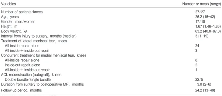
meniscal sizing technique in transverse, sagittal, and coronal views. A reference (Ref) line was created vertical to the posterior condylar line, on the trans- verse image with the LM. Anteroposterior edges of the LM were determined using the Ref line and sagit- tal and coronal views on the transverse image. The distance from the anterior to the posterior point on the Ref line was measured as the LML (Fig. 1) [22].
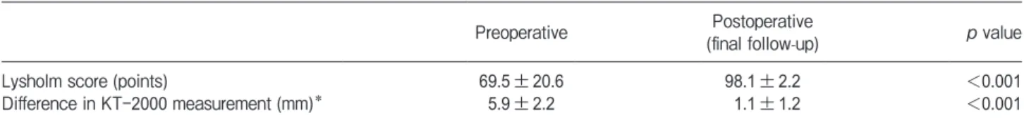
The LMW was measured from the outer border of the LM to the lateral edge of the lateral intercondylar eminence, parallel to the articular surface of the tibia, on the coronal image that crossed the center of the LML (Fig. 2). The LMBW was measured from the inner border of the LM to the outer border of the LM, and the LME was measured from the lateral margin of the tibial plateau to the outer border of the LM (Fig. 2). The LMH was measured from the inferior margin to the superior margin of the LM on the same coronal image.
The percentage of the LML to each LTP length
(i.e., the LML percentage) and the percentage of the LMW to each LTP width (the LMW percentage) were
PC line
Ref line C
D
F B
C F E
D
PC line
B E
A B C
E F
D
Fig. 1 MRI-based measurement of the LM length. Transverse (A, B), sagittal (C, E), and coronal (D, F) views. (A) A posterior condy- lar (PC) line of the femur. (B) A reference (Ref) line (dotted line) was created vertical to the PC line at the center of the LM width. The anterior edge of the LM was determined by the Ref line, sagittal (C), and coronal (D) views. The posterior margin of the LM was deter- mined on the Ref line by the sagittal (E) and coronal (F) images. The distance from the anterior to the posterior point (double-headed arrow) was measured as the LM length (LML).
LMW
LMH
LME LMBW
Fig. 2 MRI-based measurements of the lateral meniscus. A coronal image of an ACL-reconstructed knee (the right knee).
calculated [22]. Three orthopedic surgeons (NK, YK, and TT) retrospectively reviewed the radiographic images and MRI scans in a blinded manner. To test the intra- and inter-observer reliabilities, we calculated the intra- and inter-class correlation coefficients (ICCs).
The mean value of each observerʼs measurement was obtained. We compared the preoperative versus post- operative LML, LMW, LMBW, LME, LMH, LML percentage, and LMW percentage measurements.
Statistical analysis. The measurements and clinical values at the preoperative and postoperative examinations were assessed using the Wilcoxon signed-rank test. Power and statistical analyses were performed using EZR (Saitama Medical Center), which is a graphical user interface for R (The R Foundation for Statistical Computing). Data are pre- sented as the mean±standard deviation (SD). Signifi- cance was set at p<0.05.
Results
The short-term clinical evaluation. ACL reconstruction improved the patientsʼ knee-associated symptoms. No physical signs such as knee-joint swell- ing, joint-line tenderness, or locking were observed at the final follow-up evaluations. The median Lysholm score was 69.5 (range 44-73) before ACL reconstruc- tion and improved to 98.1 (range 95-100) at the final follow-up (Table 2). The mean side-to-side distance in the KT-2000 measurement was 5.9±2.2 mm (range 4-9 mm) before ACL reconstruction and decreased to 1.1±1.2 mm (range 0-3 mm) at the final follow-up examination (Table 2). Significant differences between preoperative and postoperative values were observed in these parameters (p<0.001, Table 2).
Physical features and radiographic measure- ment of the LTP. The patientsʼ mean height was 1.67±0.09 m (range 1.46-1.83 m), and their mean body weight was 63.2±11.5 kg (range 40.0-87.0 kg). The
radiograph-based LTP length and width were 38.7± 4.9 mm and 32.7±3.7 mm, respectively.
MRI-based measurement of the LM. The LML increased significantly from 33.4±3.8 mm to 33.8±4.0 mm after the lateral meniscal repair associ- ated with ACL reconstruction (p=0.048, Table 3).
The LMW increased significantly from 29.1±3.1 mm to 29.8±2.6 mm after the surgery (p=0.001, Table 3).
The LME changed significantly from 0.5±0.8 mm to 1.4±1.0 mm (p<0.001, Table 3). The LML percentage increased from 86.8±6.7 to 88.7±7.9 postopera- tively (p=0.049, Table 3). In addition, the LMW percentage increased significantly from 89.5±6.6 to 92.0±5.7 postoperatively (p<0.001, Table 3). No significant differences between the preoperative and postoperative measurements were observed in the LMBW (p=0.097) or LMH (p=0.193, Table 3).
Effective statistical powers (>0.08) were obtained in the comparisons of each parameter of LM measure- ment. There were no significant differences in the preoperative LME between the men (17 cases, 0.3± 0.9 mm) and women (10 cases, 0.8±0.6 mm). The post- operative LME of the men (1.1±1.1 mm) was similar to that of the women (1.8±0.7 mm). Concurrent MM
Table 2 Clinical results
Preoperative Postoperative
(final follow-up) p value
Lysholm score (points) 69.5±20.6 98.1±2.2 <0.001
Difference in KT-2000 measurement (mm)* 5.9±2.2 1.1±1.2 <0.001
Data are displayed as a mean±standard deviation. *Difference between the anterior cruciate ligament-injured knee and the contralateral normal knee in side-to-side distance using the KT-2000 arthrometer measurement at 134 N.
Table 3 Postoperative changes in the lateral meniscus mea- surements
Preoperative Postoperative p value
LML (mm) 33.4±3.8 33.8±4.0 0.048*
LMW (mm) 29.1±3.1 29.8±2.6 0.001*
LMBW (mm) 9.1±2.4 8.4±1.9 0.097
LME (mm) 0.5±0.8 1.4±1.0 <0.001*
LMH (mm) 6.3±1.1 6.4±1.4 0.193
LML percentage (%) 86.8±6.7 88.7±7.9 0.049* LMW percentage (%) 89.5±6.6 92.0±5.7 <0.001* Lateral meniscus length (LML), lateral meniscus width (LMW), lat- eral meniscus body width (LMBW), lateral meniscus extrusion (LME), lateral meniscus height (LMH). *p<0.05.
repair (11 cases) did not affect the increase of postop- erative LME. Both the intra- and inter-observer reli- abilities were excellent (ICC>0.91) in each measure- ment.
Second-look arthroscopic findings. Eighteen knees (67 ) among the 27 patients underwent a sec- ond-look arthroscopic evaluation at a mean of 14 months postoperatively. Of these patients, 14 (78 ) showed a complete healing of the repaired LM. One knee had an incomplete healing of the LM. The remaining 3 knees (17 ) needed additional treatments such as rasping and partial meniscectomy for an unhealed LM.
Discussion
Our present findings demonstrated that the LME increased significantly after lateral meniscal repair associated with ACL reconstruction (p<0.001, Table 3). In addition, the differences between the preopera- tive and postoperative measurements of the LMW and LMW percentage were higher than those of the LML and LML percentage (Table 3). Our results indicate that concurrent LM repair with ACL reconstruction might shift the LM laterally rather than posteriorly.
This may be caused by several factors such as menis- cal tear location, needle-penetrating direction, sutur- ing strength, leg position at suturing, suture number, the biomechanical condition of the LM, and iatrogenic injury of the LM anterior insertion during ACL reconstruction [32].
Arthroscopic all-inside repair (side-to-side repair using a suture hook) for an LM posterior root tear can reduce the LME in ACL-reconstructed knees [33].
On the other hand, meniscal treatments (inside-out repair and/or partial meniscectomy) for various types of LM tears increase radial displacement of the LM after ACL reconstruction [25]. In the present patient series, mainly the FasT-Fix all-inside suture device was used for the treatment of peripheral simple longi- tudinal tears of the LM. The FasT-Fix all-inside suture moves the meniscus to the peripheral joint capsule by tightening the meniscal body and the gap of the tear.
The inside-out suture technique may also induce the meniscal shift along with its suturing direction. A new suturing technique for stabilizing a full-thickness lon- gitudinal tear of the LM may be required in order not
to increase the LME. Both MM and LM translate posteriorly on the tibial plateau during knee flexion.
The posterior translation of the LM at 134° of knee flexion is greater than that of the MM [34]. An LM repair is often performed using the figure-of-four knee-flexed position [35]. It is thus possible that the repaired LM might shift to the posterolateral location by meniscal suturing at a knee-flexed position. Moreover, the tibia is over-constrained (posterior displacement and external rotation) after ACL reconstruction [36].
In the present study, a total of 50 N of initial ten- sion was applied for the graft fixation in anatomic double-bundle ACL reconstruction. Graft tensioning may also affect the postoperative LM shift by altering the tibial rotation. Several reports have indicated a considerable overlap between the ACL tibial footprint and LM anterior insertion on the tibial surface [13,37]. Bone tunnel reaming in the center of the ACL tibial footprint causes a decrease in the attach- ment area and ultimate strength of the LM anterior insertion [38]. An inappropriate tibial bone tunnel creation may induce a postoperative increase of the LME by damaging the LM anterior insertion [13,32].
In addition, complete radial tears of the LM produce an increase in the mean contact pressure of the knee [39]. We speculate that a postoperative increase of the LME may be caused by a mix of these factors.
Additional follow-up MRI scans in a larger sample size will be required to evaluate the relationship between the LME increase and post-traumatic knee OA.
Lateral meniscal repair with ACL reconstruction achieves high healing outcomes (89 ) [40]. Even when the LM tears remained at the time of ACL reconstruction, healing occurred in 55-74 of cases [41]. In addition, small peripheral LM tears left untreated at the time of ACL reconstruction had a low reoperation rate (2.2 ) during a minimum 6-year fol- low-up period [42]. However, the length and depth of meniscal tears were not discussed in the above studies.
In our present study, the mean length of the LM tears was 14.0 mm (range, 10-25 mm) and the full-thickness LM tears required meniscal repairs with a mean of 1.9 sutures (range, 1-4 sutures). The Lysholm score improved after concurrent LM repair with ACL reconstruction (Table 2).
There were no postoperative clinical symptoms for the repaired LM during the follow-up period. However, several remaining tears of the LM were observed at
second-look arthroscopies. We suspect that the heal- ing status of the injured LM would be affected by many factors such as the ACL reconstruction procedure, the graft tension, knee instability, the meniscal suture device, and meniscal position. Further studies are needed to optimize the treatment for lateral meniscal tears associated with an ACL injury.
This study has several limitations. This was a ret- rospective comparative study with a small sample size and short-term clinical outcomes. We evaluated the MRI-based lateral meniscal length, width, body width, extrusion, and height in a single knee flexion angle (10°) under a non-weight-bearing condition using 3-mm slice thickness. Knee motion-related differences between weight-bearing and non-weight-bearing conditions in the LM movement have been reported [43]. The postero- lateral movement of the LM in a weight-bearing condi- tion is larger than that in non-weight-bearing condition [43]. However, the relationship between ACL recon- struction and postoperative changes in LM movement remains unclear. To understand the clinical effects of the postoperative LM shift, open MRI assessments of meniscal movement using thin slices in several knee flexion angles under loading conditions are required.
In addition, three-dimensional reconstruction of the LM using dynamic-kinetic MRI may be useful to ana- lyze postoperative changes in the LM position and morphology.
In conclusion, a postoperative increase in the radial extrusion of the LM (LME) was observed after con- current LM repair with ACL reconstruction. The post-traumatic transposition of the LM may not be completely prevented by LM repair concomitant with ACL reconstruction.
Acknowledgments. We thank Drs. Shinichi Miyazawa and Hiroto Inoue for their cooperation. This work was supported by the Japanese Foundation for Research and Promotion of Endoscopy.
References
1. Jeong HJ, Lee SH and Ko CS: Meniscectomy. Knee Surg Relat Res (2012) 24:129-136.
2. Biedert RM: Treatment of intrasubstance meniscal lesions: a ran- domized prospective study of four different methods. Knee Surg Sports Traumatol Arthrosc (2000) 8: 104-108.
3. Fairbank TJ: Knee joint changes after meniscectomy. J Bone Joint Surg Br (1948) 30B: 664-670.
4. Jørgensen U, Sonne-Holm S, Lauridsen F and Rosenklint A: Long- term follow-up of meniscectomy in athletes. A prospective longitu-
dinal study. J Bone Joint Surg Br (1987) 69: 80-83.
5. Beveridge JE, Shrive NG and Frank CB: Meniscectomy causes significant in vivo kinematic changes and mechanically induced focal chondral lesions in a sheep model. J Orthop Res (2011) 29:
1397-1405.
6. Messner K and Gao J: The menisci of the knee joint. Anatomical and functional characteristics, and a rationale for clinical treatment.
J Anat (1998) 193: 161-178.
7. Turman KA and Diduch DR: Meniscal repair: indications and techniques. J Knee Surg (2008) 21:154-162.
8. Stein T, Mehling AP, Welsch F, von Eisenhart-Rothe R and Jager A: Long-term outcome after arthroscopic meniscal repair versus arthroscopic partial meniscectomy for traumatic meniscal tears.
Am J Sports Med (2010) 38:1542-1548.
9. Englund M and Lohmander LS: Risk factors for symptomatic knee osteoarthritis fifteen to twenty-two years after meniscectomy.
Arthritis Rheum (2004) 50: 2811-2819.
10. Mezhov V, Teichtahl AJ, Strasser R, Wluka AE and Cicuttini FM:
Meniscal pathology-the evidence for treatment. Arthritis Res Ther (2014) 16: 206.
11. Walter RP, Dhadwal AS, Schranz P and Mandalia V: The out- come of all-inside meniscal repair with relation to previous anterior cruciate ligament reconstruction. Knee (2014) 21: 1156-1159.
12. Fu FH, van Eck CF, Tashman S, Irrgang JJ and Moreland MS:
Anatomic anterior cruciate ligament reconstruction: a changing Paradigm. Knee Surg Sports Traumatol Arthrosc (2014) 23: 640- 13. Furumatsu T, Kodama Y, Maehara A, Miyazawa S, Fujii M, 648.
Tanaka T, Inoue H and Ozaki T: The anterior cruciate liga- ment-lateral meniscus complex: a histological study. Connect Tissue Res (2016) 57: 91-98.
14. Hussein M, van Eck CF, Cretnik A, Dinevski D and Fu FH:
Prospective randomized clinical evaluation of conventional sin- gle-bundle, anatomic single-bundle, and anatomic double-bundle anterior cruciate ligament reconstruction: 281 cases with 3- to 5-year follow-up. Am J Sports Med (2012) 40: 512-520.
15. Narazaki S, Furumatsu T, Tanaka T, Fujii M, Miyazawa S, Inoue H, Shimamura Y, Saiga K and Ozaki T: Postoperative change in the length and extrusion of the medial meniscus after anterior cru- ciate ligament reconstruction. Int Orthop (2015) 39: 2481-2487.
16. Leiter JR, Gourlay R, McRae S, de Korompay N and MacDonald PB: Long-term follow-up of ACL reconstruction with hamstring autograft. Knee Surg Sports Traumatol Arthrosc (2014) 22: 1061- 1069.
17. Ferretti A, Conteduca F, De Carli A, Fontana M and Mariani PP:
Osteoarthritis of the knee after ACL reconstruction. Int Orthop (1991) 15: 367-371.
18. Daniel DM, Stone ML, Dobson BE, Fithian DC, Rossman DJ and Kaufman KR: Fate of the ACL injured patient. A prospective out- come study. Am J Sports Med (1994) 22:632-644.
19. Nakata K, Shino K, Horibe S, Tanaka Y, Toritsuka Y, Nakamura N, Koyanagi M and Yoshikawa H: Arthroscopic anterior cruciate liga- ment reconstruction using fresh-frozen bone plug-free allogeneic tendons: 10-year follow-up. Arthroscopy (2008) 24:285-291.
20. Westermann RW, Wright RW, Spindler KP, Huston LJ, MOON Knee Group and Wolf BR: Meniscal repair with concurrent anterior cruciate ligament reconstruction: operative success and patient outcomes at 6-year follow-up. Am J Sports Med (2014) 42: 2184- 2192.
21. Potter HG, Jain SK, Ma Y, Black BR, Fung S and Lyman S: Cartilage injury after acute, isolated anterior cruciate ligament tear. Am J
Sports Med (2012) 40:276-285.
22. Furumatsu T, Miyazawa S, Tanaka T, Okada Y, Fujii M and Ozaki T: Postoperative change in medial meniscal length in con- current all-inside meniscus repair with anterior cruciate ligament reconstruction. Int Orthop (2014) 38:1393-1399.
23. Gale DR, Chaisson CE, Totterman SM, Schwartz RK, Gale ME and Felson D: Meniscal subluxation: association with osteoarthri- tis and joint space Narrowing. Osteoarthritis Cartilage (1999) 7:
526-532.
24. Wenger A, Wirth W, Hudelmaier M, Noebauer-Huhmann I, Trattnig S, Bloecker K, Frobell RB, Kwoh CK, Eckstein F and Englund M: Meniscus body position, size, and shape in persons with and persons without radiographic knee osteoarthritis. Arthritis Rheum (2013) 65:1804-1811.
25. Ichiba A and Makuya K: Radial displacement of the lateral menis- cus before and after anterior cruciate ligament reconstruction:
Arch Orthop Trauma Surg (2012) 132: 321-327.
26. Crema MD, Roemer FW, Felson DT, Englund M, Wang K, Jarraya M, Nevitt MC, Marra MD, Torner JC, Lewis CE and Guermazi A: Factors associated with meniscal extrusion in knees with or at risk for osteoarthritis: the Multicenter Osteoarthritis study. Radiology (2012) 264:494-503.
27. Stärke C, Kopf S, Petersen W and Becker R: Current concepts meniscal repair. Arthroscopy (2009) 25: 1033-1044.
28. Lysholm J and Gillquist J: Evaluation of knee ligament surgery results with special emphasis on use of a scoring scale. Am J Sports Med (1982) 10:150-154.
29. McDermott ID, Sharifi F, Bull AM, Gupte CM, Thomas RW and Amis AA: An anatomical study of meniscal allograft sizing. Knee Surg Sports Traumatol Arthrosc (2004) 12:130-135.
30. Sakata K, Furumatsu T, Abe N, Miyazawa S, Sakoma Y and Ozaki T: Histological analysis of failed cartilage repair after mar- row stimulation for the treatment of large cartilage defect in medial compartmental osteoarthritis of the knee. Acta Med Okayama (2013) 67:65-74.
31. Sakata K, Furumatsu T, Miyazawa S, Okada Y, Fujii M and Ozaki T: Comparison between normal and loose fragment chon- drocytes in proliferation and redifferentiation potential. Int Orthop
(2013) 37: 159-165.
32. Furumatsu T and Ozaki T: Iatrogenic injury of the lateral meniscus anterior insertion following anterior cruciate ligament reconstruc- tion: a case report. J Orthop Sci (2016) doi: 10.1016/j.jos.2016.
04.016.
33. Yao J, Lancianese SL, Hovinga KR, Lee J and Lerner AL:
Magnetic resonance image analysis of meniscal translation and tibio-menisco-femoral contact in deep knee flexion. J Orthop Res (2008) 26: 673-684.
34. Nishimura A, Fukuda A, Kato K, Fujisawa K, Uchida A and Sudo A:
Vascular safety during arthroscopic all-inside meniscus suture.
Knee Surg Sports Traumatol Arthrosc (2015) 23: 975-980.
35. Matsuo T, Mae T, Shino K, Kita K, Tachibana Y, Sugamoto K, Yoshikawa H and Nakata K: Tibiofemoral relationship following anatomic triple-bundle anterior cruciate ligament reconstruction.
Knee Surg Sports Traumatol Arthrosc (2014) 22: 2128-2135.
36. Ellman MB, LaPrade CM, Smith SD, Rasmussen MT, Engebretsen L, Wijdicks CA and LaPrade RF: Structural proper- ties of the meniscal roots. Am J Sports Med (2014) 42: 1881- 1887.
37. LaPrade CM, Smith SD, Rasmussen MT, Hamming MG, Wijdicks CA, Engebretsen L, Feagin JA and LaPrade RF: Consequences of tibial tunnel reaming on the meniscal roots during cruciate liga- ment reconstruction in a cadaveric model, Part 1: The anterior cruciate ligament. Am J Sports Med (2015) 43: 200-206.
38. Feng H, Hong L, Geng XS, Zhang H, Wang XS and Jiang XY:
Second-look arthroscopic evaluation of bucket-handle meniscus tear repairs with anterior cruciate ligament reconstruction: 67 con- secutive cases. Arthroscopy (2008) 24:1358-1366.
39. Pujol N and Beaufils P: Healing results of meniscal tears left in situ during anterior cruciate ligament reconstruction: a review of clinical studies. Knee Surg Sports Traumatol Arthrosc (2009) 17:
396-401.
40. Duchman KR, Westermann RW, Spindler KP, Reinke EK, Huston LJ, Amendola A, MOON Knee Group and Wolf BR: The fate of meniscus tears left in situ at the time of anterior cruciate ligament reconstruction: a 6-year follow-up study from the MOON cohort.
Am J Sports Med (2015) 43:2688-2695.