INTRODUCTION
Pancreatic cancer has been the most lethal disease of whole alimentary tract carcinomas in Japan (1). This is due to the high frequency of recurrence in the retrop-eritoneal space even after radical resection for pan-creatic carcinoma. Cancer cells easily spread to the retroperitoneal space even in case of early stage of pancreatic carcinoma. Metastasis has often occurred in the liver, and the paraaortic lymph nodes.
The reason for the high frequency of recurrence in the retroperitoneal space is that residual cancer cells remain in the pancreatic bed. These are not detected microscopically at the time of operation. Also the tumor
cells grow in to recurrence lesions in the retroperitoneal space, we have often found tumors to be widespread there.
It has been speculated that the pancreatic paren-chyma connects to the retroperitoneal space with the rich network of lymph vessels. Sometimes it has been shown that tumors of the pancreatic head invade the pancreatic duct and cause obstruction of the pancreatic duct.
We made a model of pancreatic duct obstruction in the pigs, and evaluated the lymph system changes under the special condition like pancreatic duct obstruction.
MATERIALS AND METHODS
Animals : Seven male pigs (19-40 kg body weight) were prepared for experimental animals. After all, six male pigs were used as experimental animals except one had died of intestinal obstruction. They
ORIGINAL
Changes of lymphatic flow in case of pancreatic duct obstruction
in the pig -as a model of pancreatic
cancer-Miho Kurahashi, Hidenori Miyake, Toshihide Takagi, and Seiki Tashiro
Department of Digestive and Pediatric Surgery, The University of Tokushima School of Medicine, Tokushima, Japan
Abstract : To investigate the lymph network change in pancreatic duct obstruction in pigs as a model of pancreatic cancer invading pancreatic duct, six domestic pigs weighing 17-40 kg underwent surgery as protocol. Two of them were controls, and the others underwent ligation of the pancreatic duct as a model of ductal obstruction. A CH 40 and lipiodol mixture was injected in their pancreas at 7 or 21 Days after first operation. Radiographic examination had been also performed. Five or 14 days later, they were examined radiographically, and sacrificed for histological examination. Ligation of the pancreatic duct caused experimental pancreatitis. Dilatation of the pancreatic duct and dilatation of the lymph canal in the interlobular space of pancreas was demonstrated in the ligation group. CH 40 and lipiodol showed discrepancies in the distribution. There were not distinct differences between the two groups in a route of CH 40 traveling.
Only fluoroscopic examination revealed an image of lipiodol enlargement to the caudal site in the ligated group. The congestive lymph system may have impaired flow like reflux. J. Med. Invest. 51 : 70-75, February, 2004
Keywords : lymph system, pig, obstruction, pancreatic duct, pancreatic cancer
Received for publication December 1, 2003 ; accepted December 25, 2003.
Address correspondence and reprint requests to Miho Kurahashi, M.D., Department of Digestive and Pediatric Surgery, The University of Tokushima School of Medicine, Kuramoto-cho, Tokushima, 770-8503, Japan and Fax : +81-88-631-9698.
The Journal of Medical Investigation Vol. 51 2004
were all AWAPORK, and fed nothing orally except water the day before operation.
Anesthesia. Anesthesia was induced with ketamine hydrochloride (10mg/kg) and atropin sulfate (0.01∼ 0.02 mg/kg) with intramuscular injection. The sedated pig was given bromo vecronium as a muscle relaxant and thiamilal intravenously. After positioning of en-dotracheal tube, a ventilator was connected to the tube for delivering 35% oxygen. Anesthesia was maintained with a low dose of bromo vecronium and pentobarbital intravenously.
Operation.Six pigs underwent laparotomy. Three of the six pigs underwent a second laparotomy instead of sacrifice. The pigs received an upper midline skin incision following a right subcostal skin incision .
The model of pancreatic duct obstruction was made according to the method of Waterworth et al. (2). The pancreatic duct entered the second part of duodenum at the end point of the pancreatoduodenal attachment (3). The common bile duct was independent of the pancreatic duct, it entered the upper duodenum near the pyloric ring. Duodenotomy was performed at the opposite site of the pancreatic duct orifice. The orifice was exposed and a purse string suture was performed to ligate the pancreatic duct two or three times with 5-0 non-absorbable monofilament nylon. Duodenotomy was repaired with a longitudinal incision to the hori-zontal suture, without making a narrow lumen of the duodenum. The peritoneum and abdominal wall were repaired with absorbable thread. Skin was fixed with a metal stapler. The surgical wound was covered with surgical skin aerosol. All the procedures were performed under aseptic conditions. After the operation, the pigs were fed nothing orally for 12 hours. Drip infusion was continued via the ear vein until it was spontaneously disconnected. (When the pigs stood up and walked around, the infusion line was disconnected.) Optimum temperature was maintained on the pig floor in the animal laboratory of University of Tokushima, especially after the operation. Antibiotic agents and H2-blocker
were injected on the operative day only.
LIPIODOL. Lipiodol Ultra-Fluid is ethylester of iodinated poppy-seed oil fatty acids. It is used for lym-phangiography in clinical conditions. Lipiodol and CH 40 were mixed just before injection, and it was injected manually to the pancreatic body at a rate of 0.5ml per minute.
CH40. CH40 is activated carbon particles. It is taken up selectively by the lymphatic system and regional lymph nodes. The route of CH40 travel was stained black. The lymph nodes containing CH40 were black-ened.
Experimental Protocol
Group 1. No obstruction of pancreatic duct
Two of the six pigs which did not have pancreatic duct obstruction were injected with the agent at the initial operation (Table 1). They did not undergo surgery of ligation of the pancreatic duct, and were designated no-ligation group. Case A was underwent injection with lipiodol only. The lipiodol was injected at pancreatic body just below the pancreatic capsule. Case A was received radiography at Day 7, 25, and 205. It was sac-rificed, and the organs were taken. An other pig, Case B also underwent injection with CH40 and lipiodol at the same site of case A. Case B received radiography one and two weeks after the operation, was sacrificed, and the organs were taken.
Group 2. Pancreatic duct obstruction
Four pigs, Case C,D, E, F underwent surgery as models of pancreatic duct obstruction. Case C was sacrificed at Day 7 after ligation for the purpose of examination whether pancreatic duct dilated or not under the pancreatic duct ligation. Case D was per-formed ligation, and seven days after, been injected with a mixture of CH40 and lipiodol. Case E and F were underwent a second surgery three weeks after the first operation. At that time, they were injected with a mix-ture of CH 40 and lipiodol and to the pancreatic
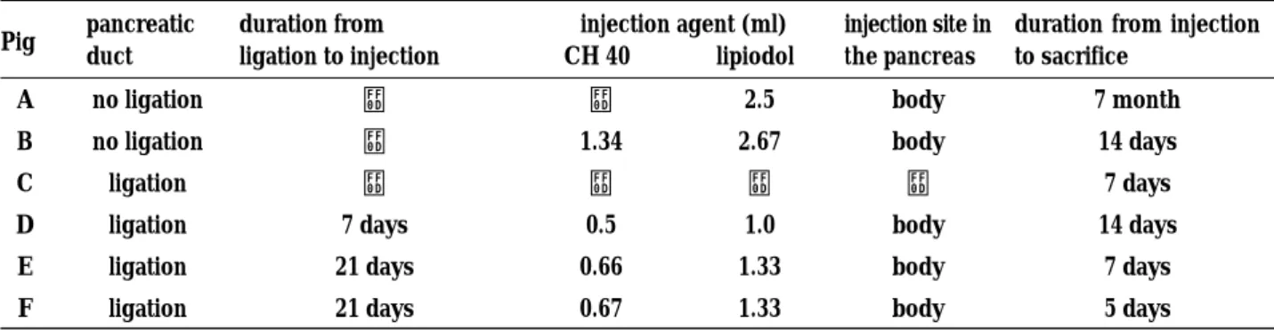
pa-Table 1. Study protocol of six pigs
Pig pancreatic duct duration from ligation to injection injection agent (ml) CH 40 lipiodol injection site in the pancreas
duration from injection to sacrifice
A no ligation − − 2.5 body 7 month
B no ligation − 1.34 2.67 body 14 days
C ligation − − − − 7 days
D ligation 7 days 0.5 1.0 body 14 days
E ligation 21 days 0.66 1.33 body 7 days
F ligation 21 days 0.67 1.33 body 5 days
renchyma. They were sacrificed at the almost sev-enth day, and consecutive extirpation was performed to take the abdominal organs.
EXTIRPATIONANDHISTOLOGICALEXAMINATION
At necropsy, for the examination of the pancreatic lymph network, not only the pancreas but also the nearby organs and additional tissue were extirpated en masse. The upper margin was the caval vein at the subphrenic level. The oral surgical margin was at the upper duodenum just below the common bile duct entrance. The portal vein was ligated at the same level. The bilateral kidney, adrenal glands and spleen were included in the resected block, and the anal margin was the small intestine at the level of the common iliac vein. The lower margin was the common iliac vein. The resected block was fixed in 10% formalin, embeded in paraffin, and stained with hematoxylin and eosin.RADIOGRAPHIC EXAMINATION
For the tracing of Lipiodol, the pigs underwent ra-diographic examination using ketamine hydroxychloride and atropin sulfate.
RESULTS
Group 1. No obstruction of the pancreatic duct
Case A was from the control group, and underwent aseptic laparotomy. The pig received the injection of the lipiodol only in the pancreas. At the seventh day after operation, the sedated pig was examined radiographically. The opacity in the abdomen was due to the lipiodol. Radiographic examination was again performed at the eighteenth day after opera-tion. The shadow of the abdomen changed in shape but had not moved very far. It seemed that the change
of shape was only due to a difference in intestinal fullness. The next radiographic examination was per-formed six months later. The opacity of the lipiodol had not moved but had become hazy.
Case B received the injection of the lipiodol-CH40 mixture in the pancreas. The seven days after operation, the pig was examined radiographically. The opacity in the abdomen had spread. However, the opacity had not moved to farther lymph nodes. Another seven days later, another exploration was performed. The radiography revealed that the shape of the shadow had enlarged slightly. After sacrifice, histological find-ings revealed that the CH40 had flowed to the paraaortic adjacent tissue at the level of the phrenic muscle and the pancreas top. However, the paraaortic lymph system did not contain CH40.There was no CH40 in the caudal paraaortic lymph nodes. The most caudal slice with CH 40 was at the pancreas end and regional fatty tissue. The slice consisted of adrenal gland, kidney, phrenic muscle, and pancreas. A particle of lipiodol was detected in the peripancreatic fatty tissue. It had not moved to any lymph nodes histologically (Table 2).
Group 2. Pancreatic duct obstruction
All pigs with pancreatic duct obstruction developed microscopic changes of acute pancreatitis at the sec-ond operation. The characteristic findings were“aciner cell homogenization”pathologically. The pancreatic duct was dilated, and the parenchyma became atro-phic, and showed inflammatory reactions, with plasma cells, lymphocytes and eosinophils. Infiltration of plasma cells, lymphocytes and eosinophils was shown as a mixture in the acini,“homogenization”. Fibrosis and proliferation of the interlobular connective tissue was also shown. Hemorrhage was slightly detected in the fatty tissue. The perilobular space was changed edematously.
Ligation of the pancreatic duct obviously caused experimental pancreatitis, and it was also considered the obstruction due to pancreatic tumor invasion. Case C underwent ligation of pancreatic duct, showed
dila-Table2. The distribution of the CH 40
Lymph node Connective tissue
pig peripancreatic paraaortic peripancreatic interlobular paraaortic
supra infra supra infra supra infra
B + + − − + + + −
D − − − − + − − −
E + − − + + + + −
F + − − − + + − −
M. Kurahashi et al. Lymphatic flow in case of pancreatic cancer 72
tation of pancreatic ducts, fibrosis, and inflammatic changes histologically.
Case D received the injection at the seventh day after the first operation of ligation of the pancreatic duct. The pancreas was reddish, but not seriously. The pig was sacrificed two weeks after the second operation. Histological findings revealed that CH40 existed only in the pancreatic connective tissue. The lymph system showed dilatation in the pancreas. CH 40 was not found in the peripancreatic lymph node nor the paraaortic lymph nodes. However, colored lymph nodes were macroscopically distinct, the picric acid process revealed the colored region with a for-malin stain not CH40. Radiographic examination of the pig revealed the opacity in the pancreas. The opacity was due to accumulation of lipiodol, though histological findings failed to detect the lipiodol. The radiographic examination after the second operation demonstrated that the lipiodol had flowed into the dilated pancreatic duct. As the experimental pancreatitis was not severe, the lipiodol may have moved to the duodenal lumen by the papilla passage. We speculated that the CH40 was also flushed out during the two weeks after the operation.
Case E and F received the injection at three weeks after the first operation. At the second operation, the
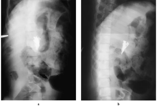
pancreas showed redness but not seriously. After successful injection, Case E was treated as the other pigs and sacrificed at the 7 days after the operation. The radiographic examination revealed that the lipiodol shadow of Case E was not changed. However, Case F, which was sacrificed at the 5 days after the second operation revealed that the lipiodol shadow was the largest in the series (Fig.1, 2). The shape of lipiodol had progressed toward even caudal site during three weeks. It was contrary to customed expectations. Histological findings of Case F revealed that CH 40 had spread into many lymph nodes and the interlobular space of the pancreas. The most cranial lymph node which contained CH40 was at the level of the pancreas top. There was phrenic muscle and abdominal aorta in the same slice. The CH40 was found in the interlobular space of the pancreas, and in the dilated lymph canal in the interlobular space. The most caudal lymph node with CH40 was at the level of the kidney, adrenal gland, and pancreas end. Lipiodol was not found in any speci-men histologically. Case E had found CH40 in the peri-pancreatic fatty tissue, periperi-pancreatic lymph nodes, and interlobular space of the pancreas histologically.
a b
Figure 1. The fluoroscopy showed the lipiodol image at the time of injection to case G.
DISCUSSION
There have been no studies of the lymph system of the pancreas under pancreatic duct obstruction. We made a model of pancreatic duct obstruction as a model of pancreatic cancer invasion. Clinically, we experienced that the contrast medium injected into the pancreas spread to the retroperitoneal space widely. One case had advanced cancer of the pancreatic head with obstruction of the pancreatic duct, with progress of recurrence to the retroperitoneal space thereafter.
Therefore, a hypothesis of the existence of a lymph network between the retroperitoneal space and the pancreas in man has been built. We speculated that the obstruction of pancreatic duct caused lymph system change, cancer cells might have spread to unexpected region. Our model of pancreatic duct obstruction as-sumed that the cancer of the pancreatic head had in-vaded the pancreatic duct. Pancreatic cancer commonly originates from the epidermal layer in the pancreatic duct, so obstructed the pancreatic duct. In general, a lymph flow has been gathered into lymph node and gone to more proximal lymph system. The most im-portant fact is that a lymph flow always goes toward proximately. Only Case F had shown a change of lymph system in the study with lipiodol. The shape of lipiodol of Case F enlarged not only upward but also downward, to caudal site. However, the results of dis-tribution of the CH40 was not unexpected. The CH40 was injected in the subcapsular space of the pancreas, flowed into a small lymph canal of interlobular space of the pancreas, moved to regional lymph nodes and reached to more proximal lymph nodes. It seemed that CH40 travel an ordinary route. Even if some were injected in peripancreatic connective tissue, they trav-eled in the loose tissue like paraaortic connective tissue in the pigs.
The discrepancy of the CH40 distribution and lipiodol location was detected in the all of the pigs. The reason
a b
Figure 2. The fluoroscopy showed the enlargement of the lipiodol image 5days after injection in the Case G. The image was enlarged upward and downward.
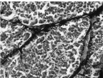
Figure 3. The CH40 was detected in the peripancreatic tissue, interlobular space, and lymph canal within the interlobular space. (H & E×200)
M. Kurahashi et al. Lymphatic flow in case of pancreatic cancer 74
for this was that CH40 had tendency to move to farther lymph nodes. In contrast, the lipiodol ultrafluid had nested in focus, as it had an affinity to oil. In this study, the histological examination could not detect lipiodol except in Case B. Macrophages with pigment granules were detected in some slices histologically. However, there was no evidence that the granules originated from lipiodol. The lipiodol ultrafluid was dissoloved by the ethanol, benzene and ether. The process of tissue staining could have effected the results. Morever, the lipiodol ultrafluid was produced for the purpose of lymphangiography. If it has not been injected into the lymph system, the lipiodol remains a lipids compound and is stored (4). Dogs which had received an injection of lipiodol to their lymph system demonstrated persis-tent images in their lymph nodes for one or two weeks radiographically (5). The lipiodol may not be a proper agent for the pancreas in this series. We can not tell whether the lipiodol succeeded in reaching the lymph canal in the pancreas, because the diameter of the par-ticle is not known. Only fluoroscopic exploration can show the enlargement of the lipiodol image moving in a caudal site object to ordinary flow. This shows the feasibility of lymph system change after pancre-atic duct ligation. The ligation can produce dilatation of the panceatic duct, and the high ductal pressure may affect the connective tissue in the interlobular space of the pancreas. The congestive lymph system may have impaired flow, like reflux. The duration of congestion did not show a difference in distribution of CH40. There are anatomical differences between pigs and man : the pancreas of the pig lies in the intes-tinal mesenterium, free from the retroperitoneum. The
orifice of the pancreatic duct is independent from the biliary system. The abdominal aorta is covered in mus-cle bundles which are concomitant to the diaphragm. The aorta and muscle bundles are separate, only con-nected with thin membranous connective tissue. Nev-erthless we can refer much anatomical differences man and pigs, the impaired flow like reflux in Case F might have occurred in man under the same condi-tion, like pancreatic duct obstruction due to cancer invasion.
REFERENCES
1. Miyazaki I, Nakagawa T : Approach to pancreas-tic Cancer. Kanehara & Co.,Ltd.,Tokyo, Japan, 1997, pp 5-18
2. Waterworth MW, Barbezart GO, Hickman R, Terblanche J : A controlled trial of glucagons in experimental pancreastitis. Br J Surg 63 : 617-620, 1976
3. Schroder T, Ramo OJ, Joffe SN : Laser Pncre-atectomy, a comparison between Dog and Pig. Res Exp Med 188 : 227-233, 1988
4. Viamote M, Soto M, Recher L : Chronic toxicity Study in dogs Following Intralymphatic injec-tions of ethiodol, ethiodol with Chlorophyl and Sterlie Non-Iodnized Poppy Seed Oil. Ind Med Surg35: 688-695, 1966
5. Schaffer B, Koehler PR, Daniel CR, Wohl GT, Rivera E, Meyers WA, Skelley JF :A critical-evaluation of Lymphangiography. Radiology 80 : 917-930, 1963