The effects of chronic testosterone administration on hypothalamic
gonadotropin-releasing hormone regulatory factors (Kiss1, NKB, pDyn, and RFRP) and their
receptors in female rats
Authors
Takeshi Iwasa, Toshiya Matsuzaki, Kiyohito Yano, Rie Yanagihara, Yiliyasi Mayila and Minoru Irahara
Department of Obstetrics and Gynecology, Institute of Biomedical Sciences, Tokushima University Graduate School, 3-18-15 Kuramoto-Cho, Tokushima 770-8503, Japan
Corresponding author
Takeshi Iwasa
Department of Obstetrics and Gynecology, Institute of Biomedical Sciences, Tokushima University Graduate School, 3-18-15 Kuramoto-Cho, Tokushima 770-8503, Japan Phone number: +81−88−633−7177
E-mail: [email protected]
Running title
Effects of testosterone injection
Keywords
This is an Accepted Manuscript of an article published by Taylor & Francis in Gynecological Endocrinology on 29/11/2017, available online: http://www.tandfonline.com/10.1080/09513590.2017.1409709.
testosterone, kisspeptin, RFRP, NKB, dynorphin
Abstract
The effects of androgens on gonadotropin-releasing hormone (GnRH) secretion in
females have not been fully established. To clarify the direct effects of androgens on hypothalamic reproductive factors, we evaluated the effects of chronic testosterone administration on hypothalamic GnRH regulatory factors in ovariectomized (OVX) female rats. Both testosterone and estradiol reduced the serum luteinizing hormone levels of OVX female rats, indicating that, as has been found for estrogen, testosterone suppresses GnRH secretion via negative feedback. Similarly, the administration of testosterone or estradiol suppressed the hypothalamic mRNA levels of kisspeptin and neurokinin B, both of which are positive regulators of GnRH, whereas it did not affect the hypothalamic mRNA levels of the kisspeptin receptor or neurokinin 3 receptor. On the contrary, the administration of testosterone, but not estradiol, suppressed the hypothalamic mRNA expression of prodynorphin, which is a negative regulator of GnRH. The administration of testosterone did not alter the rats’ serum estradiol levels, indicating that testosterone’s effects on hypothalamic factors might be induced by its androgenic activity. These findings suggest that as well as estrogen, androgens have negative feedback effects on GnRH in females and that the underlying mechanisms responsible for these effects are similar, but do not completely correspond, to the mechanisms underlying the effects of estrogen on GnRH.
Introduction
Reproductive functions are mainly regulated by the neuroendocrine system, which is known as the hypothalamic-pituitary-gonadal axis (HPG axis). Gonadotropin-releasing hormone (GnRH) functions as a central regulator of the HPG axis through its stimulatory effects on gonadotropin secretion. In the early 21st century, it was clarified that GnRH production/secretion is regulated by hypothalamic neuropeptides. Kisspeptin and its receptor (Kiss1r) act as positive regulators of GnRH, whereas RFamide-related peptides/gonadotropin inhibitory hormone (RFRP/GnIH) and its receptor G protein-coupled receptor (GPR)147 function as inhibitory regulators [1-3]. In addition, in 2007 it was shown that neurokinin B (NKB) and dynorphin (Dyn) co-localize with kisspeptin in the same neuronal populations, and therefore, these neurons are termed KNDy neurons [4,5]. NKB stimulates the activity of KNDy neurons, predominantly via its receptor tachykinin receptor 3 and increases GnRH secretion [6,7]. On the other hand, Dyn inhibits KNDy neurons and/or GnRH neurons via the kappa opioid receptor (KOR), which in turn reduces GnRH secretion [6,8]. Interestingly, although kisspeptin is found in two hypothalamic nuclei, the anteroventral periventricular nucleus (AVPV) and arcuate nucleus (ARC), its co-localization with NKB and Dyn is only observed in the ARC [6,9]. It has been well established that estrogen plays pivotal roles in the regulation of GnRH secretion via feedback effects on hypothalamic kisspeptin and RFRP/GnIH expression. Estrogen suppresses kisspeptin and NKB release from KNDy neurons in the
ARC [6], and Kiss1 (the kisspeptin encoding gene) and NKB mRNA expression are upregulated and prodynorphin (pDYN) mRNA expression is downregulated in postmenopausal women and ovariectomized animals [9,10-12]. In addition, the administration of estradiol was found to alter the activity or mRNA expression of RFRP/GnIH [13,14], although the direction of such changes varies depending on the experimental model. These changes might be related to the negative feedback effects of estrogen on GnRH. On the other hand, estrogen increases kisspeptin release in the AVPV, and the neuronal activity of RFRP/GnIH is decreased during the pre-ovulatory GnRH surge [6,15]. Therefore, these alterations are involved in the positive feedback effects of estrogen on GnRH.
In contrast to estrogen, the effects of androgens on GnRH secretion in females have not been fully established. Almost all of the previous studies that investigated the effects of androgens on female physiological functions used animal models of polycystic ovary syndrome (PCOS), which is a representative hyperandrogenism-related disorder [16]. Recently, we and other groups have investigated hypothalamic kisspeptin and/or Kiss1 mRNA levels, as well as serum gonadotropin levels, in PCOS model rodents; however, there were marked discrepancies between the results of these studies [17-19]. As ovary-intact rodents were used to produce the PCOS models, androgen-induced changes in ovarian functions might have affected their hypothalamic functions, which could explain the discrepancies among the studies’ results. Therefore, examinations performed under stable conditions are needed to clarify the effects of androgens on hypothalamic reproductive factors.
In this study, to determine the direct effects of androgens on hypothalamic reproductive factors, we evaluated the influence of chronic testosterone administration on hypothalamic GnRH regulatory factors under ovariectomized (OVX) conditions. We also assessed the effects of chronic estradiol administration on these factors, and compared them with testosterone’s effects. In addition, we measured the serum leptin level because it has been reported that leptin plays some roles in the regulation of hypothalamic GnRH regulatory factors and that the gonadal steroidal milieu affects the serum leptin level [20,21].
Materials and Methods
Animals
Eight-week-old female adult Wistar rats (200-230 g) were purchased from Charles River Laboratories Japan, Inc. (Kanagawa, Japan) and housed in a room under controlled light (12 h light, 12 h darkness; lights turned on at 0800 and turned off at 2000) and temperature (24°C) conditions. In total, 24 rats were used in this study, and all animal experiments were conducted in accordance with the ethical standards of the institutional animal care and use committee of the University of Tokushima. All surgical procedures were carried out under sodium pentobarbital- (60-80 mg/kg, intraperitoneal, i.p.) or sevoflurane-induced anesthesia.
Testosterone/estradiol administration and tissue sampling
At nine weeks of age, the rats were bilaterally OVX and housed individually after the surgery. Four weeks after the ovariectomy procedure (13 weeks of age), the rats were
randomly divided into untreated (OVX), testosterone-administered (OVX-T), and estradiol-administered (OVX-E) groups. In the estradiol-administered group, a silastic tube filled with crystalline estradiol (length of the filled part, 3 mm) was subcutaneously implanted into each rat [22]. Similarly, in the testosterone-administered group, a silastic tube filled with crystalline testosterone was subcutaneously implanted into each rat (length of the filled part, 30 mm) [23]. At 16 days after the implantation procedure, the rats were sacrificed by decapitation, and their brain and blood were collected. The rats’ serum was separated by centrifugation and stored at -20°C, and their tissues were stored at -80°C.
Hormone assay
The serum luteinizing hormone (LH) level was measured using a radioimmunoassay (rLH [I-125] RIA kit, Institute of Isotopes Co., Ltd., Tokyo, Japan). The sensitivity of the assay was 0.8 ng/ml, and the inter- and intra-assay coefficients of variation (CV) were 7.7% and 6.5%, respectively. The serum leptin level was measured using radioimmunoassay kits (multi-species leptin RIA kit, Linco Research Inc., MO, USA). The sensitivity of the assay was 1.0 ng/ml, and its inter- and intra-assay CV were 3.2% and 7.8%, respectively.
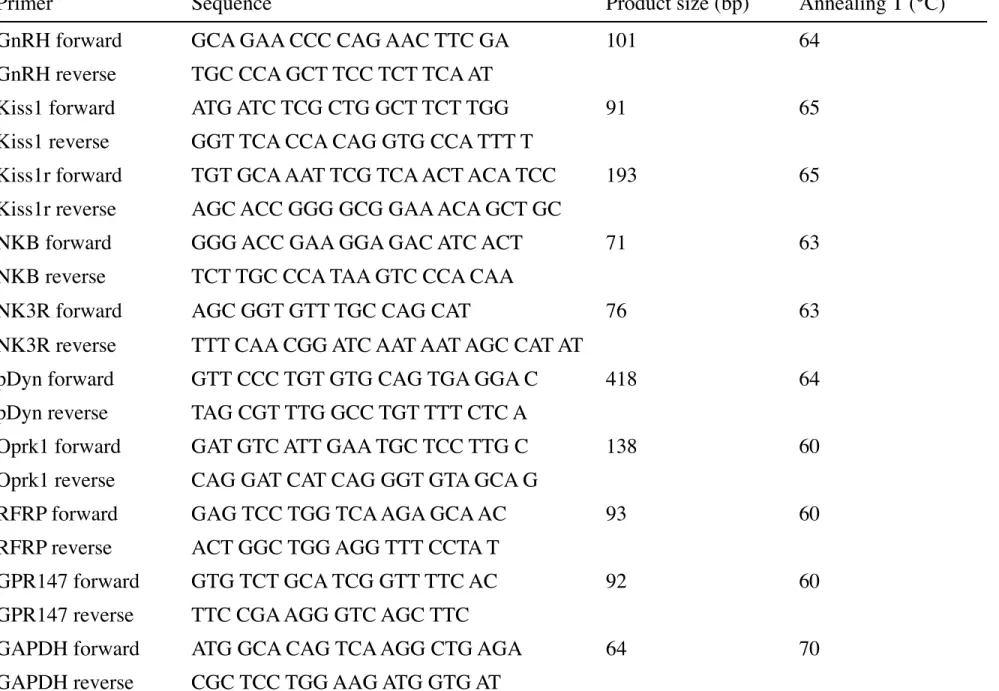
Quantitative real-time polymerase chain reaction
Whole hypothalamic explants were dissected from the frozen brains, as described previously [24]. Briefly, the brain sections were dissected out via an anterior coronal cut at the posterior border of the mammillary bodies, parasagittal cuts along the hypothalamic fissures, and a dorsal cut 2.5 mm from the ventral surface. Total RNA was isolated from
the hypothalamic explants and visceral fat using a TRIzol® reagent kit (Invitrogen Co., Carlsbad, CA, USA) and an RNeasy® mini kit (Qiagen GmbH, Hilden, Germany). cDNA was synthesized with oligo (deoxythymidine) primers at 50°C using the SuperScript III first-strand synthesis system for the real-time polymerase chain reaction (PCR; Invitrogen Co.). The PCR analysis was performed using the StepOnePlusTM real-time PCR system (PE Applied Biosystems, Foster City, CA, USA) and FAST SYBR® green. The hypothalamic mRNA levels of GnRH, Kiss1, Kiss1r, NKB, NK3R (the neurokinin 3 receptor encoding gene), pDyn, Oprk1 (the KOR encoding gene), RFRP/GnIH, and
GPR147 were measured. The mRNA expression level of each factor was normalized to
that of GAPDH. The primer sequences, product sizes, and annealing temperatures are shown in Table 1. The PCR conditions were as follows: initial denaturation and enzyme activation were performed at 95°C for 20 s, followed by 45 cycles of denaturation at 95°C for 3 s, and annealing and extension for 30 s.
Statistical analyses
All data are presented as the mean ± standard error (SE). The statistical analyses were performed using one-way analysis of variance (ANOVA) together with Dunnett’s test for multiple comparisons among the groups. Statistical significance was defined as P <0.05.
Results
Effects of testosterone/estradiol administration on serum LH and leptin levels
groups, and the serum estradiol level of the OVX-E group was higher than those of the other groups, indicating that the steroids were successfully administered (Figs. 1A and B). The rats’ serum LH levels were altered by the administration of the steroids (one-way ANOVA, F(2,23) = 76.7; P <0.01). The serum LH levels of the OVX-T and OVX-E groups were lower than that of the OVX group (Fig. 1C). Similarly, the rats’ serum leptin levels were changed by the administration of the steroids (one-way ANOVA, F(2,23) = 7.41; P <0.01). The serum leptin level of the OVX-E group was significantly lower than that of the OVX group (Fig. 1D).
Effects of testosterone/estradiol administration on the mRNA levels of hypothalamic
reproductive factors
The rats’ hypothalamic Kiss1 mRNA levels were altered by the administration of the steroids (one-way ANOVA, F(3,23) = 103.4; P <0.01). The hypothalamic Kiss1 mRNA levels of the OVX-T and OVX-E groups were lower than that of the OVX group (Fig. 2B). The administration of the steroids also changed the rats’ hypothalamic NKB mRNA levels (one-way ANOVA, F(2,23) = 15.6; P <0.01), whereas it did not affect their hypothalamic Kiss1r mRNA levels (Fig. 2C). The hypothalamic NKB mRNA levels of the OVX-T and OVX-E groups were lower than that of the OVX group (Fig. 2D). The administration of testosterone, but not estradiol, altered the rats’ hypothalamic pDYN mRNA levels (one-way ANOVA, F(2,23) = 6.93; P <0.01), but neither steroid affected their hypothalamic NK3R mRNA levels (Fig. 2E). The hypothalamic pDYN mRNA level of the OVX-T group was lower than that of the OVX group (Fig. 2G). The administration of estradiol, but not testosterone, altered the rats’ hypothalamic RFRP mRNA levels
(one-way ANOVA, F(2,23) = 8.09; P <0.01), whereas neither steroid affected their hypothalamic Oprk1 mRNA levels (Fig. 2H). The hypothalamic RFRP/GnIH mRNA level of the OVX-E group was lower than that of the OVX group (Fig. 2I). The rats’ hypothalamic GPR147 mRNA levels were not affected by the administration of the steroids (Fig. 2J).
Discussion
It has been well established that estrogen plays pivotal roles in the regulation of GnRH secretion via feedback effects on hypothalamic GnRH-regulating factors [6]. On the other hand, the effects of androgens on GnRH secretion and the mechanisms underlying these effects in females have not been fully established. Thus, we evaluated the effects of chronic testosterone, one of the most important androgens, administration on gonadotropin secretion and hypothalamic GnRH regulatory factors in the present study. As a result, we found that testosterone administration reduced the serum LH levels of OVX female rats, indicating that testosterone suppresses GnRH secretion via its negative feedback effects in females. As the serum estradiol level was not increased by the administration of testosterone, the suppressive effect of testosterone on GnRH/LH might be based on the androgenic effects of testosterone, rather than the estrogenic effects of estrogen produced from testosterone. As noted above, it has been reported that GnRH secretion is positively and negatively regulated by various hypothalamic factors [6,9-14] and that the feedback effects of sex steroid hormones on GnRH are mediated by these factors [6,13,15]. Therefore, we also examined the effects of testosterone administration
on these factors in order to investigate the underlying mechanisms responsible for their negative feedback effects on GnRH. As a result, we found that both testosterone and estradiol suppressed the hypothalamic mRNA levels of Kiss1 and NKB, whereas they did not affect the hypothalamic mRNA levels of Kiss1r or NK3R. As NKB simulates the activity of kisspeptin (KNDy) neurons and increases GnRH secretion, reductions in NKB and Kiss1 mRNA expression might be involved in the negative feedback effects of testosterone and estradiol on GnRH/LH seen in this study. Recently, it has been reported that hypothalamic kisspeptin neurons express the androgen receptor and that the numbers
of Kiss1-expressing and NKB-immunoreactive cells were decreased by
dihydrotestosterone, which is a non-aromatizable androgen, in female rats [25]. Therefore, it can be assumed that the suppression of NKB and Kiss1 mRNA expression observed in the testosterone-administered rats was induced by the androgenic actions of testosterone. On the contrary, the effects of testosterone on hypothalamic RFRP and pDyn mRNA expression did not correspond with those of estradiol in the current study. Testosterone did not affect RFRP mRNA expression, whereas estradiol decreased it. It has been reported that RFRP neurons express the estrogen receptor-α, but not the androgen receptor, in both male and female mice and that estradiol reduces the RFRP mRNA levels of OVX female mice [14]. On the other hand, as far as we know, the effects of testosterone on Dyn expression in females have not been examined, and the effects of estradiol on Dyn expression varied markedly among previous studies [12,26]. Some studies found that ovariectomy or the menopause downregulated pDyn mRNA expression, and others suggested that the administration of estradiol reduced the number of Dyn-expressing cells.
Differences in the experimental protocols, animal models, and doses of estradiol might have contributed to these discrepancies. We speculate that the estradiol and testosterone-induced reductions in the hypothalamic mRNA levels of RFRP and pDyn, respectively, observed in the present study might represent a counter-regulatory mechanism for preventing the excessive suppression of GnRH release via negative feedback effects. In this study, we also measured the serum levels of leptin, which is an anorexigenic adipocytokine, because it has been reported that leptin stimulates kisspeptin neurons and increases GnRH secretion in experimental animals [20]. Consequently, we found that the serum leptin level was reduced by the administration of estradiol, and this change might have been partially related to the reduction in the hypothalamic Kiss1 mRNA level. As noted above, the current study revealed that testosterone itself has suppressive effects on hypothalamic Kiss1 mRNA levels. On the other hand, there were marked discrepancies between the results of previous studies that examined the effects of androgens on hypothalamic kisspeptin and/or Kiss1 mRNA levels in PCOS model rodents [17-19]. We assumed that one of the causes of these discrepancies was the differences between animal models (species and age at androgen treatment) and the kinds of androgens administered (testosterone, dihydrotestosterone, or dehydroepiandrosterone). We also assumed that the interactions between endogenous estrogens and the administered androgens might have changed their effects on hypothalamic GnRH regulatory factors. Further examinations are needed to clarify the physiological roles of androgens in females under physiological hormonal conditions.
serum levels of LH, indicating that testosterone has negative feedback effects on GnRH in females. Both testosterone and estradiol suppressed hypothalamic Kiss1 and NKB mRNA expression, whereas they did not affect the hypothalamic mRNA levels of Kiss1r of NK3R. The administration of testosterone did not alter the serum estradiol level, indicating that testosterone’s effects on hypothalamic factors might be induced by its androgenic activity. On the contrary, the effects of testosterone on hypothalamic RFRP and pDyn mRNA expression did not correspond with those of estradiol. These findings indicate that the underlying mechanisms responsible for the negative feedback effects of testosterone on GnRH are similar, but do not completely correspond, to the mechanisms underlying the effects of estrogen.
Declaration of interest
The author declares no conflict of interest or financial support.
References
[1] Iwasa T, Matsuzaki T, Yano K, Irahara M. Gonadotropin-inhibitory hormone plays roles in stress-induced reproductive dysfunction. Front Endocrinol 2017;8:62 [2] Tsutsui K, Ubuka T. GnIH control of feeding and reproductive behaviors. Front
Endocrinol 2016;7:170
[3] Putteeraj M, Soga T, Ubuka T, Parhar IS. A “timed” kiss is essential for reproduction: lessons from mammalian studies. Front. Endocrinol. 2016;7:121.
[4] Cheng G, Coolen LM, Padmanabhan V, Goodman RL, Lehman MN. The kisspeptin/neurokinin B/dynorphin (KNDy) cell population of the arcuate nucleus: sex differences and effects of prenatal testosterone in sheep. Endocrinology 2010;151:301-311
[5] Goodman RL, Lehman MN, Smith JT, Coolen LM, de Oliveira CV, Jafarzadehshirazi MR, Pereira A, Iqbal J, Caraty A, Ciofi P, Clarke IJ. Kisspeptin neurons in the arcuate nucleus of the ewe express both dynorphin A and neurokinin B. Endocrinology 2007;148:5752-5760
[6] Skorupskaite K, George JT, Anderson RA. The kisspeptin-GnRH pathway in human reproductive health and disease. Hum Reprod Update 2014;20:485-500
[7] Navarro VM, Bosch MA, Leon S, Simavli S, True C, Pinilla L, Carroll RS, Seminara SB, Tena-Sempere M. The integrated hypothalamic tachykinin-kisspeptin system as a central coordinator for reproduction. Endocrinology 2015;156:627-637
[8] Lehman MN, Coolen LM, Goodman RL. Minireview: kisspeptin/neurokinin B/dynorphin (KNDy) cells of the arcuate nucleus: a central node in the control of gonadotropin-releasing hormone secretion. Endocrinology 2010;151:3479-3489 [9] Oakley AE, Clifton DK, Steiner RA. Kisspeptin signaling in the brain. Endocr Rev
2009;30:713-743
[10] Goodman RL, Coolen LM, Anderson GM, Hardy SL, Valent M, Connors JM, Fitzgerald ME, Lehman MN. Evidence that dynorphin plays a major role in mediating progesterone negative feedback on gonadotropin-releasing hormone neurons in sheep. Endocrinology 2004;145:2959-2967
[11] Rometo AM, Krajewski SJ, Voytko ML, Rance NE. Hypertrophy and increased kisspeptin gene expression in the hypothalamic infundibular nucleus of postmenopausal women and ovariectomized monkeys. J Clin Endocrinol Metab 2007; 92:2744-2750
[12] Rometo AM, Rance NE. Changes in prodynorphin gene expression and neuronal morphology in the hypothalamus of postmenopausal women. J Neuroendocrinol 2008;20:1376-1381
[13] Kriegsfeld LJ, Mei DF, Bentley GE, Ubuka T, Mason AO, Inoue K, Ukena K, Tsutsui K, Silver R. Identification and characterization of a gonadotropin-inhibitory system in the brains of mammals. Proc Acad Natl Sci USA 2006;103:2410-2415
[14] Poling MC, Kim J, Dhamija S, Kauffman AS. Development, sex steroid regulation, and phenotypic characterization of RFamide-related peptide (Rfrp) gene expression and RFamide receptors in the mouse hypothalamus. Endocrinology 2012;153:1827-1840
[15] Gibson EM, Humber SA, Jain S, Williams WP 3rd, Zhao S, Bentley GE, Tsutusi K, Kriegsfeld LJ. Alterations in RFamide-related peptide expression are coordinated with the preovulatory luteinizing hormone surge. Endocrinology 2008;149:4958-4969
[16] Walters KA, Allan CM, Handelsman DJ. Rodent models for human polycystic ovary syndrome. Biol Reprod 2012;86:149
[17] Brown RE, Wilkinson DA, Imran SA, Caraty A, Wilkinson M. Hypothalamic kiss1 mRNA and kisspeptin immunoreactivity are reduced in a rat model of polycystic
ovary syndrome (PCOS). Brain Res. 2012;1467:1-9
[18] Matsuzaki T, Tungalagsuvd A, Iwasa T, Munkhzaya M, Yanagihara R, Tokui T, Yano K, Mayila Y, Kato T, Kuwahara A, Matsui S, Irahara M. Kisspeptin mRNA expression is increased in the posterior hypothalamus in the rat model of polycystic ovary syndrome. Endocr J 2017;64:7-14
[19] Osuka S, Iwase A, Nakahara T, Kondo M, Saito A, Bayasula, Nakamura T, Takikawa S, Goto M, Kotani T, Kikkawa F. Kisspeptin in the hypothalamus of 2 rats models of polycystic ovary syndrome. Endocrinology 2017;158:367-377
[20] Castellano JM, Bentsen AH, Mikkelsen JD, Tena-Sempere M. Kisspeptins: bridging energy homeostasis and reproduction. Brain Res 2010;1364:129-138.
[21] Kimura M, Irahara M, Yasui T, Saito S, Tezuka M, Yamano S, Kamada M, Aono T. The obesity in bilateral ovariectomized rats is related to a decrease in the expression of leptin receptors in the brain. Biochem Biophys Res Commun 2002;290:1349-1353
[22] Le TY, Ashton AW, Mardini M, Stanton PG, Funder JW, Handelsman DJ, Mihailidou AS. Role of androgens in sex differences in cardiac damage during myocardial infarction. Endocrinology 2014;155:568-575
[23] De Vries GJ, Wang Z, Bullock NA, Numan S. Sex differences in the effects of testosterone and its metabolites on vasopressin messenger RNA levels in the bed nucleus of the stria terminalis of rats. J Neurosci 1994;14:1789-1794
[24] Iwasa T, Matsuzaki T, Yano K, Yanagihara R, Tungalagsuvd A, Munkhzaya M, Mayila Y, Kuwahara A, Irahara M. The effects of chronic testosterone
administration on body weight, food intake, and adipose tissue are changed by estrogen treatment in female rats. Horma Behav 2017;93:53-61.
[25] Iwata K, Kunimura Y, Matsuoka K, Ozawa H. Effect of androgen on Kiss1 expression and luteinizing hormone release in female rats. J Endocrinol 2017;223:281-292
[26] Navarro VM, Gottsch ML, Chavkin C, Okamura H, Clifton DK, Steiner RA. Regulation of gonadotropin-releasing hormone secretion by kisspeptin/dynorphin/neurokinin B neurons in the arcuate nucleus of the mouse. J Neurosci 2009;29:11859-11866
Figure legends
Fig. 1
(A) Serum testosterone, (B) estradiol, (C) LH, and (D) leptin levels of the OVX, OVX-T, and OVX-E groups
Data are expressed as mean ± SE values. ** P <0.01 vs. OVX; UD: undetectable
Fig. 2
(A-I) Hypothalamic GnRH, Kiss1, Kiss1r, NKB, NK3R, pDyn, Oprk1, RFRP, and
GPR147 mRNA levels of the OVX, OVX-T, and OVX-E groups
Data are expressed as mean ± SE values. All mRNA expression levels were normalized to the mRNA expression level of GAPDH, and the values of the OVX group were defined as 1.0. **P <0.01 vs. OVX
0.0
0.5
1.0
1.5
2.0
2.5
Serum
L
H
(
ng
/m
L
)
**
**
C
0
2
4
6
8
10
12
14
16
Serum
le
p
tin
(ng
/m
L
)
**
D
0.0
0.5
1.0
1.5
2.0
2.5
3.0
Serum
t
est
ost
erone
(ng/
m
L
)
UD
A
0
20
40
60
80
100
120
140
160
180
200
Serum
est
radi
ol
(
pg
/m
L
)
B
UD
**
Fig. 1
0.0 0.2 0.4 0.6 0.8 1.0 1.2 1.4