ORIGINAL
Clinicopathological evaluation of biological behavior of
submucosal invasive gastric carcinomas : relationship
among lymph node metastasis, mucin phenotype and
proliferative activity
Jiro Nakamoto
1, Ryusuke Torisu
1, Rika Aoki
1, Yoshitaka Kimura
1, Mitsugi Yasuda
1,
Kunihiko Shiota
2, Yousuke Yamamoto
3, and Susumu Ito
4 1Department of Gastroenterology, Kagawa Prefectural Cancer Detection Center, Kagawa, Japan,
2
Department of Surgery, Kagawa Prefectural Central Hospital, Kagawa, Japan,3
Department of Pa-thology, Kagawa Prefectural Cancer Detection Center, Kagawa, Japan ; and4
Department of Diges-tive and Cardiovascular Medicine, Institute of Health Biosciences, The University of Tokushima Gradu-ate School, Tokushima, Japan
Abstract : Background : Gastric carcinomas have been classified into the differentiated and undifferentiated type, on the basis of its tendency to gland formation. As a result of recent advances in mucin histochemistry, mucin phenotypes of gastric carcinomas have been investigated. However, no consensus on the evaluation of the grade of malignancy of early gastric carcinomas regarding mucin phenotype expression has developed. To address this issue, we evaluated the lymph node metastasis rate and proliferative activity of a submucosal invasive (sm) gastric carcinoma according to mucin phenotype expression. Methods : In resected surgical specimens from 108 patients with a single sm gastric car-cinoma, the association between clinicopathological factors and lymph node metastasis was evaluated. In all cases, immunohistochemical staining with human gastric mucin, Muc-2, and CD10 and mucin histochemical staining by paradoxical concanavalin A stain-ing were performed. The mucin phenotypes were classified into gastric-type (G-type), intestinal-type (I-type), mixed gastric and intestinal type (M-type), or a lack of mucin (LOM), using these as markers. To evaluate the cell proliferative activity of the gastric car-cinoma, proliferating cell nuclear antigen (PCNA) staining was also performed.
Results : The rate of lymph node metastasis was higher for G-type sm carcinomas. A multi-variate analysis showed that the G-type and lymphatic invasion were independent fac-tors of lymph node metastasis. However, the PCNA-labeling index (PCNA-LI) was low for G-type carcinomas irrespective of the presence or absence of lymph node metastasis. In I-type carcinomas, PCNA-LI was significantly higher in cases that were positive for lymph node metastasis than in negative cases.
Conclusion : G-type and lymphatic invasion are independent risk factors for lymph node metastasis of an sm gastric carcinoma, and proliferative activity may be a significant pa-rameter for lymph node metastasis in cases with I-type carcinomas. J. Med. Invest. 54 : 99-108, February, 2007
Keywords : submucosal invasive gastric carcinoma, mucin phenotype expression, proliferating cell nuclear
antigen, lymph node metastasis
Received for publication November 22, 2006 ; accepted December 21, 2006.
Address correspondence and reprint requests to Jiro Nakamoto, M.D., Department of Gastroenterology, Tokushima Prefectural Central Hospital, Kuramoto-cho, Tokushima 770-8539, Japan and Fax : +81-88-631-8354
The Journal of Medical Investigation Vol. 54 2007 99
INTRODUCTION
Due to recent improvements in diagnostic tech-niques, the early detection of gastric carcinomas has been increasing(1-3), and submucosal invasive (sm) gastric carcinomas have frequently been encoun-tered. However, lymph node metastasis is observed in 10-25% of patients with sm gastric carcinomas as early stage carcinomas, and previous studies have shown that endoscopic treatment alone is not ade-quate in such patients(4-9). On the other hand, it has been reported that the presence or absence of lymph node metastasis can be estimated, even in patients with sm gastric carcinomas based on his-tology and the depth of submucosal invasion, and endoscopic treatment is indicated in some of these patients(8, 9). In sm gastric carcinoma, the pres-ence of lymph node metastasis is seemed to affect not only survival after surgical or endoscopic treat-ment, but also decision of the therapy. Therefore, establishment of supplementary diagnosis of lymph node metastasis is very important.
The histology of gastric carcinomas is convention-ally classified into differentiated and undifferentiated types. The former had been regarded as intestinal-type (I-intestinal-type) carcinomas closely associated with in-testinal metaplasia, and the latter as gastric-type (G-type) carcinomas developing in the gastric mucosa (10, 11). However, due to advances in mucin histo-chemistry, the mucin phenotypes of gastric carci-noma have been investigated, and a high incidence of the G-type in differentiated-type carcinomas has been verified, and studies have extensively evalu-ated the histogenesis of gastric carcinomas(12-23). Many authors have reported that G-type carcinomas have various clinicopathological characteristics that differ from those of I-type carcinomas(14, 18, 20, 23, 24). However, the rate of lymph node metastasis according to mucin phenotype or the grade of ma-lignancy such as cell proliferative activity of early stage gastric carcinomas has not been adequately evaluated. Therefore, to clarify the association be-tween the mucin phenotypes of gastric carcinomas and the rate of lymph node metastasis or cell pro-liferative activity, we investigated mucin phenotypes by mucin histochemical and immunohistochemi-cal techniques in cases of sm gastric carcinomas and evaluated their association with the rate of lymph node metastasis and cell proliferative activity.
MATERIALS AND METHODS
Subjects
This study included 108 consecutive cases of sin-gle sm gastric carcinomas (79 differentiated- and 29 undifferentiated-type carcinomas) in which surgical resection and lymph node dissection had been per-formed between 1986 and 1996, and in which the re-sected specimens showed adequate remnants of the carcinoma nest in the mucosa. Using the resected specimens from these cases, the association between clinicopathological items (location, macroscopic type, histology, depth of submucosal invasion, venous in-vasion, and lymphatic invasion) and lymph node me-tastasis was evaluated. The gastric carcinomas were histologically classified according to Nakamura’s Classification(10) based on the extent of glandular formation into differentiated and undifferentiated types. A detailed histological classification was per-formed according to the General Rules for the Gas-tric Cancer Study(25). To classify the depth of carci-noma invasion into the submucosal layer, the sub-mucosal layer was divided into 3 equal portions, and tumors with their deepest site being in the superficial layer of the submucosal layer were defined as sm1 carcinomas, those with the deepest site close to the propria muscularis as sm3 carcinomas, and those located intermediate between sm1 and sm3 were defined as sm2 carcinomas.
Mucin histochemical and immunohistochemical stainings
The gastrectomy specimens were fixed in 10% buffered formalin for 24-120 hours, cut into 5-mm sections, and embedded in paraffin. Pathological observation was performed by routine hematoxylin/ eosin (HE) staining, and sections at the deepest site of the submucosal tumor invasion were selected for microscopic examination. Thin sections (3 μm) at the deepest site of the tumor were prepared us-ing slide glasses coated with poly-L-lysine for stain-ing. The primary antibodies used in the immuno-histochemical staining were a Human Gastric Mucin mouse monoclonal antibody (NCL-HGM-45M1 clone 45M1, Novocastra ; UK, 1 : 50) as a marker for G-type, a Muc-2 Glycoprotein mouse monoclonal an-tibody (NCL-MUC-2 clone Ccp58, Novocastra ; UK, 1 : 100) and a CD10 mouse monoclonal antibody (NCL-CD10-270 clone 56C6, Novocastra ; UK, 1 : 80) as markers for I-type, and a proliferating cell nuclear antigen (PCNA) mouse monoclonal anti-body (NCL-PCNA clone PC19, Novocastra ; UK, J. Nakamoto, et al. Clinicopathological evaluation of sm gastric carcinomas
1 : 100) as a marker for proliferative cells. For Muc-2 and CD10, for antigen enhancement, paraffin-embedded sections were deparaffinized, immersed in 0.01 M citrate buffer (pH, 6.0), and subjected to microwave irradiation (750 W) for 4 times for 5 minutes each, according to the method of Shi, et al. (26). Staining was performed by the labeled strep-tavidin biotin method using an LSAB kit (DAKO ; USA). As the primary antibodies, HGM, Muc-2, and CD10 were incubated at 25℃ for 60 minutes, and PCNA was incubated at 4℃ for 18 hours. After col-oring with a 0.02% 3,3’-diaminobenzidine (DAB) so-lution (pH, 7.6), nuclear staining was performed with Mayer’s hematoxylin. In addition, class III mucin was stained with Concanavalin A (Con A) (type IV, Sigma Chemical ; St. Louis, 1 : 1,000) by the method of Katsuyama and Spicer(27). Oxidation was per-formed by treatment with 1% periodic acid (PA) for 60 minutes, and the immersion time in the Con A so-lution was 60 minutes. Coloring and nuclear staining were performed by a method similar to the above. The Human gastric mucin antibody is specific for gastric foveolar cells and has a stable stainability(28). Paradoxical Con A staining is useful for the identifi-cation of pyloric gland-type mucin(27, 29, 30). The Muc-2 glycoprotein recognizes mucous core protein
specific to goblet cells(31-33). CD10, also known as common acute lymphocytic leukemia antigen (CALLA), is also positive for the intestinal brush border and is useful for the identification of I-type(34, 35). Proliferating cell nuclear antigen (PCNA) is use-ful for the identification of cells in the proliferative phase(36). The PCNA expression rate can be a po-tential prognostic marker in human malignancy(37). Determination of the mucin phenotype and PCNA-labeling index
For the identification of the G-type, only mucus of the cytoplasm of carcinoma cells was evaluated. The mucin phenotype is regarded as G-type when more than 5% of the entire carcinoma mucosal area is stained with G-type markers (HGM or Con A), and is regarded as I-type when 5% of the entire car-cinoma is stained by I-type markers (Muc-2 or CD 10). For CD10, only cell membrane stainability was evaluated because the brush border of absorptive intestinal enterocytes is stained. When both the G-type and I-G-type were expressed, the phenoG-type was defined as the mixed gastric and intestinal type (M-type). The absence of expression of either G-type or I-type was regarded as a lack of mucin (LOM) (Table 1). In PCNA immunostaining, more than
Table 1. Immunohistochemistry and mucin histochemistry for the classification of gastric and intestinal mucin phenotypes Immunohistochemistry
Antibody Clone Mucin histochemistry Specificity Gastric type markers
HGM 45M1 ― Peptide core of human gastric mucin ― ― PCS (Class III) Pyloric gland type mucin
Intestinal type markers
Muc-2 Ccp58 ― Muc-2 glycoprotein
CD 10 56C6 ― Brush border on luminal surface HGM : human gastric mucin ; PCS : paradoxical concanavalin A staining
Table 2. Relationship between clinicopathological factors and lymph node metastasis associated with 108 gastric carcinomas with submucosal invasion
Characteristic Value Lymph node metastasis(%)
Pvalue Age (yrs) : Mean±SD (range) 62.7±9.5 (35-82) ― ― Gender (Male : Female) 78 : 30 7.7 : 16.7 NS Location (U : M : L) 18 : 53 : 37 0 : 13.2 : 10.8 NS Macroscopic type (elevated : depressed : mixeda) 13 : 73 : 22 7.7 : 11.0 : 9.1 NS
Size (cm) : Mean±SD (range) 3.4±1.9 (0.6-9.8) ― ― Histology (differentiated : undifferentiated type) 79 : 29 8.9 : 13.8 NS Depth of submucosal invasion (sm 1 : sm 2 : sm 3) 22 : 46 : 40 13.6 : 4.3 : 15.0 NS Venous invasion (negative : positive) 101 : 7 7.9 : 42.9 <0.01 Lymphatic invasion (negative : positive) 84 : 24 3.6 : 33.3 <0.001 U, M, L : upper, middle, and lower one-thirds of the stomach, respectively. SD : standard deviation
a mixed type consists of both of elevated and depressed components.
500 carcinoma cells in areas showing relatively uni-form stainability at the invasive front were observed by microscopy, and cells in which the nucleus was stained were considered to be positive. Those, which did not show adequate stainability in the prolifera-tive zone of the non-carcinoma portion of the tis-sue section, were excluded from evaluation. Positive cells were expressed in terms of percentages as the PCNA-labeling index (PCNA-LI).
Statistical analysis
Lymph node metastasis rate was analyzed by the χ2
test and Fisher’s exact probability method, and PCNA-LI was analyzed by the unpaired t-test and ANOVA with Stat View software. A multivariate analysis by logistic regression was also performed with SAS software. The odds ratios were presented with a 95% confidence interval by multivariate analy-sis. P < 0.05 was considered to be significant.
RESULTS
Clinicopathological factors and the lymph node metastasis rate
Of the 108 cases of sm gastric carcinomas, 11
(10.2%) were positive for lymph node metastasis. Table 2 summarizes the association between clini-copathological factors for sm gastric carcinomas and lymph node metastasis. Venous invasion and lym-phatic invasion were associated with lymph node metastasis. The lymph node metastasis rate was 8.9% (7/79) for differentiated-type gastric carcinomas and 13.8% (4/29) for undifferentiated-type gastric carcinomas, with no significant difference. The rate of lymph node metastasis as a function of the depth of sm invasion was 13.6% for sm 1, 4.3% for sm2, and 15.0% for sm3, being high even for sm1 and sug-gesting no association between the depth of sm in-vasion and the rate of lymph node metastasis. The rate of lymph node metastasis was also not associ-ated with sex, location, or macroscopic type. The incidence of expression of each mucin pheno-type and its association with the lymph node me-tastasis rate
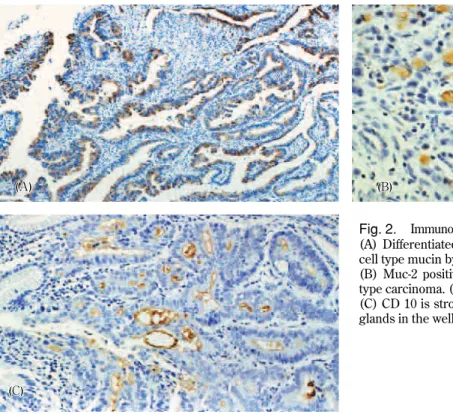
Figs. 1 A∼D and 2 A∼C show typical cases that were found to be positive for G-type and I-type mucin histochemical staining and immunohistochemical staining. Table 3 summarizes the incidences of mu-cin phenotypes and rates of lymph node metastasis, as evidenced by histology. In the differentiated-type
Fig. 1. Expression of gastric type mucin
Carcinoma cells are positive for human gastric mucin (45M-1). (A) differentiated type carcinoma (×50) (B) undifferentiated type car-cinoma (×50)
ConA III staining is partially positive in the carcinoma. (C) differentiated type carcinoma (×100) (D) undifferentiated type carci-noma (×50)
J. Nakamoto, et al. Clinicopathological evaluation of sm gastric carcinomas 102
gastric carcinomas, the I-type was the most fre-quently observed (39.2%, 31/79), followed by the M-type (31.6%, 25/79), LOM (15.2%, 12/79), and G-type (13.9%, 11/79). In the undifferentiated gastric carcinomas, the G-type was most frequently observed (41.4%, 12/29), followed by the M-type (34.5%, 10/ 29), LOM (13.8%, 4/29), and I-type (10.3%, 3/29). The rate of lymph node metastasis according to mu-cin phenotype was higher for the G-type (26.1%, 6/ 23) than for the I-type (5.9%, 2/34) or the M-type
(2.9%, 1/35), and a significant difference was rec-ognized between G-type and M-type (p < 0.05). This tendency was also observed in a separate evaluation of differentiated and undifferentiated carcinomas. Multivariate analysis of lymph node metastasis
Based on the above results on the lymph node metastasis rate, we established a hypothesis that lymphatic invasion, venous invasion, and the mu-cin phenotype are important variables as factors
as-Table 3. Relationship between mucin phenotype and lymph node metastasis Histological type
Mucin phenotype Differentiated Undifferentiated Total
G-type 3 / 11 (27.3)a 3 / 12 (25.0) 6 / 23 (26.1)c I-type 2 / 31 (6.5) 0 / 3 (0) 2 / 34 (5.9) M-type 0 / 25 (0)b 1 / 10 (10.0) 1 / 35 (2.9)d LOM 2 / 12 (6.7) 0 / 4 (0) 2 / 16 (12.5) Total 7 / 79 (8.9) 4 / 29 (13.8) 11 / 108 (10.2) ( ) : % a vs b, c vs d : p<0.05
Fig. 2. Immunohisitochemical expression of intestinal type mucin (A) Differentiated type adenocarcinoma, expression of goblet cell type mucin by Muc-2 glycoprotein (×25)
(B) Muc-2 positive cells are scattered in the undifferentiated type carcinoma. (×100)
(C) CD 10 is strongly positive along the luminal surface of the glands in the well differentiated adenocarcinoma. (×50)
Table 4. Multivariate logistic regression analysis of lymph node metastasis
Characteristic Odds ratio 95% CI Pvalue G-type 5.128 0.552 - 47.656 0.151 I-type 0.565 0.048 - 6.648 0.650 M-type 0.221 0.013 - 3.869 0.302 Venous invasion 9.266 0.894 - 96.081 0.062 Lymphatic invasion 12.942 2.481 - 67.502 0.002 95% CI : 95% confidence interval
sociated with lymph node metastasis. To confirm this hypothesis, multivariate analysis using a logistic model was performed. The results of analysis us-ing 5 variables first selected (lymphatic invasion, venous invasion, G-type, I-type, and M-type) are shown in Table 4. Subsequently, variable selection by the backward elimination procedure was per-formed, and two variables (lymphatic invasion and G-type) remained as significant variables in the final model (Lymphatic invasion : odds ratio, 16.846 ; 95% confidence interval, 3.454-82.171 ; p < 0.001 G-type : odds ratio, 7.690 ; 95% confidence interval, 1.609-36.748 ; p = 0.011).
Clinicopathological factors and PCNA-LI
To evaluate proliferative activity, PCNA staining was performed in all 108 cases. As a result, only 53 of 108 cases could be immunohistochemically evalu-ated in the point of their stainability. It was
consid-ered that PCNA immunoreactivity possibly had been greatly reduced or abolished because the time of fixation in formalin solution was too long in many cases(36). The association between the clinicopa-thological factors and PCNA-LI in 53 cases in which it was possible to immunohistochemically evaluate is shown in Table 5. PCNA-LI was significantly higher (p < 0.05) in cases that were positive for lymph node metastasis (43.4 ± 27.4%) than in negative cases
(27.7±19.0%). PCNA-LI according to the depth of
sm invasion was significantly higher for sm3 (45.1
±24.3%) than for sm1 (21.3±15.0%) or sm2 (25.5 ±17.5%), and that according to histology was
sig-nificantly higher (p < 0.01) for papillary adenocar-cinomas (58.8 ± 26.2%) than for the other
histo-logical types. PCNA-LI according to mucin pheno-type did not significantly differ among I-pheno-type (36.2
± 24.0%), M-type (27.1 ± 21.4%), LOM (41.9 ±
23.2%), and G-type (22.7±15.1%) but was the
low-Table 5. Relationship between clinicopathological factors and PCNA-LI Clinicopathological factors Number of cases PCNA-LI, % Mean±SD Pvalue Lymph node metastasis
+ 11 43.4±27.4 <0.05 − 42 27.7±19.0 Submucosal invasion sm1 15 21.3±15.0a sm2 20 25.5±17.5b <0.01 (c vs a,b) sm3 18 45.1±24.3c Histological type pap 7 58.8±26.2d well 14 25.8±14.4e moderate 12 28.0±24.2f <0.01 (d vs e,f,g,h) por 8 25.9±17.1g sig 12 27.2±16.3h Mucin phenotype G-type 12 22.7±15.1 I-type 12 36.2±24.0 NS M-type 19 27.1±21.4 LOM 10 41.9±23.2 Lymphatic invasion + 24 36.4±23.1 NS − 29 26.4±19.8 Venous invasion + 5 40.6±33.9 NS − 48 30.0±20.3
pap : papillary adenocarcinoma ; well : well differentiated adenocarcinoma ; moderate : moderately differentiated adenocarcinoma por : poorly differentiated adenocarcinoma ; sig : signet ring cell carcinoma
J. Nakamoto, et al. Clinicopathological evaluation of sm gastric carcinomas 104
est for G-type. PCNA-LI according to mucin phe-notype in the presence or absence of lymph node metastasis was evaluated. In cases positive for lymph node metastasis, PCNA-LI for the G-type (23.5 ±
15.0%) was significantly lower (p < 0.05) than that for the I-type (75.2±18.8%) or LOM (64.7±23.3%).
PCNA-LI for the G-type was similar between cases that were positive (23.5±15.0%) and those that were
negative (21.9 ± 16.7%) for lymph node
metasta-sis, while that for the I-type was significantly higher in cases positive for lymph node metastasis (75.2±
18.8%) and negative cases (28.4±16.0%) (Table 6).
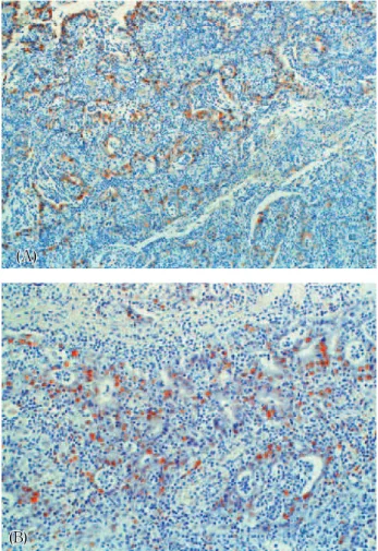
Fig. 3 A, B shows Muc-2 and PCNA staining mi-crographs in the case of an I-type differentiated ade-nocarcinoma that was positive for lymph node me-tastasis.
DISCUSSION
Due to recent advances in mucin histochemical techniques, a considerable number of differentiated-type carcinomas showing G-differentiated-type and undifferenti-ated carcinomas showing I-type have been reported (12, 14, 17-23). In this study, according to the mucin phenotypes, the incidence of G-type was 13.9% (11/ 79) in differentiated-type adenocarcinomas, and that of the I-type was 10.3% (3/29) in undifferentiated-type adenocarcinomas. As previously reported(14, 20, 23, 38), there is no consistency between mucin phenotype expression and histological type.
The rate of lymph node metastasis for sm G-type carcinomas was 26.1%, clearly higher than that for the other mucin phenotypes, suggesting its high grade of malignancy. A multivariate analysis showed that the G-type is an independent factor associated with lymph node metastasis. Noda(38) analyzed 18 cases of early gastric carcinomas showing lymph node metastasis and observed a high incidence of G-type (15/18, 84%). Oya, et al.(39) identified the mucin phenotype in 47 cases of early gastric carci-nomas that were positive for lymph node metasta-sis and 91 not showing lymph node metastametasta-sis, and suggested that early gastric carcinomas showing the G-type in an early stage are biologically aggressive. Koseki, et al.(24) evaluated cases of differentiated sm gastric carcinomas and observed a high rate of lymph node metastasis for G-type sm carcinomas (11/29, 37.9%) and the highest correlation between lymph node metastasis and the G-type. The results of this study support their findings.
Kushima, et al.(18) examined 1-2 cm diameter intramucosal carcinomas and reported that only 30% of the gastric differentiated adenocarcinomas were I-type from the initial stage, and most were the G-I-type or M-type. Tatematsu, et al.(17) compared mucin phenotype expression between m carcinomas and carcinomas showing sm or deeper invasion and observed a significantly higher incidence of the I-type in the latter. Egashira, et al., who studied dif-ferentiated gastric microcarcinomas (< 5 mm), also observed the G-type in 41.1% and the addition of the I-type with an increase in tumor diameter(21).
Fig. 3. Moderately differentiated adenocarcinoma with lymph node metastasis, intestinal type
(A) Carcinoma cells are positive for Muc-2. (×25)
(B) PCNA-positive cells were clearly identified in the invasive front of the cancer, as evidenced by brown nuclear staining. PCNA-LI is 61.9%. (×50)
Table 6. Relationship between mucin phenotype and PCNA-LI associated with lymph node metastasis
Lymph node metastasis
Mucin phenotype + ‐ Total G-type 23.5±15.0a(n=6) 21.9±16.7(n= 6) 22.7±15.1 (n=12)
I-type 75.2±18.8b(n=2) 28.4±16.0d(n=10) 36.2±24.0 (n=12)
M-type 56.7(n=1) 25.5±20.7(n=18) 27.1±21.4 (n=19) LOM 64.7±23.3c(n=2) 36.3±20.7(n= 8) 41.9±23.2 (n=10)
a vs b : p<0.01 ; a vs c : p<0.05 ; b vs d : p<0.01
On the other hand, in undifferentiated-type carci-nomas, changes in the mucin phenotype have also been reported. Various studies have shown com-plete preservation of the G-type in the early stage of signet ring cell carcinomas but acquisition of the I-type in the process of progression(13, 16, 19). Thus, it has been gradually clarified that the mu-cin phenotype can change during the process of progression. Considering these changes, lymph node metastasis may tend to develop in cases showing the G-type from the initial stage and preservation of the G-type without changes in the mucin pheno-type, even after sm invasion.
To clarify the reason for the high incidence of lymph node metastasis in G-type carcinomas, we used PCNA-LI as a parameter for proliferative ac-tivity. PCNA-LI was significantly higher in cases that were positive for lymph node metastasis, showing its usefulness for evaluating proliferative activity. However, despite the high rate of lymph node metastasis for G-type sm gastric carcinomas, their proliferative activity was generally and unexpect-edly low. These results suggest that proliferative activity may not be a parameter of lymph node metastasis in type sm gastric carcinomas. In G-type carcinomas, even when the proliferative activ-ity is low, lymph node metastasis can occur. In the G-type, no difference in carcinoma cell proliferative activity was found between metastasispositive and -negative cases. In contrast, in the case of the I-type, proliferative activity significantly differed between metastasis-positive and-negative cases. Kushima, et al.(18) examined gastric intramucosal carcinomas and reported that the number of PCNA-positive cells tended to be lower in G-type adenocarcinomas than in other-type adenocarcinomas. They speculated that this is because many cancer cells in G-type ade-nocarcinomas contain foveolar-epithelium-type or pyloric-gland -type mucin and have lost the ability to proliferate, or that they have mimicked the differen-tiation mode of normal cells in the gastric proper mucosa, and most cancer cells drop out of the cell cycle and differentiate and mature into cells that resemble surface mucous cells and pyloric gland cells. Saito, et al.(22) reported in their study of early gastric carcinomas that differentiated adenocarci-nomas with the G-type histologically transform into undifferentiated-type adenocarcinomas with an in-crease in tumor size. Certain factors other than pro-liferative activity may be involved in the lymph node metastasis of G-type sm gastric carcinomas. Such factors were not clarified in this study. However, we
speculate that G-type sm gastric carcinomas, de-spite their low proliferative activity, transform into the undifferentiated type with the progression of invasion, which facilitates carcinoma cell separa-tion from the glands, leading to vascular invasion and lymph node metastasis. In this study, in I-type sm gastric carcinomas, proliferative activity was significantly higher in cases that were positive for lymph node metastasis than in negative cases, and proliferative activity can be a parameter for lymph node metastasis. Thus, it would be of interest to determine if the incidence of lymph node metastasis and proliferative behavior differed among mucin phenotypes.
In conclusion, G-type expression in sm carcino-mas may be an important risk factor for lymph node metastasis, and attention should be paid to G-type carcinomas even if they are clinically of the differ-entiated type. Furthermore, it is seemed that pro-liferative activity is a significant parameter for lymph node metastasis in cases with I-type carcinomas. Therefore, in addition to the conventional exami-nation of histological type, a preoperative examina-tion of the mucin phenotype may be an important supplementary diagnosis.
ACKNOWLEDGEMENTS
We wish to express our gratitude to all the mem-bers of our laboratory for their technical support.
REFERENCES
1. Sano T, Kobori O, Muto T : Lymph node me-tastasis from early gastric cancer : Endoscopoic resection of tumor. Br J Surg 79 : 241-244, 1992 2. Yanai T , Noguchi S, Mizumachi H, Tokiyama H, Nakamura H, Tada M : A blind comparison of the effectiveness of endoscopic ultrasonogra-phy and endoscopy in staging early gastric cancer. Gut 44 : 361-365, 1999
3. Ono H, Kondo T, Gotoda T : Endoscopic mu-cosal resection for treatment of early gastric cancer. Gut 48 : 225-229, 2001
4. Oguro Y : Endoscopic treatment of early gas-tric cancer. Diagn Endosc 3 : 3-15, 1991 5. Yasuda K, Mizuna Y, Kawai K : Endoscopic laser
treatment for early gastric cancer. Endoscopy 25 : 451-454, 1993
6. Maehara Y, Orita H, Okuyama T, Moriguchi J. Nakamoto, et al. Clinicopathological evaluation of sm gastric carcinomas
S, Tsujitani S, Korenaga D, Sugimachi K : Predictors of lymph node metastasis in early gastric cancer. Br J Surg 79 : 245-247, 1992 7. Inoue K, Tobe T, Kan N, Nio Y, Sasaki M,
Takeuchi E, Sugiyama T : Problems in the definition and treatment of early gastric cancer. Br J Surg 78 : 818-821, 1991
8. Nishida T, Tanaka S, Haruma K, Yoshihara M, Sumii K, Kajiyama G : Histologic grade and cellular proliferation at the deepest invasive portion correlate with the high malignancy of submucosal invasive gastric carcinoma. Oncol-ogy 52 : 3340-346, 1995
9. Yasuda M, Niki M, Torisu R, Bandou G, Hayashi T, Sakashita O, Takahashi Y, Yamanoi A, Takeuchi Y, Yamamoto Y : Indications of en-doscopic mucosal resection (EMR) for submu-cosal invasive gastric cancer with special ref-erence to hisitopathologic findings. -Research by using Logistic regression analysis method. Gatroenterol Endosc 45 (in Japanese with Eng-lish abstract) : 233-240, 2003
10. Nakamura K, Sugano H, Takagi K : Carcinoma of the stomach in icipient phase : Its histogene-sis and histological appearances. GANN 59 : 251-258, 1968
11. Lauren P : The two histological main types of gastric carcinoma : Diffuse and so-called intestinal-type carcinoma. Acta Pathol Microbiol Scand 64 : 31-49, 1965
12. Hattori T : Morphological range of hyperplas-tic polyps and carcinomas arising in hyper-plastic polyps of the stomach. J Clin Pathol 38 : 622-630, 1985
13. Sugihara H, Hattori T, Fukuda M, Fujita S : Cell proliferation and differentiation in intra-mucosal and advanced signet-ring cell carci-nomas of the human stomach. Virchows Arch [A]411 : 117-127, 1987
14. Tatematsu M, Ichinose M, Miki K, Hasegawa R, Kato T, Ito N : Gastric and intestinal pheno-typic expression of human stomach cancers as revealed by pepsinogen immunochemistry and mucin histochemistry. Acta Pathol Jpn 40 : 494-504, 1990
15. Ficca R, Villani L, Tenti P, Cornaggia M, Finzi G, Riva C, Capella C, Bara J, Samloff I.M., Solcia E : The foveolar cell component of gas-tric cancer. Hum Pathol 21 : 260-270, 1990 16. Akamatsu T, and Katsuyama T :
Histochemi-cal demonstration of mucins in the intramu-cosal laminated structure of human gastric
sig-net ring cell carcinoma and its relation to sub-mucosal invasion. Histochem J 22 : 416-425, 1990
17. Tatematsu M, Hasegawa R, Ogawa K, Kato T, Ichinose M, Miki K, Ito N : Histogenesis of human stomach cancers based on assessment of differentiation. J Clin Gastroenterol (suppl 14): S1-7, 1992
18. Kushima R, Hattori T : Histogenesis and charastereristics of gastric-type adenocarcino-mas in the stomach. J Cancer Res Clin Oncol 120 : 102-111, 1993
19. Kushima R, Jancic S, Hattori T : Association between expression of sialosyl-Tn antigen and intestinalization of gastric carcinomas. Int J Cancer 55 : 904-908, 1993
20. Egashira Y : Mucin histochemical study of differentiated adenocarcinoma of stomach. Jpn J.Gastroenterol 91 (in Japanese with English ab-stract) : 839-848, 1994
21. Egashira Y, Shimoda T, Ikegami M : Mucin histochemical analysis of minute gastric differ-entiated adenocarcinoma. Pathol Int 49 : 55-61, 1999
22. Saito A, Shimoda T, Nakanishi Y, Ochiai A, Toda G : Histologic heterogeneity and mucin phenotype expression in early gastric cancer. Pathology International 51 : 165-171, 2001 23. Shimoda T, Fujisaki J, Kashimura H :
Hisologi-cal type of gastric carcinoma in relation to the mode of intramural spreading of cancer cells. Stomach and Intestine 26 (in Japanese with Eng-lish abstract) : 1225-1134, 1991
24. Koseki K, Takizawa T, Koike M, Ito M, Nihei Z, Sugihara K : Distinction of differentiated type early gastric cancer with gastric type mucin ex-pression. Cancer 89 : 724-732, 2000
25. Japanese Research Society for Gastric Cancer. The General Rules for the Gastric Cancer Study. Kanehara, Tokyo, 1993
26. Shi SR, Key ME, Kaira K : Antigen retrieval in formalin-fixed, paraffin-embedded tissues an enhancement method for immunohistochemi-cal staining based on microwave oven heating of tissue sections. J Histochem Cytochem 39 : 741-748, 1991
27. Katsuyama T, Spicer SS : Histochemical dif-ferentiation of complex carbohydrates with vari-ants of the concanavalin A horseradish peroxi-dase method. J Histochem Cytochem 26 : 223-250, 1987
28. Bara J, Gautier R, Mouradian P, Decaens C, The Journal of Medical Investigation Vol. 54 February 2007 107
Daher N : Oncofetal mucin M 1 epitope family : Characterization and expression during colonic carcinogenesis. Int J Cancer 47 : 304-310, 1991 29. Tatematsu M, Katsuyama T, Fukushima S, Takahashi M, Shirai T, Ito N, Nasu T : Mucin histochemistry by paradoxical concanavalin A staining in experimental gastric cancer in-duced in Wistar rats by N-methyl-N’-nitro-N-Nitrosoguanidine or 4-Nitroquinoline-1-oxide. J Natl Cancer Inst 64 : 835-843, 1980
30. Tatematsu M, Katsuyama T, Mutani M : Pylo-ric gland phenotypic expression of gastPylo-ric can-cers developing in the rat fundic glandular stom-ach. Carcinogenesis 10 : 1033-1039, 1989
¨
31. Tygat KM, Buller HA, Opdam FJ, Kim YS, Einerhand AW, Dekker J : Biosynthesis of hu-man colonic mucin : Muc2 is the prominent se-cretory mucin. Gastroenterology 107 : 1352-1363, 1994
32. Ajioka Y, Allison LJ, Jass JR : Significance of MUC 1 and MUC 2 mucin expression in col-orectal cancer. J Clin Pathol 49 : 560, 1996 33. Ajioka Y, Watanabe H, Jass JR : MUC 1 and
MUC 2 mucin in flat and polypoid colorectal adenomas. J Clin Pathol 50 : 417, 1997
34. Ronco P, Allegri L, Melcion C, Pirotsky E, Appay M, Bariety J, Pontillon F, Verroust P : A monoclonal antibody to brush border and passive Heymann nephritis. Clin Exp Immuno 55 : 319-332, 1984
35. Trejdosiewicz LK, Malizia G, Oakes J, Losowsky MS, Janossy G : Expression of the common acute lymphoblastic leukemia antigen (CLALLA gp100) in the brush border of normal jejunum of patients with coeliac disease. J Clin Pathol 38 : 1002-1006, 1985
36. Hall PA, Levinson DA, Woods AL, Yu CC-W, Kellock DB, Watkins JA, Barnes DM, Gillett CE, Camplejohn R, Dover R, Waseem NH, Lane DP : proliferating cell nuclear antigen (PCNA) immunolocalization in paraffin sections : An index of cell proliferation with evidence of de-regulated expression in some neoplasms. J Pa-thol 162 : 285-294, 1990
37. Robbins B.A., Vega D, Ogata K, Tan EM, Nakamura RM : Immunohistochemical detec-tion of proliferating cell nuclear antigen in solid human malignancies. Arch Pathol Lab Med 111 : 841-845, 1987
38. Noda M : Characteristics of early gastric cancer accompanied by lymph node metastasis. Jpn J Gastroenterol 92 (in Japanese with English ab-stract) : 742-750, 1995
39. Oya M, Yao T, Tsuneyoshi M : A correlation of intramural tumor invasion with mucin histo-chemistry, and immunohistochemical expres-sions of oncogene-related proteins in early gas-tric carcinomas. Stomach and Intestine 32 (in Japanese with English abstract) : 31-39, 1997 J. Nakamoto, et al. Clinicopathological evaluation of sm gastric carcinomas