Development of Intra-Orbital Follicular
Lymphoma and Intra-Oral MALT-Type Lymphoma in
a Single Patient
著者
NAKAMINE Hirokazu, NISHINO Eisei, KURIBAYASHI
Koichi, TAKENAKA Toru, KOHNAMI Shinji, TAMURA
Shinji, TSUDA Tadaaki, OKAMOTO Yukiharu, OHTA
Kiichiroh, TAKAGISHI Chika, SAIKA Shizuya,
KAWASHIMA Toshihiro, OKAMOTO Keiichiroh,
MORITA Nobuo
journal or
publication title
鹿児島大学医学雑誌=Medical journal of
Kagoshima University
volume
47
number
Suppl. 2
page range
133-136
URL
http://hdl.handle.net/10232/18331
Med. J. Kagoshima Univ., Vol. 47, Suppl. 2. 133-136, November, 1995
Case Report
Development of
Intra-Orbital Follicular Lymphoma and
Intra-Oral MALT-Type Lymphoma
in a Single Patient
Hirokazu NAKAMINE1,
Eisei NISHINO1, Koichi KURIBAYASHI1, Toru TAKENAKA1,
Shinji KOHNAMI2, Shinji TAMURA3,
Tadaaki TSUDA4, Yukiharu OKAMOTO4, Kiichiroh OHTA4,
Chika TAKAGISHI5, Shizuya SAIKA5,
Toshihiro KAWASHIMA6, Keiichiroh OKAMOTO6, and Nobuo MORITA6
Departments of laboratory Medicine, Pediatrics, 3Otorhinolarygology, 4Transfusion Medicine,
5Ophthalmology, and 6Oral and Maxillofacial Surgery, Wakayama Medical University, Wakayama, Japan
Abstract
An 81-year-old woman with intra-orbital follicular large-cell lymphoma and intra-oral low-grade B-cell lymphoma, compatible with MALT-type lymphoma, is reported. Several hematopathologic features indicated that the two morphologically different lymphomas were originated from a single clone of cells, but no definite evidence to confirm this possibility could be obtained. Result of chemotherapy indicated difference in sensi tivity of lymphoma cells of the two lesions to cytotoxic drugs.
Key words: Follicular lymphoma, MALT-type lym
phoma, Histogenesis, T cell-rich variant.
Introduction
Non-Hodgkin's lymphoma arising from the mucosa-associated lymphoid tissue (MALT lymphoma), first
described by Isaacson and Wright1^ in 1983, has been
reported with an increasing frequency to date. Although it is generally accepted that MALT-type
Address for Correspondence: Hirokazu NAKAMINE, Department of Laboratory Medicine, Wakayama Medical University, 27, 7-Bancho, Wakayama 640, Japan
lymphoma, which was difficult to characterize in the past, does exist, whether MALT lymphoma is a distinct clinicopathologic entity or not, is still a matter of
debate \ because a lymphoma with similar hemato
pathologic features, i.e., monocytoid B-cell lymphoma(MBCL), can develop de novo in the lymph node5'6).
One of the feature of MBCL is a relatively frequentassociation of follicular lymphoma6"10).
In this report, we describe a patient who developed intra-orbital follicular large-cell lymphoma and intra oral MALT-type lymphoma.
Case summary
An 81-year-old woman first noticed bilateral con junctival swelling 19 months before admission. Because of the progressive nature of the process, she was admitted to our hospital in June, 1993. Neither loss of body weight, night drenching, nor fever was noted. Physical examination revealed bilateral intra-orbital masses, but there was no peripheral lymphadenopathy or hepatosplenomegaly. Complete blood count, serum analysis, bone marrow aspiration, and abdominal CT scan showed no abnormalities. She was diagnosed as having one of follicular large-cell lymphoma on the basis of a biopsy of the right intra-orbital mass. The tumors disappeared completely two and a half months after the initiation of chemotherrapy consisting of
(134) Med. J. Kagoshima Univ., Vol. 47, Suppl. 2, November, 1995
vincristine, cyclophosphamide, and prednisolone. However, an intra-oral mass at the buccal region was found at that time. The mass was totally resected and it was histopathologically characterized as low-grade B-cell lymphoma, compatible with MALT-type lympho
m a .
The patient was discharged from the hospital three months after admission. So far, no findings indicative of recurrence of the tumors were noted.
Hematopathologic findings
Histopathology
The orbital mass showed nodular proliferation of lymphoid cells in the collagenous tissue. Each nodule
Fig.
isy
'MM:
mwmm \ '♦* y a..,
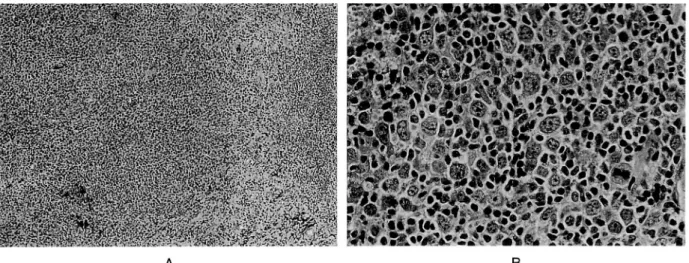
-consisted of large lymphoid cells admixed with small cleaved cells and small lymphocytes. The large cells had round or oval-shaped nuclei with either finely dispersed or somewhat clumped chromatin and amphophilic cytoplasm (Fig. 1).
The oral mass was composed of diffuse proliferation of lymphoid cells with round or cleaved nuclei and fairly abundant pale cytoplasm. Large lymphoid cells were occasionally seen. Ductal structures with periduc tal hyalinization were present among the lymphoprolif-eration, but the lymphoepithelial lesion did not appear to be a significant feature. In addition to the nests of ductal epithelial cells, numerous vessels were filled with neoplastic lymphoid cells (Fig. 2).
A B
Intra-orbital tumor. A (left): Follicular proliferation of lymphoid cells is seen. B (right): Proliferation of large noncleaved cells admixed with small lymphoid cells is evident in the center of each neoplastic follicle. (H & E. A. x 64; B, x320)
A B
Fig. 2. Intra-oral tumor. A (left): The tumor is composed of diffuse proliferation of lymphoid cells. Ductal structures are present at the left. B (right): The proliferating cells have small, but irregular, nuclei and pale cytoplasm. Intravascular involvement is seen at the right. (H & E. A. x 64; B, x320)
Follicular and MALT-Type Lymphomas [135]
Immunohistochemistry
In frozen sections of the orbital mass, the proliferat ing cells were positive for surface Ig * , CD 19, CD20, and CD22, and were negative for surface Ig A, CD10, CD21, CD5, CD2, CD3, CD4, CD8, and CD30. Ki-67-positive cells were occasionally present. The large cells were positive for CD20-cy and cytoplasmic Ig K, and were negative for CD45RO and cytoplasmic IgA in paraffin sections.
In paraffin sections of the oral mass, cells with CD20-cy and those with CD45RO were equally present, but
the size of the latter was smaller than that of the
former. The larger cells were positive for cytoslasmic Ig
* and negative for cytoplasmic Ig A. Frozen tissue of the oral mass was not available for study.
Gene amplification study
DNA sample was prepared from paraffin-embedded tissue of both orbital and oral masses. By semi-nested
PCRn) using consensus oligonucleotide primers for the
framework 3 portion of the variable region (FR3A) and of the 3' portion of the joining region (LJH and VLJH)of the Ig heavy-chain gene (IgH) 2\ the rearranged
V-D-J fragments were clonally amplified in the orbital mass, but the amplified fragment in the oral mass was equivocal with regard to clonality. Nested PCR using
primers for IgH (JH) and bcl-2 (both mbr and mcr)1^
showed no amplification in both samples.
Discussion
There are several hematopathologic features which indicate that the two lymphomas in the current case were originated from a single clone of cells. These include; 1) presence of intravascular neoplastic cells in the oral lesion (Fig 2B), 2) numerous reactive-appear ing T cells in both lesions, 3) absence of CD 10 expression in the orbital lesion, and 4) absence of amplification of rearranged bcl-2 in orbital as well as oral lesions. This possibility is supported by the reported findings that MALT-type lymphoma may mimic follicular lymphoma due to "follicular
colonization"14), that MBCL, which is closely related
to, but may not be identical with, MALT-type lymphoma, is associated with follicular lymphoma with
a relatively high frequency6""l0), and that follicular
lymphoma can show partial monocytoid
differentiation15). However, failure of definite clonal
amplification of IgH in the oral lesion and lack of the oral frozen tissue humpered final determination of the common or different clonality of the two lymphomas. Proliferation of large noncleaved cells without "centrocyte-like" features in any part of the orbital lesion indicate the follicular growth patten not to be a result of "follicular colonization". Furthermore, sensi tivity of lymphoma cells of the two lesions to cytotoxic drugs was different, because the oral mass was present at the time when the orbital lesion had completely
regressed by chemotherapy,
Numerous reactive-appearing T cells in the oral lesion can lead to the interpretation that the lesion is a T cell-rich vatiant of MALT-type lymphoma. B-cell lymphomas with abundant reactive T cells have been
reported as either pseudo T-cell lymphoma16) or T
cell-rich B-cell lymphoma17). It is stressed, however, that
these B-cell lymphomas including "T cell-rich MALT-type lymphoma", while important in the diagnosis, should not be characterized as a disease entity until distinctiveness of their clinicopathologic features are confirmed.
Acknowledgments
The authors thank Drs. K. Lennert and A.C. Feller for their helpful suggestions on this case. We are also grateful to Dr. E. Sato, the president of the Seminar, for providing us an opportunity to present this case.
References
1) Isaacson P, Wright DH: Malignant lymphoma of mucosa-associated lymphoid tissue. A distinctive type of B-cell lymphoma. Cancer 1983; 52: 1410-6. 2) Harris NL. Low-grade B-cell lymphoma of mucosa-associated lymphoid tissue and monocytoid B-cell lymphoma. Related entities that are distinct from other low-grade B-cell lymphomas. Arch Pathol Lab Med 1993; 117: 771-5.
3) Nakamine H, Takenaka T, Nishino E, Tamura S, Kohnami S, Aoki J, et al. Monocytoid B-cell lymphoma (MBCL). Report of 47 Japanese patients and review of the literature with regard to the relation between MBCL and low-grade B-cell lymphoma arising from the mucosa-associated lymphoid tissue (MALT lymphoma). J Jpn Soc RES 1993; 33: 331-46 (in Japanese with English abstract). 4) Nakamine H, Takenaka T, Nishino E, Nanba K. Monocytoid B-cell lymphoma. A study of nodal and extranodal disease. Mod Pathol 1994; 7: 117A.
5) Sheibani K, Sohn CC, Burke JS, Winberg CD, Wu AM, Rappaport H: Monocytoid B-cell lymphoma. A novel B-cell neoplasm. Am J Pathol 1986; 124: 310-8.
6) Sheibani K. Monocytoid B cell lymphoma. In: KnowlesDM, editor. Neoplastic Hematopathology. Baltimore, MD, Williams & Wilkins, 1992: 629-44. 7) Ngan BY, Warnke RA, Wilson M, Takagi K, Cleary ML, Dorfman RF. Monocytoid B-cell lymphoma. A study of 36 cases. Hum Pathol 1991; 22: 409-21.
8) Nathwani BN, Mohrmann RL, Brynes RK, Taylor CR, Hansmann ML, Sheibani K. Monocytoid B-cell lymphoma. An assessment of diagnostic criteria and a perspective on histogenesis. Hum Pathol 1992; 23: 1061-71.
[136] Med. J. Kagoshima Univ., Vol. 47, Suppl. 2, November, 1995
in malignant lymphomas of diverse histologic type. Lab Invest 1987; 56: 50A.
10) Hernandez AM, Nathwani BN, Nguyen D, Shibata D, Chuan W, Nichols P, et al. Nodal benign and malignant monocytoid B cells with and without follicular lymphomas. A comparative study of follicular colonization, light chain restric tion, bcl-2, and t(14;18) in 39 cases. Hum Pathol 1995; 26: 625-32.
11) Wan JH, Trainor KJ, Brisco MJ, Morley AA. Monoclonality in B cell lymphoma detected in paraffin wax embedded sections using the polymerase chain reaction. J Clin Pathol 1990; 43: 888-90.
12) Brisco MJ, Tan LW, Orsborn AM, Morley AA. Development of a highly sensitive assay, based on the polymerase chain reaction, for rare B-lympho-cyte clones in a polyclonal population. Br J Haematol 1990; 75: 163-7.
13) Gribben JG, Freedman AS, Woo SD, Blake K, Shu RS, Freeman G, et al. All advanced stage
non-Hodgkin's lymphomas with a polymerase chain reaction amplifiable breakpoint of bcl-2 have residual cells containing the bcl-2 rearrangement at
evaluation and after treatment. Blood 1991; 78: 3275-80.
14) Isaacson PG, Wotherspoon AC, Diss T, Pan L. Follicular colonization in B-cell lymphoma of mucosa-associated lymphoid tissue. Am J Surg
Pathol 1991; 15: 819-28.
15) Chan JKC, Hui PK. An unusual morphological variant of follicular lymphomas. Report of two cases. Histopathology 1988; 12: 649-58.
16) Jaffe ES, Longo DL, Cossman J, Hsu SM, Arnold A, Korsmeyer SJ. Diffuse B cell lymphomas with T cell predominance in patients with follicular lymphoma or "pseudo T cell lymphoma". Lab Invest 1984; 50: 27A-8A.
17) Ramsay AD, Smith WJ, Isaacson PG. T-cell-rich B-cell lymphoma. Am J Surg Pathol 1988; 12: 433-43.