IRUCAA@TDC : Osteoma of Mandibular Condyle as Cause of Acute Pain and Limited-mouth-opening: Case Report
6
0
0
全文
(2) 193. Bull Tokyo Dent Coll (2007) 48(4): 193–197. Case Report. Osteoma of Mandibular Condyle as Cause of Acute Pain and Limited-mouth-opening: Case Report Hakubun Yonezu, Mamoru Wakoh*, Takamichi Otonari*, Tsukasa Sano*, Sadamitsu Hashimoto** and Takeshi Uchiyama Department of Oral and Maxillofacial Surgery, Tokyo Dental College, 1-2-2 Masago, Mihama-ku, Chiba 261-8502, Japan * Department of Oral and Maxillofacial Radiology, Tokyo Dental College, 1-2-2 Masago, Mihama-ku, Chiba 261-8502, Japan ** Department of Oral Pathology, Tokyo Dental College, 1-2-2 Masago, Mihama-ku, Chiba 261-8502, Japan. Received 3 August, 2007/Accepted for publication 3 December, 2007. Abstract In benign tumors in the mandibular condyle such as osteoma and osteochondroma, symptoms such as pain and limited-mouth-opening are rarely observed. Therefore, these tumors are often detected after the development of changes in occlusion and mandibular midline deviation. We encountered a very rare patient with mandibular condyle osteoma who showed acute pain and markedly limited-mouth-opening. Key words:. Osteoma—Condyle—Acute pain —Limited-mouth-opening. Introduction Osteoma is a benign tumor composed of both cortical and cancellous bone that increases in size by continuous formation of bone1,6). It is essentially restricted to the maxilla and mandible, and is rarely, if ever, diagnosed in other bones4,5). In Gardner syndrome, multiple osteomas are present in the alveolar bone of the maxilla or mandible. Osteoma is a slow-growing, asymptomatic, usually solitary lesion which affects mainly young adults2,8). Osteoma occurring in either the condyle or condylar process may result in morphologic and functional disturbances, including facial asymmetry and temporomandibular joint. (TMJ) dysfunction. It is rare for osteoma of the condyle to cause acute pain and limitedmouth-opening.. Case Report A 5X-year-old man noticed spontaneous pain in the left temporomandibular joint on September 1, 200X and visited a local dental clinic on September 3. Soft laser therapy was performed, but no relief of pain was achieved. On September 5, he was referred to our hospital. Diffuse swelling and marked pain were observed in the left preauricular area. However, tenderness and heat sensation were. 193.
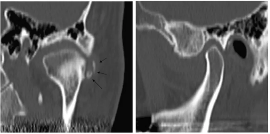
(3) 194. Yonezu H et al.. absent in this area, and skin color was normal. Interincisal mouth-opening was 15 mm. Although no treatment was given at this time, both swelling and pain disappeared 4 days later.. the lateral aspect of the left mandibular condyle (Fig. 1). This did not appear, however, in the orthopantomogram. Non-contrast CT images obtained at initial examination showed a poorly-delineated mass lesion in contact with the lateral margin of the left mandibular condyle, with well-delineated,. Image Findings Orbito-ramus projection radiography revealed an area of well-delineated dense opacity on. Fig. 1 Orbito-ramus projection radiograph Some well-delineated radiopaque images on the lateral side of the left mandibular condyle (arrows).. Fig. 2 Non-contrast CT (axial image) A poorly-delineated mass lesion in contact with the lateral margin of the left mandibulr condyle and well-delineated and maviously-sized high density compartment inside the lesion (arrows).. Fig. 3 Multiplaner reformation (MPR) image of CT Left photograph is coronal view. Right photograph is sagittal view. A coronal MPR images revealed well-delineated and variously-sized high density compartment on the lateral side of the left mandibulr condyle (arrows). Sagittal MPR images showed neither a high-density area in the joint space nor definite abnormal findings in bone inside the condyle..
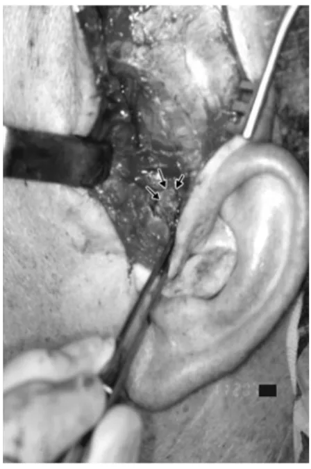
(4) Osteoma of Mandibular Condyle. high density compartments of various sizes inside. The lateral pterygoid muscle and masseter muscle on the left side were swollen compared with those on the right side (Fig. 2). No contrast-CT examination was performed. Coronal multiplanar reformation (MPR) images also revealed well-delineated, high density compartments of various sizes on the lateral aspect of the left mandibular condyle. Sagittal MPR images, however, revealed neither these compartments in the joint space, nor definite abnormal findings in bone inside the condyle (Fig. 3). MR images obtained 4 days after initial examination revealed the disappearance of the swelling in the lateral pterygoid and masseter muscles observed by CT at the first visit, and the presence of a hypointense mass with internal hyperintense elements on the lateral aspect of the left mandibular condyle (Fig. 4).. 195. had been observed by preoperative imaging. Part of the hard tissue was in contact with the mandibular condyle and upper joint compartment (Fig. 5). Figure 6 shows osseous hard tissue resected from the lateral aspect of the mandibular condyle.. Operation Findings On November 20, the tumor was resected under endotracheal general anesthesia. As the photo taken during the operation shows, there was osseous hard tissue capsulated by connective tissue in the area where the lesion. Fig. 4 MR image: T1W (TR/TE 380/11) Presence of a mass lesion on the lateral side of the left mandibular condyle (arrows).. Fig. 5 Operation view Osseous hard tissue capsulated by connective tissue (arrows).. Fig. 6 Resected osteoma Osseous hard tissue resected from the lateral side of the mandibular condyle..
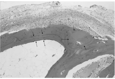
(5) 196. Yonezu H et al.. Histopathological Findings. Discussion. Figure 7 shows a pathological specimen after H-E staining. The lesion consisted of spongy osseous hard tissue contained in a capsule composed of coarse fibrous connective tissue. This tissue showed cell components in the lacunae, a clear lamellar structure, and trabeculae formation. The medullary cavity was dilated and contained a mature fat medulla. At the trabecular base on one side, slightly basophilic fibrous cartilage containing small cells with a round nucleus was observed, and this was continuous with the trabeculae. In one area, collagen fiber bundles formed a tendonlike structure, showing continuous transfer. Based on these findings, a diagnosis of cancellous osteoma was made.. Osteoma of the condyle may cause a slow, progressive shift in occlusion, with deviation of the midline of the chin toward the unaffected side7). This results in facial asymmetry and malocclusion such as cross-bite2). However, no facial asymmetry or malocclusion was observed in this patient. In osteoma and osteochondroma in the mandibular condyle, acute pain or limited-mouth-opening is rarely observed. In our patient, CT at initial examination revealed swelling of the lateral pterygoid and masseter muscles. This suggests that the acute symptoms seen here resulted from inflammatory changes in the TMJ. Osteomas in the bones of the jaw are uncommon. They may arise from the surface of bone as a polypoidal or sessile mass (periosteal osteoma), or may be located within the medullary bone (endosteal osteoma)4). There are 2 types of osteoma: central osteoma, arising from increase in cancellous bone, and peripheral osteoma, arising from increase in cortical bone, with the latter being the most common3). Accordingly, osteomas occurring in the condylar process can be classified into. Postoperative Course At postoperative week one, the pain disappeared, and interincisal mouth opening exceeded 50 mm. The patient did not develop facial palsy. No recurrence was observed, and recovery was good at one year postoperatively.. Fig. 7 Findings on histopathological specimen (H-E Stain) Spongy osseous hard tissue showed cell components in the lacunae, a clear lamellar structure, and trabeculae formation (arrows)..
(6) 197. Osteoma of Mandibular Condyle. 2 types: those which proliferate and cause replacement of the condyle by osteoma, and those which appear as a pedunculated or osseous mass on the condyle or neck of the mandible2,6). In the case reported here, some osteoid hard tissue encapsulated by connective tissue was observed in the lateral aspect of the condyle. Radiographic images revealed that the osteomas were circumscribed masses, of similar density to normal bone, and of approximately 5 mm in size. At their centers, such masses may exhibit a mixed radiolucent-radiopaque appearance, depending on the amount of marrow tissues present. Smaller endosteal osteomas are difficult to differentiate from foci of condensing osteitis, focal chronic sclerosing osteomyelitis, or idiopathic osteosclerosis4). A large osteoma of the condyle resulting in pain, facial asymmetry and malocclusion such as cross-bite may require condylectomy, whereas small, asymptomatic lesions may only require periodic observation. In the present case, even though no condylectomy was performed, the patient’s pain and limitation of mouth-opening disappeared. Histologically, an osteoma consists of either a normal-appearing dense mass of lamellar bone with minimal marrow tissue (compact osteoma), or of trabeculae of mature lamellar bone, with intervening fatty or fibrous marrow (cancellous osteoma). This case represents the latter entity, as evidenced by the wide spread of the bone marrow cavity and mature fatty marrow inside1).. References 1) Cawson RA, Binnie WH, Speit PM, Barret AW, Wright JM (1998) Lucas’s Pathology of Tumors of Oral Tissue, pp.169–171, Churchill Livingstone, Edinburgh. 2) Kondoh T, Seto K, Kobayashi K (1998) Osteoma of the mandibular condyle: report of a case with a review of the literature. J Oral Maxillofac Surg 56:972–979. 3) Mancini JC, Woltmann M, Felix VB, Frites RR (2005) Peripheral osteoma of the mandibular condyle. Int J Oral Maxillofac Surg 34:92–93. 4) Neville BW, Damm DD, Allen CM, Bouquot JE (2002) Oral & Maxillofacial Pathology, pp.566–567, WB Saunders, Philadelphia. 5) Odell EW, Morgan P (1998) Biopsy Pathology of the Oral Tissues, p.266, Chapman and Hall Medical, London. 6) Schneider LC, Dolinsky HB, Grodjesk JE (1980) Solitary peripheral osteoma of the jaws: report of a case and review of literature. J Oral Surg 38:452–455. 7) Siar CH, Jalil AA, Ram S, Ng KH (2004) Osteoma of the condyle as the cause of limitedmouth opening: a case report. J Oral Sci 46: 51–53. 8) Syan NB, Ucok C, Karasu HA, Gunhan O (2002) Peripheral osteoma of the oral and maxillofacial region: a study of 35 new cases. J Oral Maxillofac Surg 60:1299–1301. Reprint requests to: Dr. Hakubun Yonezu Department of Oral and Maxillofacial Surgery, Tokyo Dental College, 1-2-2 Masago, Mihama-ku, Chiba 261-8502, Japan E-mail: hayonezu@tdc.ac.jp.
(7)
図
関連したドキュメント
Standard domino tableaux have already been considered by many authors [33], [6], [34], [8], [1], but, to the best of our knowledge, the expression of the
Giuseppe Rosolini, Universit` a di Genova: rosolini@disi.unige.it Alex Simpson, University of Edinburgh: Alex.Simpson@ed.ac.uk James Stasheff, University of North
This is a consequence of a more general result on interacting particle systems that shows that a stationary measure is ergodic if and only if the sigma algebra of sets invariant
We solve by the continuity method the corresponding complex elliptic kth Hessian equation, more difficult to solve than the Calabi-Yau equation k m, under the assumption that
We show that a discrete fixed point theorem of Eilenberg is equivalent to the restriction of the contraction principle to the class of non-Archimedean bounded metric spaces.. We
In [9], it was shown that under diffusive scaling, the random set of coalescing random walk paths with one walker starting from every point on the space-time lattice Z × Z converges
We describe a generalisation of the Fontaine- Wintenberger theory of the “field of norms” functor to local fields with imperfect residue field, generalising work of Abrashkin for
Shen, “A note on the existence and uniqueness of mild solutions to neutral stochastic partial functional differential equations with non-Lipschitz coefficients,” Computers
