Chem-Bio Informatics Journal, Vol.21, pp.28–38 (2021)
Web Server with a Simple Interface for Coarse-grained Molecular
Dynamics of DNA Nanostructures
Yudai Yamashita
1, Kotaro Watanabe
1, Satoshi Murata
1, Ibuki Kawamata
1, 2* 1Department of Robotics, Graduate School of Engineering, Tohoku University, 6-6-01, Aramaki AzaAoba Aoba-ku, Sendai, Miyagi 980-8579, Japan
2Natural Science Division, Faculty of Core Research, Ochanomizu University, 2-1-1 Ohtsuka, Bunkyo-ku, Tokyo
112-8610, Japan
*E-mail: ibuki.kawamata@tohoku.ac.jp
(Received January 5, 2021; accepted April 5, 2021; published onlineApril 30,2021)
Abstract
We introduce an automated procedure of coarse-grained molecular dynamic simulation for DNA nanostructure that has great potential for realizing molecular robotics. As DNA origami is now a standardized technology to fabricate DNA nanostructures with high precision, various computer-aided design software has been developed. For example, a design tool called caDNAno with a simple and intuitive interface is widely used for designing DNA origami structures. Further, a simulation tool called oxDNA is used to predict the behavior of such nanostructures based on coarse-grained molecular dynamics. These tools, however, are not linked directly; thus, repeating the cycle of design and simulation is cumbersome to the user. Moreover, the computer skills required to setup, launch, and run an oxDNA simulation are a potential barrier for non-experts. In our proposal, oxDNA simulation can be launched on a web server simply by providing a caDNAno file; the web server then analyzes the simulation results and provides a visual response. The validity of the proposal is demonstrated using an example. The advantages of our proposed method compared with other conventional methods are also described. This simple-to-use interface for user-friendly simulation of DNA origami eliminates stress to users and accelerates the design process of complicated DNA nanostructures such as wireframe architecture.
Key Words: DNA origami, DNA wireframe architecture, Molecular robotics, Coarse-grained molecular dynamics, Web service with simple interface, Automated procedure, caDNAno, oxDNA
Chem-Bio Informatics Journal, Vol.21, pp.28–38 (2021)
1. Introduction
DNA nanotechnology is a novel field involving the fabrication of nanoscale structures using DNA as building blocks [1, 2]. Standardized geometry and the well-characterized chemical properties of DNA double helices are exploited for fabricating these structures [3, 4]. One of the biggest innovations in DNA nanotechnology is the invention of DNA origami, which utilizes short single-stranded DNAs to fix a circular long DNA into the desired shape [5]. This method allows utilization of the designed DNA nanostructures for applications such as nano-mechanical devices [6, 7], drug-delivery [8], pore-forming structures [9], and so on [10].
DNA origami and its variations typically require a two-step design process of modeling a structure with correct geometry using DNA helices as building blocks, and subsequent design validation through simulation [11]. To obtain potentially good design candidates for a laboratory experiment, trial and error in the design-simulation cycle are usually necessary. Many computer-aided software packages have been developed to accelerate the design process [12]. Among them, caDNAno [13] is one of the most popular tools for modeling 2D and 3D structures of DNA origami. Complicated structures have been designed using this software; for instance, rotaxane and a heptagonal module were developed in our laboratory [14, 15]. One of the advantages of caDNAno is that the design information of DNA origami is abstracted as a 2D blueprint where DNA strands are represented as simple lines. However, in caDNAno, the section of DNA helices must be aligned in square or honeycomb lattices, which hinders the designing of lattice-free DNA origami, such as wireframe structures [16, 17, 18]. Further, the software is not equipped with a simulation tool; thus the feasibility of the designed structure cannot be tested directly.
As standard tools that can import caDNAno files, CanDo [19, 20] and oxDNA [21, 22] have been widely used to predict the behavior of DNA origami through simulation. CanDo simplifies DNA origami based on a finite element model and provides visualization of the simulation result through a simple-to-use online graphical user interface (GUI) [23]. It can also predict a mechanically stable shape of the structure with a relatively short computation time. In contrast, oxDNA is based on coarse-grained molecular dynamics, in which DNA molecules are represented with higher precision compared to those in CanDo. For example, oxDNA predicts time-course development with the accuracy of nanometers and tens of femtoseconds [24]. However, to obtain an executable file for oxDNA, it is necessary to compile the source code and to run it using a command line user interface (CUI).
Although both simulation tools are practically useful, some drawbacks are known. First, the model of CanDo does not have an exclusion volume effect among molecules, and sometimes shows an unrealistic prediction where a DNA helix overlaps with other helices. Second, although the mechanical parameters of DNA double helices such as bending and torsional stiffnesses can be manually specified in CanDo, it is not possible to specify non-mechanical parameters such as ion concentration. Third, as CanDo is dedicated to predicting the relaxed state of a DNA origami structure, the concept of time is not considered, and only thermal fluctuation under 25°C is considered. Finally, the original version of the CanDo server is not the best for wireframe DNA origami simulation based on the caDNAno format. In contrast, oxDNA can be used to predict the behavior of wireframe structures in a more general and realistic way owing to its detailed model of DNA molecules. However, running oxDNA and analyzing the simulation results requires several computer skills, such as compiling the source code, writing a script for file conversions, mastering a command line interface, and programming a script for data extraction. Moreover, because oxDNA simulation is computationally heavy, it is important to take advantage of general-purpose computing on graphics processing units (GPGPU).
Chem-Bio Informatics Journal, Vol.21, pp.28–38 (2021)
run an oxDNA simulation using GPGPU, which then provides fully visualized simulation results. The user interface is prepared as a web page asking a user to input a caDNAno file along with parameters such as temperature, salt concentration, and time to simulate. Once a user launches a session, the server returns a new web page that summarizes the simulation results. After the simulation is terminated, the result of the simulation is automatically analyzed through a set of preprogrammed scripts. For example, in the result page, graphs of the total energy and root mean square deviation (RMSD) are shown. The structure is further visualized in the XYZ and perspective views from the first to the last time frame and movies that show the complete time development. The root mean square fluctuation (RMSF) of each entity is also visualized by rendering the value as a color code. RMSD and RMSF are useful values to evaluate the characteristic of molecules in atomistic scale [25]. The feasibility of our simulation server is demonstrated using a simple example of a square wireframe DNA module. Finally, a comparison with other tools is described to elucidate the advantages of our web server.
2. Construction of the oxDNA remote execution system on a web server
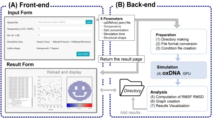
We constructed an execution environment for oxDNA simulation on a web server that provides a GUI of simple online forms and returns the visualized simulation results. The web server was built on Apache Tomcat software for communicating via a hypertext transfer protocol (HTTP) and bridging the user input to the execution of simulations. The concept of our system is shown in Fig. 1. The front-end, the user side of our system, provides an input form for selecting the simulation conditions and sending design files to the server, and provides a result page for checking and downloading the analysis results. The back-end or the server side of the system executes a batch program operating the entire simulation and analysis process, including file import, data conversion, execution of oxDNA, visual rendering, and graph plotting. Hereafter, the detailed implementation of the web system is explained systematically, starting from the front-end.
2.1 Input form in front-end
The front-end program runs on the user’s web browser, and is written in three languages: HTML, CSS, and JavaScript. A user can specify five parameters through the input form: the JSON-formatted caDNAno file, temperature of the solution, concentration of monovalent salt, simulation time, and lattice type of DNA origami. The default parameters of temperature and the salt concentration are 25°C and 0.1 mol/L, respectively, minimizing the user’s operation.
When a user presses the execute button (Run oxDNA button) on the input form, the caDNAno file and simulation conditions are sent to the server side by the POST procedure of HTTP. The server responds with a redirect command to the browser, resulting in an immediate display of the result page to the user. The transition from the input form to the result page occurs asynchronously with the execution of the time-consuming simulation, which considerably reduces user stress. Each time the users reload the result page, the contents are updated along with the simulation progress. Empirically, launching a simulation using our server requires less than 1 minutes if a caDNAno JSON file is ready because a user is supposed to specify only 5 parameters before pressing the “run” button.
2.2 Processes in back-end
As shown in Figure 1, there are seven processes in the back-end of the simulation. The first three are pre-processes including (1) making an empty target directory, (2) converting the file format of
Chem-Bio Informatics Journal, Vol.21, pp.28–38 (2021)
caDNAno to oxDNA, and (3) creating an input file for the simulation. The file conversion (2) is performed using a Python script that is distributed as a utility program for oxDNA. The input file (3) for the simulation is obtained by overwriting an example file of oxDNA, replacing parameters such as simulation time, ion concentration, and temperature with the values specified by the user. In addition, the unoccupied GPGPU of the server to run the simulation is assigned automatically, whereas an error is returned to the user if all the GPGPU are occupied.
(4) The actual oxDNA simulation is run on a GPGPU of the server, generating intermediate files in the target directory made in (1). The results of the simulation are then passed to three post-processes including (5) computation of RMSF and RMSD, (6) creating transition graphs of RMSD and energy, and (7) visualization. The computation algorithm for RMSF and RMSD is coded in a homemade script that runs on a general-purpose molecular dynamics viewer called VMD [26]. The graphs are plotted using a standard software called gnuplot and are reshaped using Inkscape. Visualization of the simulation space from the front, side, top, and perspective views is rendered using VMD and Cogli-1 [27] by choosing representative 100 frames from the simulation. Because processes (4) – (7) are computationally heavy, a user can check their progress on the result page as mentioned in the previous section.
Following is the specification of the computer, operating system (OS), and the driver used for the server. Intel Core i9-9900X, 32GiB memory, 256GB SSD (for OS and software), RAID10 4TB HDD (for data storage), NVIDIA GeForce RTX2080 and RTX2080Ti, Ubuntu 18.04.5, CUDA driver version 450.102.04.
Figure 1. Diagram of the entire system
(A) There are two web pages in the front-end: the input form and result page. Once the simulation is launched, 5 parameters set by a user are sent to the back-end, redirecting a user to the result page. (B) There are 7 processes taking place systematically in the back-end: (1) making a new directory, (2) converting file formats, (3) creating a condition file, (4) running the oxDNA simulation, (5) computing RMSF and RMSD, (6) creating graphs, and (7) visualizing the results. Processes (2–7) make files in the directory. The result page shows the contents of the directory.
Chem-Bio Informatics Journal, Vol.21, pp.28–38 (2021)
2.3 Results page in the front-end
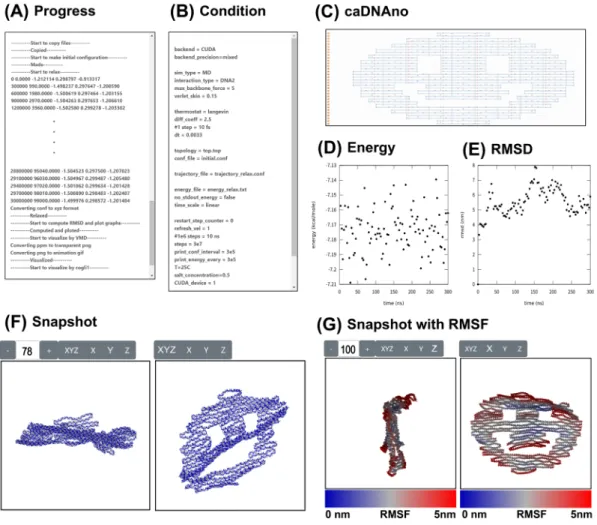
The simulation results are displayed on the result page. A program on the result page coded in JavaScript dynamically rewrites the HTML of the page to show the rendered images and plotted graphs of the current results. The simulation results are shown in seven blocks arranged concisely in a single page for the user’s convenience (Fig. 2).
The “Progress” block displays a text message indicating the simulation progress (Fig. 2A). “Condition” shows all the parameters specified by the user in the simulation (Fig. 2B), and “caDNAno” shows the blueprint of the nanostructure to be analyzed (Fig. 2C). “Energy calculation” plots the time-based evolution of the sum of mechanical energies of the structure. We can evaluate the thermal stability of the structure by the convergence of this value (Fig. 2D). “RMSD” is a plot of the time-based evolution of averaged deviation from the initial position (Fig. 2E). Like “Energy,” the kinetic stability of the structure depends on the convergence of this index. “Snapshot” shows the frames taken from the simulation (Fig. 2F) as a movie. “Snapshot with RMSF” shows the last part of the movie with the RMSF value by a color code (Fig. 2G). It provides visual information about the unstable part of the structure.
Figure 2. Result page
(A) Progress of simulation. (B) List of simulation parameters. (C) Design blueprint using caDNAno software. (D) Graph of time-based evolution of the calculated sum of mechanical energies. (E) Graph of time-based evolution of the average RMSD of the whole entity. (F) Snapshot. 101 frames taken at every 1/100 time of the simulation. (G) Snapshot with RMSF. The last 26 frames from the above 101 frames with RMSF values represented by a color code.
Chem-Bio Informatics Journal, Vol.21, pp.28–38 (2021)
2.4 Advantages of proposed web server
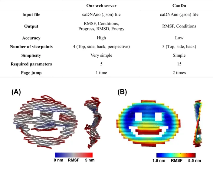
Table 1 shows a comparison between our web server and CanDo. In both systems, the input is given in the JSON format, and the commonly available visual output is RMSF. In addition to RMSF, our server provides RMSD and total mechanical energy, which are not available in CanDo. As an example, Fig.3 shows the end-point snapshots from both simulations of a smiley-face shaped DNA origami. Both results agree with each other in terms of the RMSF.
Although the user interface is designed to be as simple as possible to minimize the necessary input parameters and page transitions, our server provides rich information from the simulation results. Moreover, its usability is improved by providing the current status of the system, which realizes a stress-free simulation environment.
Table 1. Comparison of our server and CanDo
Our web server CanDo
Input file caDNAno (.json) file caDNAno (.json) file
Output Progress, RMSD, Energy RMSF, Conditions, RMSF, Conditions
Accuracy High Low
Number of viewpoints 4 (Top, side, back, perspective) 3 (Top, side, back)
Simplicity Very simple Simple
Required parameters 5 15
Page jump 1 time 2 times
Figure 3. Comparison of RMSF analysis by web server and CanDo
(A) oxDNA simulation with conditions: Time was 3 × 10⁷ steps equivalent to 300 nanoseconds; Temperature was 25°C; Salt concentration was 0.5 mol/L. (B) CanDo simulation.
Chem-Bio Informatics Journal, Vol.21, pp.28–38 (2021)
3. Demonstration: wireframe structure analysis on the web server
To demonstrate the performance of our web server, we analyzed a simple wireframe structure. As an example, a square lattice structure consisting of four rigid double-stranded DNA edges joined by flexible single-stranded DNA hinges was designed using caDNAno (Fig. 4A).
Chem-Bio Informatics Journal, Vol.21, pp.28–38 (2021)
Figure 4. Analysis of a wireframe structure on the web server and CanDo
(A) Left: schematic diagram of the example structure. Right: a blueprint of caDNAno. (B) Rendered images obtained from the simulation results using oxDNA at each temperature. The colors represent RMSF. Blue is 0 nm, and red is 5 nm or larger. From left to right, the temperatures are −20°C, 25°C, 65°C, and 80°C. (C) Histogram of the RMSF distribution at each temperature. The horizontal axis represents the RMSF (nm), whereas the vertical axis represents the number of entities. (D) Rendered image obtained from simulation results of the lattice structure incorporating the bridge. From left to right, the length of the brace is 0, 15, 30, and 45 bp. (E) Graph of the lattice angle with respect to brace length. The horizontal axis represents the length of the brace (bp), whereas the vertical axis represents the angle (deg). The dashed line shows the result of linear fitting, and the error bars show the standard deviation over different frames. (F) Finite element simulation with CanDo. The colors represent the RMSF. Blue indicates 3.8 nm or less, red indicates 15.1 nm or larger. Simulations are as follows: (B) Time was 3 × 10⁶ steps, equivalent to 30 nanoseconds, and salt concentration was 0.1 mol/L, whereas in (D) Time was 3 × 10⁶ steps, equivalent to 30 nanoseconds, temperature was 25°C, and salt concentration was 0.1 mol/L.
First, we analyzed the stability of the structure. Usually, we assume room temperature (typically 25°C) in the simulation. However, when considering enzymatic reactions [28] or strand displacement reactions, the temperature should be increased to some extent. In these cases, it is important to investigate the heat resistance of the structure. For a simple double-stranded DNA, the denaturation temperature can be predicted from the base sequence; however, for structures such as DNA origami, such prediction is difficult without thermodynamic simulation. In the CanDo simulation, the solution temperature cannot be changed; therefore, oxDNA is needed to investigate thermal stability. We examined the stability of the structure by performing simulations at temperatures ranging from −20°C to 80°C (Fig. 4B). At −20°C, the RMSFs of most of the entities in the structure were close to 0 nm, indicating that the structure was quite stable. As expected, the distribution of RMSF values shifted to the right due to thermal fluctuations as the temperature increased to 25°C or 65°C. When the temperature reached 80°C, the structure could not maintain its shape, and denaturation of DNA was observed. All simulation data could be downloaded, enabling further analysis. For example, a histogram of the RMSFs could be created (Fig. 4C) based on the rmsf.txt file.
Along with the temperature, small structural differences result in large changes in shape. To show this through simulation, we added braces made of double-stranded DNA with adjustable length at two diagonal corners of the lattice. We simulated the structures with brace lengths ranging from 0 to 45 bp (Fig. 4D). The results confirmed that the shape became narrow and squashed when the brace length was 0 bp, whereas it maintained a shape close to a square when the brace length was 45 bp. To investigate this quantitatively, we extracted the coordinates of the four vertices of the lattice and calculated the average angle. By plotting the results against the brace length, we confirmed that the angle was increased in proportion to the brace length (Fig. 4E).
The results of the CanDo simulation for a structure without braces are shown in Figure 4F. While the simulation was completed without errors, the initial placement on a lattice lingered. Although the new version of CanDo can handle wireframe structures [23], the current online version that only accepts a lattice-based caDNAno format is not suitable for simulating such lattice-free structures.
4. Conclusion
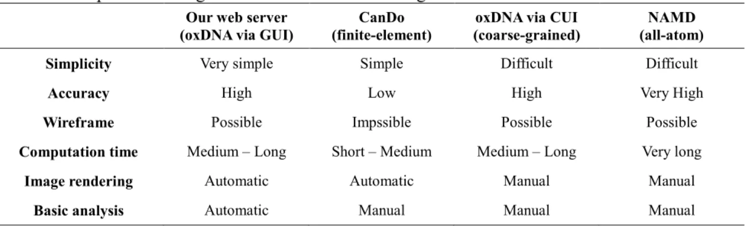
To highlight the merits of our proposal, a comparison of our web server with conventional DNA origami simulation tools is shown in Table 2. Here, our proposal, the conventional command-line oxDNA, CanDo web server, and command-line NAMD [29] are listed from the left to right. The criteria include “Simplicity” (i.e., number of parameters and page jumps), “Accuracy” (i.e., resolution
Chem-Bio Informatics Journal, Vol.21, pp.28–38 (2021)
in time and space), simulation of “Wireframe,” “Computation time,” visualization by “Image rendering,” and automated “Basic analysis” (i.e., RMSD and energy calculation).
Our web server has advantages in terms of simplicity, automated image rendering, and computation of RMSD, RMSF, and energy transition compared with conventional command-line oxDNA. The benefit of GUI over CUI is that a user does not need to acquire computer skills such as programming, writing scripts, mastering commands, and so on. This comparison clarifies that our web server is easy to use for beginners in simulation as well as for experts conducting laboratory experiments.
Since the GeForce license does not allow to use the server as a data center, the website is not open to the public at this moment. Those who want to test their own DNA nanostructure need to contact the authors. If one wants to test our interface by their own computer, it is possible to download the source codes necessary to setup the server [30]. Nevertheless, the concept of using the web server with an easy-to-use interface for oxDNA simulation is clearly shown by the developed prototype that is explained throughout this paper. In the future, it will be meaningful to open our prototype as a service available by public and maintain it with a reasonable financial and technical supports.
A trade-off between accuracy and computation time always exists. The oxDNA has a higher computational cost but higher accuracy than CanDo, whereas it has less accuracy but a shorter computation time than NAMD. Owing to the versatility of oxDNA, it can deal with classical lattice-based DNA structures as well as wireframe-lattice-based structures. Overall, our web server has great potential to accelerate the design process of DNA nanostructures for general users.
Table 2. Comparison among simulation tools of DNA origami
Our web server
(oxDNA via GUI) (finite-element) CanDo (coarse-grained) oxDNA via CUI (all-atom) NAMD
Simplicity Very simple Simple Difficult Difficult
Accuracy High Low High Very High
Wireframe Possible Impssible Possible Possible
Computation time Medium – Long Short – Medium Medium – Long Very long
Image rendering Automatic Automatic Manual Manual
Basic analysis Automatic Manual Manual Manual
Acknowledgement
The authors thank Sho Aradachi for helping build the web server, and Shoji Iwabuchi for giving feedback and constructive comments. This project is supported by JSPS KAKENHI Grant Numbers 18K18144, 19KK0261, 20H00618, 20K20979, 20H05969, 20H05971, and Program for Creation of Interdisciplinary Research of Frontier Research Institute for Interdisciplinary Sciences Tohoku University.
References
[1] Seeman, N. C.; Sleiman, H. F. DNA Nanotechnology. Nat. Rev. Mater. 2017, 3, 17068.
Chem-Bio Informatics Journal, Vol.21, pp.28–38 (2021)
[2] Nummelin, S.; Kommeri, J.; Kostiainen, M. A.; Linko, V. Evolution of Structural DNA Nanotechnology. Adv. Mater. 2018, 30 (24), 1703721. doi:10.1002/adma.201703721
[3] Watson, J. D.; Crick, F. H. C. Molecular Structure of Nucleic Acids. Nature 1953, 171 (4356),
737–738. doi:10.1097/BLO.0b013e3181468780
[4] Seeman, N. C. Nucleic Acid Junctions and Lattices. J. Theor. Biol. 1982, 99 (2), 237–247.
doi:10.1016/0022-5193(82)90002-9
[5] Rothemund, P. W. K. Folding DNA to Create Nanoscale Shapes and Patterns. Nature 2006, 440
(7082), 297–302. doi:10.1038/nature04586
[6] Suzuki, Y.; Kawamata, I.; Mizuno, K.; Murata, S. Large Deformation of a DNA‐Origami Nanoarm Induced by the Cumulative Actuation of Tension‐Adjustable Modules. Angew. Chem.
Int. Ed. 2020, 59 (15), 6230–6234. doi:10.1002/anie.201916233
[7] Tomaru, T.; Suzuki, Y.; Kawamata, I.; Nomura, S. M.; Murata, S. Stepping Operation of Rotary DNA Origami Device. Chem. Commun. 2017, 53, 7716–7719. doi:10.1039/C7CC03214E
[8] Douglas, S. M.; Bachelet, I.; Church, G. M. A Logic-Gated Nanorobot for Targeted Transport of Molecular Payloads. Science 2012, 335 (6070), 831–834. doi:10.1126/science.1214081
[9] Krishnan, S.; Ziegler, D.; Arnaut, V.; Martin, T. G.; Kapsner, K.; et al. Molecular Transport through Large-Diameter DNA Nanopores. Nat. Commun. 2016, 7, 12787.
doi:10.1038/ncomms12787
[10] Shen, H.; Wang, Y.; Wang, J.; Li, Z.; Yuan, Q. Emerging Biomimetic Applications of DNA Nanotechnology. ACS Appl. Mater. Interfaces 2018, 11 (15), 13859–13873.
doi:10.1021/acsami.8b06175
[11] Andersen, E. S. Prediction and Design of DNA and RNA Structures. New Biotechnol. 2010, 27
(3), 184–193. doi:10.1016/j.nbt.2010.02.012
[12] Kekic, T.; Barisic, I. In Silico Modelling of DNA Nanostructures. Comput. Struct. Biotechnol.
J. 2020, 18, 1191–1201. doi:10.1016/j.csbj.2020.05.016
[13] Douglas, S. M.; Marblestone, A. H.; Teerapittayanon, S.; Vazquez, A.; Church, G. M.; et al. Rapid Prototyping of 3D DNA-Origami Shapes with CaDNAno. Nucleic Acids Res. 2009, 37
(15), 5001–5006. doi:10.1093/nar/gkp436
[14] Uchida, T.; Abe, K.; Endo, Y.; Ichiseki, S.; Akita, S.; et al. Revolving Vernier Mechanism Controls Size of Linear Homomultimer. Small 2017, 13 (41), 1702158.
doi:10.1002/SMLL.201702158
[15] Liu, S.; Murata, S.; Kawamata, I. DNA Ring Motif with Flexible Joints. Micromachines 2020,
11 (11), 987. doi:10.3390/mi11110987
[16] Simmel, S. S.; Nickels, P. C.; Liedl, T. Wireframe and Tensegrity DNA Nanostructures. Acc.
Chem. Res. 2014, 47 (6), 1691–1699. doi:10.1021/ar400319n
[17] Benson, E.; Mohammed, A.; Gardell, J.; Masich, S.; Czeizler, E.; et al. DNA Rendering of Polyhedral Meshes at the Nanoscale. Nature 2015, 523 (7561), 441–444.
doi:10.1038/nature14586
[18] Watanabe, T.; Sato, Y.; Otaka, H.; Kawamata, I.; Murata, S.; et al. DNA Origami “Quick” Refolding inside of a Micron-Sized Compartment. Molecules 2019, 25 (1), 8.
doi:10.3390/molecules25010008
[19] Castro, C. E.; Kilchherr, F.; Kim, D.N.; Shiao, E. L.; Wauer, T.; Wortmann, P.; Bathe, M.; Dietz, H. A Primer to Scaffolded DNA Origami. Nat. Methods 2011, 8 (3), 221–229.
doi:10.1038/nmeth.1570
[20] Kim, D.N.; Kilchherr, F.; Dietz, H.; Bathe, M. Quantitative Prediction of 3D Solution Shape and Flexibility of Nucleic Acid Nanostructures. Nucleic Acids Res. 2012, 40 (7), 2862–2868.
doi:10.1093/nar/gkr1173
Coarse-Chem-Bio Informatics Journal, Vol.21, pp.28–38 (2021)
Grained Model of DNA. Phys. Rev. Lett. 2010, 104 (17), 178101.
doi:10.1103/PhysRevLett.104.178101
[22] Ouldridge, T. E. Coarse-Grained Modelling of DNA and DNA Self-Assembly. J. Chem. Inf.
Model. 2012, 53 (9), 1689–1699. doi:10.1007/978-3-642-30517-7
[23] Pan, K.; Kim, D. N.; Zhang, F.; Adendorff, M. R.; Yan, H.; et al. Lattice-Free Prediction of Three-Dimensional Structure of Programmed DNA Assemblies. Nat. Commun. 2014, 5, 5578.
doi:10.1038/ncomms6578
[24] Doye, J. P. K.; Ouldridge, T. E.; Louis, A. A.; Romano, F.; Šulc, P.; et al. Coarse-Graining DNA for Simulations of DNA Nanotechnology. Phys. Chem. Chem. Phys. 2013, 15 (47), 20395.
doi:10.1039/c3cp53545b
[25] Roodhuizen, J. A. L.; Hendrikx, P. J. T. M.; Hilbers, P. A. J.; de Greef, T. F. A.; Markvoort, A. J. Counterion-Dependent Mechanisms of DNA Origami Nanostructure Stabilization Revealed by Atomistic Molecular Simulation. ACS Nano, 2019, 13 (9), 10798–10809, doi:
10.1021/acsnano.9b05650
[26] Humphrey, W.; Dalke, A.; Schulten, K. VMD: Visual Molecular Dynamics. J. Mol. Graphics
1996, 14 (1), 33–38. doi:10.1016/0263-7855(96)00018-5
[27] cogli1, simple tool for the visualization of coarse-grained systems, https://sourceforge.net/projects/cogli1/
[28] Kageyama, R.; Kawamata, I.; Tanabe, K.; Suzuki, Y.; Nomura, S.; et al. Construction of T-Motif-Based DNA Nanostructures Using Enzymatic Reactions. ChemBioChem 2018, 19 (8),
873–876. doi:10.1002/cbic.201700682
[29] Phillips, J. C.; Hardy, D. J.; Maia, J. D. C.; Stone, J. E.; Ribeiro, J. V.; et al. Scalable Molecular Dynamics on CPU and GPU Architectures with NAMD. J. Chem. Phys. 2020, 153 (4), 044130.
doi:10.1063/5.0014475 [30] https://ibuki-kawamata.org/