RESEARCH ARTICLE
Short-term and long-term comparisons of
laparoscopy-assisted proximal gastrectomy
with esophagogastrostomy by the double-flap
technique and laparoscopy-assisted total
gastrectomy for proximal gastric cancer
Tomoko Tsumura1, Shinji KurodaID1,2*, Masahiko Nishizaki1, Satoru Kikuchi1,3, Yoshihiko Kakiuchi1, Nobuo Takata1, Atene Ito1,4, Megumi Watanabe1,5, Kazuya Kuwada1,6, Shunsuke KagawaID1,3, Toshiyoshi Fujiwara11 Department of Gastroenterological Surgery, Okayama University Graduate School of Medicine, Dentistry and Pharmaceutical Sciences, Okayama, Japan, 2 Center for Innovative Clinical Medicine, Okayama University Hospital, Okayama, Japan, 3 Minimally Invasive Therapy Center, Okayama University Hospital, Okayama, Japan, 4 Department of Surgery, Tsuyama Chuo Hospital, Tsuyama, Japan, 5 Department of Surgery, Iwakuni Clinical Center, Iwakuni, Japan, 6 Department of Gastroenterological Surgery, Japanese Red Cross Okayama Hospital, Okayama, Japan
Abstract
Background
Although proximal gastrectomy (PG) is a recognized surgical procedure for early proximal gastric cancer, total gastrectomy (TG) is sometimes selected due to concern about severe gastroesophageal reflux. Esophagogastrostomy by the double-flap technique (DFT) is an anti-reflux reconstruction after PG, and its short-term effectiveness has been reported. How-ever, little is known about the long-term effects on nutritional status and quality of life (QOL).
Methods
Gastric cancer patients who underwent laparoscopy-assisted PG (LAPG) with DFT or lapa-roscopy-assisted TG (LATG) between April 2011 and March 2014 were retrospectively ana-lyzed. Body weight (BW), body mass index (BMI), and prognostic nutritional index (PNI) were reviewed to assess nutritional status, and the Postgastrectomy Syndrome Assess-ment Scale (PGSAS)-45 was used to assess QOL.
Results
A total of 36 patients (LATG: 17, LAPG: 19) were enrolled. Four of 17 LATG patients (24%) were diagnosed with Stage�II after surgery, and half received S-1 adjuvant chemotherapy. BW and PNI were better maintained in LAPG than in LATG patients until 1-year follow-up. Seven of 16 LATG patients (44%) were categorized as “underweight (BMI<18.5 kg/m2)” at 1-year follow-up, compared to three of 18 LAPG patients (17%; p = 0.0836). The PGSAS-45 showed no significant difference in all QOL categories except for decreased BW (p = a1111111111 a1111111111 a1111111111 a1111111111 a1111111111 OPEN ACCESS
Citation: Tsumura T, Kuroda S, Nishizaki M,
Kikuchi S, Kakiuchi Y, Takata N, et al. (2020) Short-term and long-Short-term comparisons of laparoscopy-assisted proximal gastrectomy with
esophagogastrostomy by the double-flap technique and laparoscopy-assisted total gastrectomy for proximal gastric cancer. PLoS ONE 15(11): e0242223.https://doi.org/10.1371/ journal.pone.0242223
Editor: Aleksandra Barac, Clinic for Infectious and
tropical diseases, Clinical centre of Serbia, SERBIA
Received: May 28, 2020 Accepted: October 28, 2020 Published: November 12, 2020
Copyright:© 2020 Tsumura et al. This is an open access article distributed under the terms of the Creative Commons Attribution License, which permits unrestricted use, distribution, and reproduction in any medium, provided the original author and source are credited.
Data Availability Statement: All relevant data are
within the manuscript.
Funding: The authors received no specific funding
for this work.
Competing interests: The authors have declared
0.0132). Multivariate analysis showed that LATG was the only potential risk factor for severe BW loss (odds ratio: 3.03, p = 0.0722).
Conclusions
LAPG with DFT was superior to LATG in postoperative nutritional maintenance, and can be the first option for early proximal gastric cancer.
Introduction
Proximal gastrectomy (PG) is described in the Japanese gastric cancer treatment guideline as a surgical procedure that can be considered for proximal tumors where more than half of the dis-tal stomach can be preserved [1]. Though PG is preferable in terms of organ preservation and function preservation, no standard reconstruction method after PG has yet been established, mainly due to the problem of gastroesophageal reflux after surgery [2]. Esophagogastrostomy (EG) with no additional anti-reflux procedure often causes severe reflux esophagitis, and even alternative procedures, such as jejunal interposition (JI), jejunal pouch interposition (JPI), and a double-tract (DT), do not resolve this problem satisfactorily. Unsuccessful reconstruction after PG causes substantial decline in quality of life (QOL) after surgery [3], and for this reason, total gastrectomy (TG) is sometimes selected as the first option at the expense of stomach preserva-tion, although the prognosis after PG is reportedly comparable to that after TG [4,5].
Our first-choice reconstruction method for proximal gastric cancer is PG with the double-flap technique (DFT) reconstruction. DFT reconstruction, also known as the Kamikawa proce-dure, is an anti-reflux EG after PG, first reported by Kamikawa et al in 1998, in which the distal esophagus and the anastomosis are embedded into the submucosal space of the gastric rem-nant and covered by the seromuscular double-flap [6,7]. While DFT reconstruction has been reported to show acceptable short-term outcomes in preventing reflux esophagitis after sur-gery in a multicenter, retrospective study [8], there is little evidence of the long-term outcomes, including patients’ QOL.
In the present study, the feasibility of laparoscopy-assisted PG (LAPG) with DFT recon-struction was compared with that of laparoscopy-assisted TG (LATG) in terms of short-term and long-term effects on body weight (BW) and QOL after surgery. This study will help estab-lish a standard surgical procedure for early proximal gastric cancer.
Methods
Patients
The medical records of patients with gastric cancer who underwent LAPG with D1+ lymph node dissection followed by DFT reconstruction or who underwent LATG with D1+ lymph node dissection followed by Roux-en-Y reconstruction between April 2011 and March 2014 at Okayama University Hospital were reviewed. This study was reviewed and approved by the institutional review board (IRB) of Okayama University Hospital (#1505–022). The IRB waived the requirement to obtain informed consent for this study.
Surgical procedures of LATG and LAPG
In both LATG and LAPG, gastrectomy and lymph node dissection were performed laparosco-pically in a conventional 5-port setting, and reconstruction was performed under direct vision through a small incision placed in the epigastrium. Esophago-jejunal anastomosis and
jejuno-jejunal anastomosis in LATG were conducted using a circular stapler and a linear stapler, respectively. DFT reconstruction was conducted by a complete hand-sewn suturing process, as previously reported [6].
Clinical data
Patients’ characteristics included age, sex, height, BW, body mass index (BMI), preoperative co-morbidities, American Society of Anesthesiologists physical status (ASA-PS) classification, vital capacity (VC), forced expiratory volume 1.0 (FEV1.0), prognostic nutritional index (PNI) and presence or absence of sarcopenia. Histological findings such as histological type, pathological depth of tumor (pT), pathological lymph node metastasis (pN), and pathological stage (pStage) were described according to the 3rd English edition of the Japanese Classification of Gastric Carci-noma [9]. Surgical outcomes included operation time, blood loss, length of skin incision, presence or absence of concurrent cholecystectomy, postoperative complications classified according to the Clavien-Dindo classification, highest body temperature (BT) after surgery, duration of BT �37.5˚C, the number of days until the first flatus after surgery, and the length of hospital stay after surgery. BW was recorded at 1-month, 6-month, and 1-year follow-ups, and PNI was recorded at 6-month and 1-year follow-ups as well. At 1-year follow-up, reflux esophagitis was evaluated by endoscopic examination and classified according to the Los Angeles (LA) classification [10].
QOL survey
At approximately 3 years after surgery, a QOL survey was carried out by mail using the ques-tionnaire of the Postgastrectomy Syndrome Assessment Scale (PGSAS)-45, which consists of a total of 45 questions [11,12]. Based on this questionnaire, the total symptom score and the 7 symptom subscales (SS) for esophageal reflux, abdominal pain, meal-related distress, indiges-tion, diarrhea, constipaindiges-tion, and dumping were calculated. In addiindiges-tion, decreased BW, ingested amount of food per meal, need for additional meals, quality of ingestion, ability to work, dissatisfaction with symptoms, dissatisfaction with meals, dissatisfaction with working, dissatisfaction with daily life, physical component summary, and mental component summary were also assessed. PGSAS-45 was proved to be a valid and reliable questionnaire for assess-ment of living status and QOL in postgastrectomy patients in a previous study [11].
Statistical analysis
Statistical analysis was conducted using JMP software (SAS Institute, Cary, NC, USA). Stu-dent’st-test was used to assess the continuous variables of age, BW, BMI, VC, FEV1.0, and
PNI. The Wilcoxon signed-rank test was used for the other continuous variables of operation time, blood loss, length of skin incision, highest BT, duration of BT �37.5˚C, the number of days until the first flatus after surgery, and the length of hospital stay after surgery. Pearson’s χ2
test was used for the categorical variables of sex, presence of preoperative co-morbidities, ASA-PS, histological type, pT, pN, pStage, concurrent cholecystectomy, postoperative compli-cations, and BMI <18.5 kg/m2. QOL was also compared by the Wilcoxon signed-rank test. Overall survival and recurrence-free survival curves were evaluated using the Kaplan-Meier method. Ap value <0.05 was considered significant.
Results
Patients’ characteristics
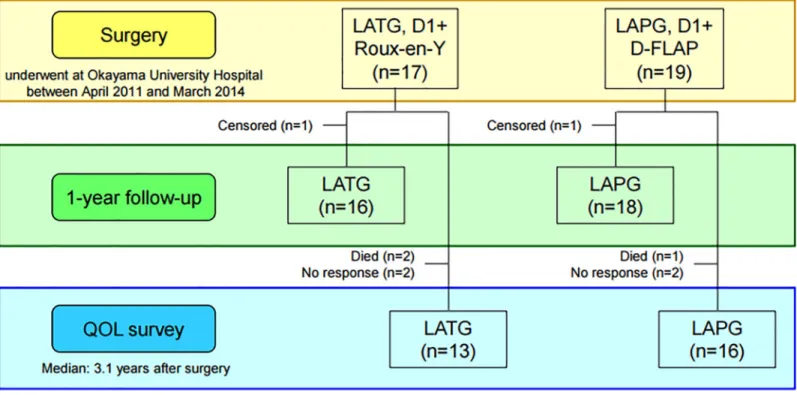
A total of 36 patients were enrolled, 17 patients who underwent LATG and 19 patients who underwent LAPG (Fig 1). In the patients’ characteristics and histological findings (Table 1),
there were no significant differences between LATG and LAPG except for histological type and pStage. The proportion of undifferentiated type was significantly higher in LATG (44%) than in LAPG (5%) (p = 0.0069). All 19 patients who underwent LAPG were diagnosed with
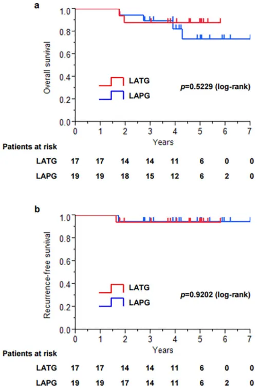
pStage I after surgery, whereas four of 17 patients (24%) who underwent LATG were diag-nosed with pStage II or higher after surgery, although all 17 patients were diagdiag-nosed with clini-cal T1 and N0 before surgery, and 2 of the 4 patients received adjuvant chemotherapy with S-1 after surgery according to the Japanese gastric cancer treatment guidelines 2010 (ver.3) [1]. The Kaplan-Meier survival analysis (median follow-up period, 4.2 years) showed that there were no significant differences between LATG and LAPG in overall survival (p = 0.5229) and
recurrence-free survival (p = 0.9202) (Fig 2).
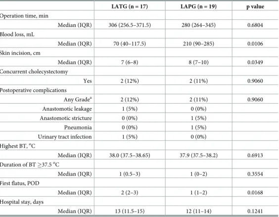
Surgical outcomes
In surgical outcomes (Table 2), operation time was not significantly different between LATG (306 min) and LAPG (280 min) (p = 0.6804), whereas blood loss was significantly higher in
LAPG (210 mL) than in LATG (70 mL) (p = 0.0106). The skin incision placed in the
epigas-trium for reconstruction was significantly longer in LAPG (8 cm) than in LATG (7 cm) (p = 0.0349), which may have been influenced by complexity of reconstruction procedure.
There was no significant difference in the incidence of postoperative complications
(p = 0.9060). In detail, the incidence of postoperative complications (CD any grade) in LATG
was 12% (two of 17 patients) including an anastomotic leakage (5%), while that in LAPG was 11% (two of 19 patients), including an anastomotic stricture (5%). In the postoperative courses, there were no significant differences except for the number of days until the first flatus after surgery, which was significantly higher in LATG (2 days) than in LAPG (1 day)
(p = 0.0168). This meant earlier recovery of bowel function in LAPG and may have led to
Fig 1. CONSORT diagram.
shorter hospital stay in LAPG (12 days) than in LATG (13 days) although difference was not statistically significant (p = 0.1241).
One-year follow-up
Overall, 16 patients who underwent LATG and 18 patients who underwent LAPG were moni-tored for at least until one year after surgery (Fig 1). BW change at 1, 6, and 12 months after surgery (LATG vs LAPG) was -10.4% vs -8.2%, -17.1% vs -10.4%, and -17.8% vs -9.8%, respec-tively; BW loss was significantly smaller in LAPG at every time point (Fig 3A). The same applied to the change in PNI. PNI change at 6 and 12 months after surgery (LATG vs LAPG)
Table 1. Patients’ characteristics and histological findings.
LATG (n = 17) LAPG (n = 19) p value
Age, y Mean± SD 68.9± 12.1 71.2± 8.0 0.5063 Sex Male 11 (65%) 16 (84%) 0.1773 Female 6 (35%) 3 (16%) BMI, kg/m2 Mean± SD 23.6± 3.3 23.5± 3.5 0.9363 Preoperative co-morbidities Yes 11 (65%) 15 (79%) 0.3409 ASA-PS 1 3 (18%) 2 (11%) 0.8266 2 9 (53%) 11 (58%) �3 5 (29%) 6 (32%) VC, % Mean± SD 105.4± 18.2 99.5± 18.2 0.3390 FEV1.0, % Mean± SD 73.1± 9.3 73.1± 10.9 0.9925 PNI Mean± SD 51.3± 6.0 51.1± 5.6 0.9049 Sarcopenia Yes 3 (20%) 4 (24%) 0.8096 Histological type Differentiated 9 (56%) 18 (95%) 0.0069 Undifferentiated 7 (44%) 1 (5%) Pathological T status (pT) 1 14 (82%) 17 (89%) 0.5374 �2 3 (18%) 2 (11%) Pathological N status (pN) 0 14 (82%) 18 (95%) 0.2379 �1 3 (18%) 1 (5%)
Pathological Stage (pStage)
IA 13 (76%) 16 (84%) 0.0270
IB 0 (0%) 3 (16%)
�II 4 (24%) 0 (0%)
BMI, body mass index; VC, vital capacity; FEV1.0, forced expiratory volume 1.0; PNI, prognostic nutritional index; SD, standard deviation. https://doi.org/10.1371/journal.pone.0242223.t001
was -7.3% vs +1.4% and -9.8% vs -3.7%, respectively; PNI was significantly higher in LAPG at every time point (Fig 3B). When BMI<18.5 kg/m2was defined as “underweight”, seven of 16 LATG patients (44%) and three of 18 LAPG patients (17%) were categorized as “underweight” at 1-year follow-up (p = 0.0836), though no patient in both procedures was categorized as
“underweight” before surgery (Fig 3C). Endoscopic examination at 1-year follow-up showed that no LAPG patient had reflux esophagitis, whereas one LATG patient had reflux esophagitis (LA grade C) (Fig 3D).
Fig 2. Kaplan-Meier survival analysis. a. Overall survival. b. Recurrence-free survival.
Postoperative QOL assessment with the PGSAS-45
Twenty-nine of 36 patients responded to the QOL survey by mail (collection rate, 81%), including 13 of 17 LATG patients and 16 of 19 LAPG patients in, and the median time after surgery of these patients at the time of this QOL survey was 3.1 years (Fig 1). There were no significant differences in all categories between LATG and LAPG patients except for decreased BW (p = 0.0132), which was 15.1% in LATG patients and 9.6% in LAPG patients (Table 3). The esophageal reflux SS of LAPG was 1.3, which was as good as the 1.5 of LATG. When BW loss �15% was defined as “severe BW loss”, univariate and multivariate analyses showed that LATG was a potential risk factor for “severe BW loss” (odds ratio: 3.03,p = 0.0722) (Table 4).
Discussion
In the present study, the advantages of LAPG with DFT over LATG were evaluated in terms of short-term and long-term nutritional maintenance. BW and PNI were actually better main-tained after LAPG with DFT than after LATG until 1-year follow up, which was consistent with many previous reports. Sugiyama et al reported that laparoscopic PG (LPG) with DT reconstruction maintained BW and skeletal muscle better than laparoscopic TG (LTG) at 1 year after surgery [13], and Kosuga et al reported that nutritional status, such as changes in BW and blood chemistries including hemoglobin, serum albumin, and total lymphocyte count, was consistently better in LPG with EG than LTG at 6 months and 1 and 2 years after surgery [14]. BMI is a simple value calculated from weight and height, and it is a convenient
Table 2. Surgical outcomes.
LATG (n = 17) LAPG (n = 19) p value
Operation time, min
Median (IQR) 306 (256.5–371.5) 280 (264–345) 0.6804 Blood loss, mL Median (IQR) 70 (40–117.5) 210 (90–285) 0.0106 Skin incision, cm Median (IQR) 7 (6–8) 8 (7–10) 0.0349 Concurrent cholecystectomy Yes 2 (12%) 2 (11%) 0.9060 Postoperative complications Any Gradea 2 (12%) 2 (11%) 0.9060 Anastomotic leakage 1 (5%) 0 (0%) Anastomotic stricture 0 (0%) 1 (5%) Pneumonia 0 (0%) 1 (5%)
Urinary tract infection 1 (5%) 0 (0%)
Highest BT,oC
Median (IQR) 38.0 (37.5–38.65) 37.9 (37.5–38.2) 0.6913
Duration of BT �37.5oC
Median (IQR) 1 (0.5–3) 1 (0–2) 0.3554
First flatus, POD
Median (IQR) 2 (2–3) 1 (1–2) 0.0168
Hospital stay, days
Median (IQR) 13 (11.5–15) 12 (11–14) 0.1241
BT, body temperature; POD, postoperative day; IQR, interquartile range.
aaccording to the Clavien-Dindo classification.
measure representing nutritional status. BMI <18.5 kg/m2is defined as “underweight” in the World Health Organization (WHO) criteria, and being “underweight” is in general associated with a variety of health risks. In gastric cancer as well, “underweight” patients were reported to have the worst overall survival and disease-specific survival among six categories (“under-weight”, “normal-(“under-weight”, “over(“under-weight”, “mildly obese”, “moderately obese”, and “severely obese”) divided according to BMI [15]. In the present study, seven of 16 patients (44%) were “underweight” 1 year after LATG, though no patient was “underweight” before surgery. Two patients died after LATG in the present study, both of whom were “underweight” 1 year after surgery, and the causes of death were gastric cancer death and multiple organ failure. In con-trast, only three of 18 patients (17%) were “underweight” 1 year after LAPG, but one of 4 patients who died after LAPG was “underweight” 1 year after surgery, and the cause of death was pneumonitis. Based on these results, severe BW loss, especially to BMI <18.5 kg/m2, is critical, and, therefore, PG, which can maintain BW better than TG, will be recommended for early proximal gastric cancer from the standpoint of nutritional maintenance and health risks. However, to select PG as a standard procedure for early proximal gastric cancer, a recon-struction method that can effectively maintain the patients’ postoperative QOL is definitely
Fig 3. Follow-up to one year after surgery. a. Change in body weight.�,p<0.05. b. Change in PNI.�,p<0.05. c. Change in BMI. BMI � 18.5 kg/m2is defined as
“underweight”. POY, postoperative year. d. Incidence of reflux esophagitis. https://doi.org/10.1371/journal.pone.0242223.g003
necessary. DFT, one of the EGs with an anti-reflux procedure, has recently been becoming increasingly commonly performed in Japan based on its capability of effectively preventing
Table 3. Postoperative QOL assessment with PGSAS-45.
LATG (n = 13) LAPG (n = 16) p value
Median IQR Median IQR
Symptoms Esophageal reflux 1.5 (1.1–2.5) 1.3 (1.0–2.4) 0.3531 Abdominal pain 1.3 (1.0–1.7) 1.7 (1.0–2.2) 0.5112 Meal-related distress 2.7 (2.0–4.0) 2.3 (1.7–2.7) 0.2509 Indigestion 2.0 (1.8–2.6) 1.9 (1.0–2.7) 0.6419 Diarrhea 2.7 (1.5–3.7) 2.0 (1.2–3.3) 0.5511 Constipation 2.0 (1.3–2.8) 2.5 (1.8–3.3) 0.2607 Dumping 2.0 (1.0–2.7) 1.0 (1.0–2.1) 0.1999 Total symptom 1.9 (1.7–2.8) 1.9 (1.5–2.4) 0.6930 Living status
Decrease in body weight (%) 15.1 (14.5–24.6) 9.6 (5.0–14.4) 0.0132
Ingested amount of food per meal 6.0 (5.0–8.0) 6.5 (5.0–8.0) 0.5314
Necessity for additional meals 2.0 (1.5–2.5) 2.0 (1.0–2.0) 0.3275
Quality of ingestion 4.0 (3.3–4.7) 3.8 (3.0–4.9) 0.7557
Ability for work 2.0 (1.5–2.5) 2.0 (1.0–3.8) 0.6313
QOL
Dissatisfaction with symptoms 1.0 (1.0–2.0) 1.0 (1.0–1.8) 0.5368
Dissatisfaction at the meals 3.0 (1.0–3.0) 3.0 (1.0–3.0) 0.6005
Dissatisfaction at working 1.0 (1.0–2.0) 1.5 (1.0–3.0) 0.4388
Dissatisfaction for daily life 2.0 (1.0–2.5) 1.5 (1.0–2.8) 0.9283
Physical component summary 52.2 (49.5–52.9) 47.9 (41.4–54.3) 0.1604
Mental component summary 51.6 (44.5–55.2) 53.2 (45.6–54.9) 0.9650
https://doi.org/10.1371/journal.pone.0242223.t003
Table 4. Univariate and multivariate analyses of risk factors for severe body weight loss.
Univariate Multivariate p value OR p value Background Age (�80 y) 0.3414 Sex (Male) 0.1552 BMI (�25 kg/m2) 0.0795 0.71 0.3872 Comorbidity (+) 0.1343 ASA-PS (�3) 0.7394 PNI (<50) 0.2920 Sarcopenia (+) 0.9399
Pathological Stage (pStage �II) 0.5714
Operation Operation procedure (LATG) 0.0462 3.03 0.0722
Operation time (�360 min) 0.7064
Blood loss (�300 mL) 0.6569
Postoperative complications 0.4911
Clavien-Dindo (�II) 1.0000
Postoperative chemotherapy (+) 0.6402
BMI, body mass index; PNI, prognostic nutritional index; CI, confidence interval; OR, odds ratio. https://doi.org/10.1371/journal.pone.0242223.t004
gastroesophageal reflux after surgery, leading to a large decline in postoperative QOL [16]. In the present study, reflux esophagitis was not observed in any patients on endoscopic examina-tion at 1-year follow-up after LAPG with DFT, whereas the incidence of reflux esophagitis (LA grade B or higher) after DFT investigated in a previous multicenter retrospective study was 6.0%, which was considered acceptable [8]. The PGSAS-45 questionnaire, in which the severity of gastroesophageal reflux is assessed by the esophageal reflux SS, was used for the QOL survey in the present study. The range of this scale is 1.0 to 5.0, and a smaller value means less symp-toms. In the present study, the esophageal reflux SS of LAPG was 1.3, which was considered relatively good and comparable to that of LATG, in which esophageal reflux is fundamentally unlikely to happen. When the present data of the esophageal reflux SS of LAPG were compared with the previous national multi-institutional data of this scale of the PGSAS-45 after PG [17], in which 115 cases of EG, 34 cases of JI, and 44 cases of JPI were included, by using the PGSAS Statistics Kit available on the web, the esophageal reflux SS of the present study (mean: 1.6, standard deviation [SD]: 0.8) was better than that of the national data (mean: 2.0, SD: 1.0) (p = 0.063, Cohen’s d = 0.41) [18], which may show that DFT has some advantage over a vari-ety of reconstruction methods after PG.
While the present study produced some interesting outcomes, it does have several limita-tions. First, this was a retrospective, single-center study with a limited number of cases. Sec-ond, four LATG patients were diagnosed with advanced stage gastric cancer, and two of them received adjuvant chemotherapy with S-1 for a year after surgery, which may have affected postoperative nutritional status. Third, nutritional status was evaluated only by BW and PNI, and other major nutritional indices, such as the Glasgow Prognostic Score (GPS), the neutro-phil/lymphocyte ratio (NLR), and Controlling Nutritional Status (CONUT score), were not assessed in this study. Then, considering the surgical outcomes, the reason why blood loss was significantly higher in LAPG than in LATG was actually hard to explain, but one possibility might be that gastric fluid overflowing during the DFT procedure was counted as blood loss.
Although a comparison of DFT with other EGs or other types of reconstruction methods such as DT was the most intriguing topic in the evaluation of appropriate reconstruction methods after PG, the present study demonstrated that LAPG with DFT was superior at least to LATG in terms of short-term and long-term nutritional maintenance after surgery, without decreasing QOL. Focusing on BW loss after surgery, this study showed that TG was associated with a higher risk of “underweight” (BMI <18.5 kg/m2) after surgery, which leads to overall health risks irrespective of cancer, and was the only potential risk factor for severe BW loss (>15%) on univariate and multivariate analyses. In conclusion, LAPG with DFT reconstruc-tion can be the first opreconstruc-tion for early gastric cancer located in the proximal stomach from a standpoint of postoperative nutritional maintenance although QOL may not be much different from LATG.
Author Contributions
Conceptualization: Shinji Kuroda.
Data curation: Atene Ito, Megumi Watanabe, Kazuya Kuwada.
Formal analysis: Shinji Kuroda, Satoru Kikuchi, Yoshihiko Kakiuchi, Nobuo Takata. Supervision: Masahiko Nishizaki, Shunsuke Kagawa, Toshiyoshi Fujiwara.
Writing – original draft: Tomoko Tsumura. Writing – review & editing: Shinji Kuroda.
References
1. Japanese Gastric Cancer A. Japanese gastric cancer treatment guidelines 2010 (ver. 3). Gastric Can-cer. 2011; 14: 113–23.https://doi.org/10.1007/s10120-011-0042-4PMID:21573742
2. Ueda Y, Shiroshita H, Etoh T, Inomata M, Shiraishi N. Laparoscopic proximal gastrectomy for early gas-tric cancer. Surg Today. 2017; 47: 538–47.https://doi.org/10.1007/s00595-016-1401-xPMID:
27549773
3. Nakamura M, Nakamori M, Ojima T, Katsuda M, Iida T, Hayata K, et al. Reconstruction after proximal gastrectomy for early gastric cancer in the upper third of the stomach: an analysis of our 13-year experi-ence. Surgery. 2014; 156: 57–63.https://doi.org/10.1016/j.surg.2014.02.015PMID:24799083 4. Ichikawa D, Komatsu S, Kubota T, Okamoto K, Shiozaki A, Fujiwara H, et al. Long-term outcomes of
patients who underwent limited proximal gastrectomy. Gastric Cancer. 2014; 17: 141–5.https://doi.org/ 10.1007/s10120-013-0257-7PMID:23558459
5. Jung DH, Ahn SH, Park DJ, Kim HH. Proximal Gastrectomy for Gastric Cancer. J Gastric Cancer. 2015; 15: 77–86.https://doi.org/10.5230/jgc.2015.15.2.77PMID:26161281
6. Kuroda S, Nishizaki M, Kikuchi S, Noma K, Tanabe S, Kagawa S, et al. Double-Flap Technique as an Antireflux Procedure in Esophagogastrostomy after Proximal Gastrectomy. J Am Coll Surg. 2016; 223: e7–e13.https://doi.org/10.1016/j.jamcollsurg.2016.04.041PMID:27157920
7. Muraoka A, Kobayashi M, Kokudo Y. Laparoscopy-Assisted Proximal Gastrectomy with the Hinged Double Flap Method. World J Surg. 2016; 40: 2419–24.https://doi.org/10.1007/s00268-016-3510-5 PMID:27094564
8. Kuroda S, Choda Y, Otsuka S, Ueyama S, Tanaka N, Muraoka A, et al. Multicenter retrospective study to evaluate the efficacy and safety of the double-flap technique as antireflux esophagogastrostomy after proximal gastrectomy (rD-FLAP Study). Ann Gastroenterol Surg. 2019; 3: 96–103.https://doi.org/10. 1002/ags3.12216PMID:30697614
9. Japanese Gastric Cancer A. Japanese classification of gastric carcinoma: 3rd English edition. Gastric Cancer. 2011; 14: 101–12.https://doi.org/10.1007/s10120-011-0041-5PMID:21573743
10. Lundell LR, Dent J, Bennett JR, Blum AL, Armstrong D, Galmiche JP, et al. Endoscopic assessment of oesophagitis: clinical and functional correlates and further validation of the Los Angeles classification. Gut. 1999; 45: 172–80.https://doi.org/10.1136/gut.45.2.172PMID:10403727
11. Nakada K, Ikeda M, Takahashi M, Kinami S, Yoshida M, Uenosono Y, et al. Characteristics and clinical relevance of postgastrectomy syndrome assessment scale (PGSAS)-45: newly developed integrated questionnaires for assessment of living status and quality of life in postgastrectomy patients. Gastric Cancer. 2015; 18: 147–58.https://doi.org/10.1007/s10120-014-0344-4PMID:24515247
12. Isozaki H, Matsumoto S, Murakami S, Takama T, Sho T, Ishihara K, et al. Diminished Gastric Resection Preserves Better Quality of Life in Patients with Early Gastric Cancer. Acta Med Okayama. 2016; 70: 119–30.https://doi.org/10.18926/AMO/54191PMID:27094837
13. Sugiyama M, Oki E, Ando K, Nakashima Y, Saeki H, Maehara Y. Laparoscopic Proximal Gastrectomy Maintains Body Weight and Skeletal Muscle Better Than Total Gastrectomy. World J Surg. 2018; 42: 3270–6.https://doi.org/10.1007/s00268-018-4625-7PMID:29691620
14. Kosuga T, Ichikawa D, Komatsu S, Okamoto K, Konishi H, Shiozaki A, et al. Feasibility and Nutritional Benefits of Laparoscopic Proximal Gastrectomy for Early Gastric Cancer in the Upper Stomach. Ann Surg Oncol. 2015; 22 Suppl 3: S929–35.https://doi.org/10.1245/s10434-015-4590-4PMID:25986867 15. Lee JH, Park B, Joo J, Kook MC, Kim YI, Lee JY, et al. Body mass index and mortality in patients with
gastric cancer: a large cohort study. Gastric Cancer. 2018; 21: 913–24. https://doi.org/10.1007/s10120-018-0818-xPMID:29651648
16. Hayami M, Hiki N, Nunobe S, Mine S, Ohashi M, Kumagai K, et al. Clinical Outcomes and Evaluation of Laparoscopic Proximal Gastrectomy with Double-Flap Technique for Early Gastric Cancer in the Upper Third of the Stomach. Ann Surg Oncol. 2017; 24: 1635–42.https://doi.org/10.1245/s10434-017-5782-x PMID:28130623
17. Takiguchi N, Takahashi M, Ikeda M, Inagawa S, Ueda S, Nobuoka T, et al. Long-term quality-of-life comparison of total gastrectomy and proximal gastrectomy by postgastrectomy syndrome assessment scale (PGSAS-45): a nationwide multi-institutional study. Gastric Cancer. 2015; 18: 407–16.https://doi. org/10.1007/s10120-014-0377-8PMID:24801198
18. Matsuhashi N, Kudo M, Yoshida N, Murakami K, Kato M, Sanuki T, et al. Factors affecting response to proton pump inhibitor therapy in patients with gastroesophageal reflux disease: a multicenter prospec-tive observational study. J Gastroenterol. 2015; 50: 1173–83. https://doi.org/10.1007/s00535-015-1073-0PMID:25851931