Abbreviations: AIDS, acquired imunodeficiency syndrome; CAEBV, chronic active EBV infection; CTL, cytotoxic T lymphocyte; EA, early antigen; EBNA, EBV-determined nuclear antigens; EBV, Epstein-Barr virus; EBV-AHS, EBV-associated hemophagocytic syndrome; HHV, human herpesvirus; IM, infectious mononucleosis; LMP, latent membrane proteins; LP, leader protein; LPD, lymphoproliferative disease; MHV, murine gammahersesvirus; NK, natural killer; PAL, pyothorax-associated lymphoma; VCA, viral capsid antigen
EBV-associated diseases in humans
Epstein-Barr virus (EBV) is one of eight known human herpesviruses (HHVs) and a member of the gamma herpesvirus family (lymphocryptovirus). EBV was the first human tumor virus identified from cultured lymphoblasts of Burkittʼs lymphoma (Epstein et al., 1964), and its potential role as a causative agent of EBV-associated tumors has been
EBV-Associated Diseases in Humans and their Animal
in vivo
Models: Part I
Kazuhiko Hayashi
Division of Molecular Pathology, Department of Microbiology and Pathology, School of Medicine, Tottori University Faculty of Medicine, Yonago 683-8503 Japan
Epstein-Barr virus (EBV) is one of human herpesviruses and a member of the gamma herpesvirus family (lymphocryptovirus). Infectious mononucleosis, Burkittʼs lymphoma and nasopharyngeal carcinoma are well-known EBV-associated diseases. The range of EBV-associated diseases has recently expanded to include Hodgkinʼs lymphoma, T-cell lymphoma, pyothorax-associated or methotrexate-associated B-cell lymphoma, primary effusion lymphoma and lymphoepithelioma-like carcinoma of the stomach, thymus and salivary gland, lymphoproliferative disorders (LPDs) or leiomyosarcomas from im-munocompromized host, oral hairy leukoplakia and EBV-associated hemophagocytic syndrome. Animal models of human EBV-associated diseases are essential to elucidate the pathogenesis of EBV-infection and EBV-associated diseases. However, only several reports on the animal models of EBV infection have been reported. Here I review the summary of EBV-associated diseases in humans and those previous animal models using EBV or EBV-like herpesviruses and describe some details on our two newly developed rabbit models of LPD induced by simian EBV-like viruses and a mouse model with mu-rine gammaherpesvirus. These animal models are useful and inexpensive alternative experimental model systems for studying the biology and pathogenesis of EBV, and pro-phylactic and therapeutic regimens.
Key words: animal model; EBV-associated disease; human; lymphocryptovirus
the important subject of such investigation for the last 40 years. EBV preferentially infects human B cells, T cells, natural killer (NK) cells, epithe-lial cells and smooth muscle cells (Rickinson and Kieff, 2001). Latent EBV infection occurs in the oropharyngeal epithelium, where EBV virions are replicated and released from the epithelial cells to saliva. The EBV-infected cells express a differ-ent array of EBV-associated antigens depending on lytic or latent infection (Baer et al., 1984; Rowe
et al., 1992), and these viral antigens are targeted by EBV-specific cytotoxic T lymphocytes (CTLs) (Catalina et al., 2001). The CTL responses to EBV infections induce a variety of inflammatory systemic symptoms. On the other hand, the lack of CTLs such as in patients with congenital immu-nodeficiencies or acquired immunodeficiency syn-drome (AIDS) or in recipients receiving a potent immunosuppressant, allows EBV-infected cells to proliferate and result in the development of lethal lymphoproliferative diseases (LPDs). EBV is clas-sically associated with infectious mononucleosis (IM), Burkittʼs lymphoma in equatorial Africa and nasopharyngeal carcinoma (Rickinson and Kieff, 2001). The range of EBV-associated diseases has recently expanded to include oral hairy leukoplakia, leiomyosarcoma from AIDS patients, Hodgkinʼs lymphoma, T-cell lymphoma, Ki-1 lymphoma, pyothorax-associated B-cell lymphoma, methotrex-ate-associated B-cell lymphoma, primary effusion lymphoma, LPDs of primary and secondary im-munodeficiency, and lymphoepithelioma-like car-cinoma of the stomach, thymus, lung and salivary gland (Weiss et al., 1989; Chang et al., 1992; Weiss and Chang, 1996; Anagnostopoulos and Hummel, 1996; Kawa, 2000; Iwatuski et al., 2004) (Table 1, Fig. 1).
Primary EBV infection and its associated diseases
Infectious mononucleosis
Acute IM is clinically characterized by fever, lymphadenopathy, tonsillitis, pharyngolaryngitits and hepatosplenomegaly, and increased IgM and IgG antibodies to EBV-viral capsid antigens (VCAs) and early antigens (EAs). Acute IM is fairly com-mon in the United States and Western Europe, where a primary infection often occurs during adolescence. Asymptomatic primary infections are common in Asia and developing countries because primary EBV infections occur early in life. EBV virions bearing gp350/220 infect B cells via CD21 (CR2) or a receptor for C3d, and form an episomal EBV in the nucleus (Fingeroth et al., 1984). A complex of gp85(gH)/gp25(gL)/gp42 binds to HLA
class II molecules to induce cell membrane fusion in B cells (Molesworth et al., 2000). Binding of the gp42 molecule to HLA class II is essential for virus entry into B cells. Following an incubation pe-riod of 2 to 7 weeks, EBV-infected B cells increase in number during acute IM. The EBV-infected B cells, however, are quickly abrogated by cellular immune responses mediated by NK cells, activated T cells and antibody-dependent cell-mediated cyto-toxicity (Rickinson and Moss, 1997). CD8+, HLA-DR+ activated T cells increase in peripheral blood, and are defined as “mononucleosis” by hemograms when systemic symptoms manifest. During IM as primary EBV infection, EBV is capable of express-ing all viral antigens of lytic cycle: immediate early, early and late proteins, and all antigens of la-tent infection genes: six EBV-determined nuclear antigens (EBNAs) consisting of EBNA-1, -2, -3A, -3B, -3C and -leader protein (LP), and three latent membrane proteins (LMPs), including LMP-1, -2A and -2B (latency type III, Table 1). All viral anti-gens of lytic cycle, all EBNAs and LMPs, except for EBNA-1, are target molecules for EBV-specific cytotoxic T cells. Therefore, CTLs direted to these EBV antigens suppress the EBV-infected cells in immunocompetent hosts.
EBV-associated hemophagocytic syndrome
Hemophagocytic syndrome is an unusual syndrome characterized by fever, splenomegaly, jaundice, pancytopenia, disseminated intravascular coagula-tion, and features of increased hemophagocytic macrophages in the bone marrow and other tissues (Imashuku, 2002). EBV-associated hemophago-cytic syndrome (EBV-AHS) may be associated with acute IM, and EBV-associated LPD, lympho-mas, cancers and autoimmune diseases, and com-monly occurs in children and adolescents in Asia, but is rarely seen in Western countries. Laboratory examinations reveal pancytopenia, liver dysfunc-tion, coagulopathy, elevated levels of lactate dehy-drogenase, ferritin, β2-microglobulin, and serum cytokines including IFN-γ, IL-6, IL-10, sIL-2R, sFas and Fas L (Imashuku, 2002). These cytokines increased in the process of CTL responses directed against EBV-infected CD8+ T cells may induce
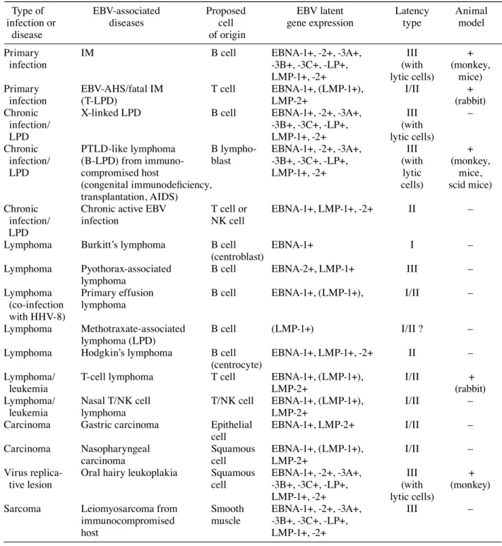
he-Table 1. Comparative overview of EBV-associated diseases (tumors) in humans and their compat-ible animal models
Type of EBV-associated Proposed EBV latent Latency Animal
infection or diseases cell gene expression type model
disease of origin
Primary IM B cell EBNA-1+, -2+, -3A+, III +
infection -3B+, -3C+, -LP+, (with (monkey,
LMP-1+, -2+ lytic cells) mice) Primary EBV-AHS/fatal IM T cell EBNA-1+, (LMP-1+), I/II +
infection (T-LPD) LMP-2+ (rabbit)
Chronic X-linked LPD B cell EBNA-1+, -2+, -3A+, III –
infection/ -3B+, -3C+, -LP+, (with
LPD LMP-1+, -2+ lytic cells)
Chronic PTLD-like lymphoma B lympho- EBNA-1+, -2+, -3A+, III + infection/ (B-LPD) from immuno- blast -3B+, -3C+, -LP+, (with (monkey,
LPD compromised host LMP-1+, -2+ lytic mice,
(congenital immunodeficiency, cells) scid mice)
transplantation, AIDS)
Chronic Chronic active EBV T cell or EBNA-1+, LMP-1+, -2+ II – infection/ infection NK cell
LPD
Lymphoma Burkittʼs lymphoma B cell EBNA-1+ I –
(centroblast)
Lymphoma Pyothorax-associated B cell EBNA-2+, LMP-1+ III – lymphoma
Lymphoma Primary effusion B cell EBNA-1+, (LMP-1+), I/II – (co-infection lymphoma
with HHV-8)
Lymphoma Methotraxate-associated B cell (LMP-1+) I/II ? – lymphoma (LPD)
Lymphoma Hodgkinʼs lymphoma B cell EBNA-1+, LMP-1+, -2+ II – (centrocyte)
Lymphoma/ T-cell lymphoma T cell EBNA-1+, (LMP-1+), I/II +
leukemia LMP-2+ (rabbit)
Lymphoma/ Nasal T/NK cell T/NK cell EBNA-1+, (LMP-1+), I/II –
leukemia lymphoma LMP-2+
Carcinoma Gastric carcinoma Epithelial EBNA-1+, LMP-2+ I/II – cell
Carcinoma Nasopharyngeal Squamous EBNA-1+, (LMP-1+), I/II –
carcinoma cell LMP-2+
Virus replica- Oral hairy leukoplakia Squamous EBNA-1+, -2+, -3A+, III +
tive lesion cell -3B+, -3C+, -LP+, (with (monkey)
LMP-1+, -2+ lytic cells)
Sarcoma Leiomyosarcoma from Smooth EBNA-1+, -2+, -3A+, III – immunocompromised muscle -3B+, -3C+, -LP+,
host LMP-1+, -2+
This table was modified from the tables in the textbook (Rickinson and Kieff, 2001).
EBV, Epstein-Barr virus; EBV-AHS, EBV-sassociated hemophagocytic syndrome; HHV-8, human herpesvirus-8; IM, infectious mononucleosis; LPD, lymphoproliferative disease; PTLD, post-transplant lymphoproliferative dis-ease.
mophagocytosis by activated macrophages. Latent
against EBV antigens, which may allow the survival of EBV-infected NK or T cells.
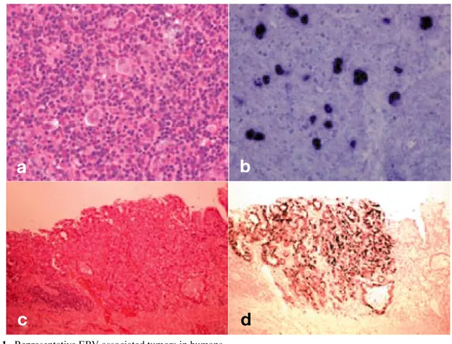
EBV latent infection and tumor development Most EBV-associated LPDs are of B cell lineage, but T cell neoplasms and Hodgkinʼs lymphoma may occur. Latent EBV infection plays a pivotal role in the occurrence of African Burkittʼs lym-phoma, pyothorax-associated lymphomas (PALs), Hodgkinʼs lymphoma (Figs. 1a and b), primary ef-fusion lymphoma induced by HHV-8 coinfection, and various types of B cell lymphomas (Iwatsuki et al., 2004). The association of latent EBV infection with NK/T cell lymphomas is less common than B cell lymphomas. Extranodal NK/T-cell lymphoma, nasal type is more prevalent in Asia, Mexico and Central and South America, and a disease char-acterized by histological features of angiocentric and angiodestructive infiltration of lymphoma cells with severe ulceration or necrosis in nasal cavity. All those LPD except PAL have latency type I or II, whereas latency type III is detected in PAL. PAL is considered to have a localized immunodeficiency in pyothorax lesions.
EBV-associated carcinomas such as nasopha-ryngeal carcinoma and stomach carcinoma (Figs. 1c and d) show latency type I/II. Entry of EBV into epithelial cells that do not express CD21 or HLA class II is mediated by gp85(gH)/gp25(gL) complexes without gp42 (Borza and Hutt-Fletcher, 2002). The restriction of EBV gene expression such as latency type I (or II) showing only EBNA-1 (or LMPs) allows EBV-infected cells to evade im-mune surveillance, and persists throughout latent infection. EBNA-1 is essential for replicating EBV episomes during latency by binding to OriP, a cis-acting element of the EBV genome, and by promot-ing the replication of viral episomes by host cell DNA polymerase. LMP-1 shows oncogene activity with the upregulation of cellular gene expression in various cell types, and prevents apoptosis by the induction of bcl-2 (Rickinson and Kieff, 2001). EBV-AHS, CAEBV and NK/T cell lympho-mas are common in Asia (Iwatsuki et al., 2004). Since EBV gene expression is restricted in these disorders (latency II), only LMPs expressed by EBV-associated LPD or leiomyosarcomas
in immunocompromized indivisuals
In patients with congenital immunodeficiencies or AIDS or in recipients of organ transplantaions receiving a potent immunosuppressant, the lack or suppression of CTLs caused the EBV-associated B-cell LPD with latency type III infection of EBV. EBV is also involved in patients with methotrexate-associated LPD. Leiomyosarcomas may also de-velop in the immunocompromized hosts, although mechanism of EBV infection to smooth muscles has not been clarified (Rickinson and Kieff, 2001). Chronic and latent EBV infection and their associated diseases
X-linked lymphoproliferative syndrome (Duncan disease)
In patients with X-linked lymphoproliferative syn-drome, an inherited immunodeficiency character-ized by increased susceptibility to EBV and B-cell LPD (Nichols et al., 1998), the presence of small deletions and mutations in DSHP/SH2DIA/SLAM-associated protein (SAP) gene was detected. Chronic active EBV infection
Chronic active EBV infection (CAEBV) is a dis-ease of CD4+ T-cell or NK cell LPD characterized by chronic or recurrent infectious mononucleosis– like symptoms persisting over a long time and by abnormally high titers of anti-EBV antibodies and increased levels of EBV-DNA in the peripheral blood (Okano, 1991). CAEBV is also characterized by a high mortality and high morbidity with life-threatening complications, such as virus-associated hemophagocytic synrome, interstitial pneumonia and malignant lymphomas several years after dis-ease onset. Patients with T-cell CAEBV had a shorter survival time than those with NK-cell type of disease (Kimura et al., 2001). Latency type II in-fection of EBV expressing EBNA-1 and LMPs pro-teins is demonstrated in cases with CAEBV. How-ever, patients with CAEBV may have congenital or acquired immunological defects in CTL responses
Fig. 1. Representative EBV-associated tumors in humans. a: Hodgkinʼs lymphoma, hematoxylin and eosin stain.
b: EBER-1+ Hodgkin and Reed-Sternberg cells observed in Hodgkinʼs lymphoma. EBV-EBER-1 ISH. c: Gastric adenocarcinoma, hematoxylin and eosin stain.
d: EBER-1 expression in adenocarcinoma of stomach. EBV-EBER-1 ISH.
the EBV-infected cells are targeted by host CTLs. Despite the absence of overt immunodefi ciency, EBV-infected cells are insuffi ciently abrogated by CTLs in these disorders. Therefore, selective im-munological defects to LMPs as genetic predisposi-tion can be considered in those patients, including an HLA-restricted low response of CTLs to LMPs, immunological tolerance to LMPs and selective de-letion of LMP-specifi c CTLs.
The release of virokines such as vIL-10 and the down-regulation of cell adhesion molecules are additional strategies for EBV-infected cells with latency type I/II to evade the host immune system. vIL-10 inhibits the synthesis of IFN-γ from lympho-cytes and NK cells, and suppresses IFN-γ-mediated cellular events such as the up-regulation of the MHC class I expression and CTL responses. Low levels
of intercellular adhesion molecule 1 and leukocyte function-associated antigen 3 expression are as-sociated with an impaired ability to interact with EBV-specifi c CTL (Iwatsuki et al., 2004).
Most EBV-associated tumors arise with a very long latency in long-term EBV carriers. This sug-gests the multistep oncogenesis through malignant transformation from a single cell within the EBV-infected pool (Rickinson and Kieff, 2001). EBV may require certain risk factors to induce malig-nancy in humans. These risk factors are immuno-logic risk factor, genetic risk factors such as racial predisposition and personal predisposition, and environmental risk factors like Euphorbia tirucalli and malarial infection for African Burkittʼs lym-phoma (Osato, 1998). The translocation of c-myc proto-oncogene and mutation or deletion of genes
b
a
d
like p53 are needed for the development of the most EBV-associated tumors in addition to EBV infec-tion.
Despite intensive investigations on the role of EBV infection in the pathogenesis of EBV-associated tumors, a causal relationship between EBV and these tumors has not been established except for LPD arising in immunosuppressed indi-viduals. Therefore, animal models of human EBV-associated diseases are essential to elucidate the pathogenesis of EBV-infection and EBV-associated diseases.
References
1 Anagnostopoulos I, Hummel M. Epstein-Barr virus in tumours. Histopathology 1996;29:297–315. 2 Baer R, Bankier AT, Biggin MD, Deininger PL,
Farrell PJ, Gibson TJ, et al. DNA sequence and ex-pression of the B95-8 Epstein-Barr virus genome. Nature 1984;310:207–211.
3 Borza CM, Hutt-Fletcher LM. Alternate replication in B cells and epithelial cells switches tropism of Epstein-Barr virus. Nat Med 2002;8:594–599. 4 Catalina MD, Sullivan JL, Bak KR, Luzuriaga
K. Differential evolution and stability of epitope-specific CD8+ T cell responses in EBV infcetion. J Immunol 2001;167:4450–4457.
5 Chang KL, Chen YY, Shibata D, Weiss LM. De-scription of an in situ hybridization methodology for detection of Epstein-Barr virus RNA in paraffin-embedded tissues, with a survey of normal and neo-plastic tissues. Diagn Mol Pathol 1992;1:246–255. 6 Epstein MA, Achong BG, Barr YM. Virus particles
in cultured lymphoblasts from Burkittʼs lymphoma. Lancet 1964;1:702–703.
7 Fingeroth JD, Weis JJ, Tedder TF, Strominger JL, Biro PA, Fearon DT. Epstein-Barr virus receptor of human B lymphocytes is the C3d receptor CR2. Proc Natl Acad Sci USA 1984;81:4510–4514. 8 Imashuku S. Clinical features and treatment
strate-gies of Epstein-Barr virus-associated hemophago-cytic lymphohistiocytosis. Crit Rev Oncol Hematol 2002;44:259–272.
9 Iwatsuki K, Yamamoto T, Tsuji K, Suzuki D, Fujii K, Matsuura H, Oono T. A spectrum of clinical mani-festations caused by host immune responses against Epstein-Barr virus infections. Acta Med Okayama 2004;58:169–80.
10 Kasahara Y, Yachie A, Takei K, Kanegane C, Okada
K, Ohta K, et al. Differential cellular targets of Epstein-Barr virus (EBV) infection between acute EBV-associated hemophagocytic lymphohistio-cytosis and chronic active EBV infection. Blood 2001;98:1882–1888.
11 Kawa K. Epstein-Barr virus-associated diseases in humans. Int J Hematol 2000;71:108–117.
12 Kimura H, Hoshino Y, Kanegane H, Tsuge I, Okamura T, Kawa K, et al. Clinical and virologic characteristic of chronic active Epstein-Barr virus infection. Blood 2001;98:280–286.
13 Molesworth SJ, Lake CM, Borza CM, Turk SM, Hutt-Fletcher LM Epstein-Barr virus gH is essen-tial for penetration of B cells but also plays a role in attachment of virus to epithelial cells. J Virol 2000;74:6324–6332.
14 Nichols KE, Harkin DP, Levitz S, Krainer M, Kolquist KA, Genovese C, et al. Inactivating muta-tions in an SH2 domain-encoding gene in X-linked lymphoproliferative syndrome. Proc Natl Acad Sci USA 1998;95:13765–13770.
15 Okano M, Matsumoto S, Osato T, Sakiyama Y, Thiele GM, Purtilo DT. Severe chronic active Epstein-Barr virus infection syndrome. Clin Micro-biol Rev 1991;4:129–135.
16 Osato T. Epstein-Barr virus infection and onco-genesis. In: Osato T, Takada K, Tokunaga M, eds. Epstein-Barr virus and human cancer. Tokyo: Ja-pan Scientific Societies Press; 1998. p. 3–16 (Gann monograph on cancer research, vol 45).
17 Rickinson AB, Kieff E. Epstein-Barr virus. In: Knipe DM, Howley PM, eds. Fields virology. Philadelphia: Lippincott, Williams & Wilkins; 2001. p. 2575–2627.
18 Rickinson AB, Moss DJ. Human cytotoxic T lym-phocyte responses to Epstein-Barr virus infection. Annu Rev Immunol 1997;15:405–431.
19 Rowe M, Lear AL, Croom-Carter D, Davies AH, Rickinson AB. Three passways of Epstein-Barr vi-rus gene activation from EBNA 1-positive latency in B lymphocytes. J Viol 1992;66:122–131.
20 Weiss LM, Chang KL. Association of the Epstein-Barr virus with hematolymphoid neoplasia. Adv Anat Pathol 1996;3:1–15.
21 Weiss LM, Movahed LA, Warnke R, Sklar J. De-tection of Epstein-Barr viral genomes in Reed-Sternberg cells of Hodgkinʼs disease. N Engl J Med 1989;320:502–506.
Received and accepted January 17, 2005 Corresponding author: Kazuhiko Hayashi, MD