Abbreviations: ADEM, acute disseminated encephalomyelitis; CSF, cerebrospinal fluid; GBS, Guillain-Barré syn-drome; Ig, immunoglobulin; MER, meningo-encephalo-radiculitis; MRI, magnetic resonance imaging
Urinary Retention as an Initial Symptom of Acute
Meningo-Encephalo-Radiculitis in Epstein-Barr Virus Infection
Yasumasa Asai, Ryosuke Fujita, Hiroyuki Nakayasu, Kaori Suzuki, Taizo Kaneto* and Kenji Nakashima†
Clinic of Neurology, Tottori Prefectural Central Hospital, Tottori 680-0901, *Clinic of Neurol-ogy, Tottori Medical Center, Tottori 689-0203 and †Department of NeurolNeurol-ogy, Institute of Neuro-logical Sciences, Tottori University Faculty of Medicine, Yonago 683-8503 Japan
A 48-year-old man presented with urinary retention followed by disturbance of con-sciousness, areflexia, ophthalmoplegia, muscle weakness, and atrophy. Epstein-Barr virus DNA by PCR was positive in his cerebrospinal fluid. The cerebrospinal fluid re-vealed elevated myelin basic protein and an oligoclonal band. Magnetic resonance imag-ing showed high signal intensity in the pons, basal ganglia, and cerebral white matter on T2-weighted imaging and fluid-attenuated inversion recovery imaging. His conscious-ness, ophthalmoplegia, and muscle weakness almost full recovered. In this patient, the inflammation is thought to have begun as sacral radiculitis, and then extended to en-cephalitis, and brachial and lumbar radiculoneuritis.
Key words: acute disseminated encephalitis; Guillain-Barré syndrome; meningo-encephalo-radiculitis; urinary retention
Acute disseminated encephalomyelitis (ADEM) is a monophasic inflammatory demyelinating disor-der of the central nervous system, typically occur-ring after infection or vaccination. The disease is characterized by multifocal white matter lesions on neuroimaging. The polysymptomatic presenta-tion reflects the demyelinating lesions and consists of various combinations of motor, sensory, visual, and cognitive symptoms.
Guillain-Barré syndrome (GBS), which is an acute immune-mediated polyneuropathy with a monophasic course, is classified as acute inflam-matory demyelinating polyneuropathy and acute motor axonal neuropathy. The cardinal clinical features of GBS are weakness, paresthesia, and hyporeflexia. Albuminocytological dissociation is one of the diagnostic findings in GBS. Central nervous system involvement in GBS has been
reported (Baker, 1943; Morley and Reynolds, 1966; Blennow et al., 1968; Gamstorp, 1974; Amit et al., 1986; Laarman et al., 1987; Amit et al., 1992; Okumura et al., 2002; Pfausler et al., 2002; Tsugawa et al., 2004; Chikakiyo et al., 2005). Blennow et al. introduced the term “encephalo-myelo-radiculo-neuropathy” to describe this combined involvement. However, there are a few reports of both GBS and ADEM occurring si-multaneously (Amit et al., 1986, 1992). In these reports, the term “acute severe central and pe-ripheral nervous system combined demyelination” was used to replace the combination of GBS and ADEM. Guillain (1936) has refused to recognize this syndrome as belonging to any condition with a spinal fluid pleocytosis (Pfausler et al., 2002). However, Baker (1943) proposed that GBS might involve any part of the peripheral or the central
nervous system, which would include “encephalo-myelo-radiculitis”. We use the term encephalo-radiculitis (MER), because meningo-encephalitis and radiculitis was obvious and my-elitis was uncertain in our patient.
During the course of ADEM and GBS, mic-turitional disturbance is commonly seen. Lum-bosacral myelitis in ADEM and lumLum-bosacral ra-diculoneuritis in GBS appear to be the main causes of micturitional disturbance. Local lumbosacral radiculitis can cause micturitional disturbance, a condition known as Elsberg syndrome (Hemrika et al., 1985). Even in the presence of Elsberg syn-drome, micturitional disturbance as a first symp-tom of MER is rare (Laarman et al., 1987; Tsugawa et al., 2004; Sasaki et al., 2006). We report a case of MER with Epstein-Barr virus (EBV) infection presenting urinary retention as an initial symptom.
Patient Report
A 48-year-old Japanese man had joint pain in his knee and elbow, lumbago and headache in mid-March, 2006. Five days after the onset, he had urinary retention, and a urinary catheter was inserted intermittently. He was taken to hospital because of the presenting urinary retention and high fever nine days after onset. He developed consciousness disturbance on Day 2 after hospi-talization (10 days after onset).
On physical examination, his body tem-perature was 38.5°C, blood pressure was 155/80 mmHg, and pulse was regular (100/min). There were no cardiac murmurs, rhonchus or crackles. Neurological examination revealed disorientation and a stiff neck. He also could not walk because of muscle weakness in his lower limbs. Deep tendon reflexes were absent in his lower limbs. Neither sensory loss nor paresthesia was observe d. A urinary catheter was inserted because of dif-ficult micturition.
A hematological investigation showed an elevated white blood cell count of 15,440/μL. The blood urea nitrogen level was 65.6 mg/dL, creatinine level was 1.71 mg/dL, natrium level was 131 mEq/L, and C-reactive protein level was 2.78 mg/dL. Titers of antibody herpes simplex virus (HSV), varicella-zoster virus (VZV) and cytomegalovirus (CMV) were not elevated in his serum. Titers of Epstein-Barr virus (EBV) virus capsid antigen immunoglobulin M (IgM) were < 10 (normal range < 10), virus capsid antigen IgG 80 (normal range < 10), early anti-gen IgG < 10 (normal range < 10), Epstein-Barr nuclear antigen 20 (normal range < 10) in his serum. Results of cerebrospinal fluid (CSF) ex-amination revealed the following: 492 cells/mm3
(polymorphonuclear : mononuclear = 1:122), pro-tein level of 299 mg/dL, IgG index 0.79, myelin basic protein (343 pg/mL, control < 102). Oligo-clonal band was positive. Viorogical analysis of
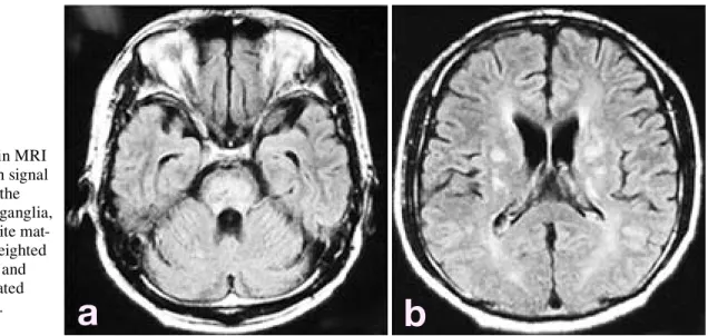
Fig. 1. Brain MRI
showed high signal intensity in the pons, basal ganglia, cerebral white mat-ter on T2 weighted imaging (a) and fluid attenuated
CSF revealed the presence of EBV DNA by PCR, but no HSV, VZV and CMV DNA in the CSF. Magnetic resonance imaging (MRI) showed a normal brain on Day 9 after hospitalization (17 days after onset). Gadolinium enhancement was not done. Single photon emission tomography did not show hypoperfusion or hyperperfusion. Electroencephalography showed a 6- to 7-Hz slow wave. On Day 2 (10 days after onset), viral en-cephalitis was the tentative diagnosis, and vidara-bine was administered intravenously. However, he developed consciousness disturbance and present-ed tonic seizure and tetraplegia, so steroid pulse therapy (methylprednisolone 1000 mg × 3 days) and immunoglobulin (2.5 g × 5 days) were added. He was incubated and required mechanical venti-lation. On Day 5 (13 days after onset), total oph-thalmoplegia presented, and deep tendon reflexes in all limbs and bilateral oculocephalic reflexes were absent. Immunoglobulin (27.5 g × 5 days) was administered because of the suspected Fisher syndrome on Day 12 (20 days after onset). In the clinical course, diffuse muscle atrophy in the up-per and lower limbs was present. On Day 34 (42 days after onset), his consciousness gradually re-covered. Ophthalmoplegia and muscle weakness were almost full recovered and he could walk a few steps. However, he performed intermittent self-catheterization because of voiding difficulties. Orthostatic hypotension was not shown in a head-up tilt test. Coefficient of variation of RR
inter-vals was 1.22%. A sweat test indicated no sweat on his trunk and lower limbs. Phenylephrine eye drops test showed mild mydriasis. Antibody activities against ganglioside GM1, GM2, GM3, GD1a, GD1b, GD3, GT1b, GQ1b, GA1, Gal-C and GalNAc-GD1a were all negative. Fifty days after onset, an electrophysiological examination was performed. Distal complex motor action potential amplitudes were 11.7/8.3 (right/left) mV in his me-dian nerve and 0.8/0.7 mV in his tibial nerve. Mo-tor nerve conduction velocities were 56.5/57.5 mV in his median and 48.6/45.0 mV in his tibial nerve. Terminal latencies were 3.9/4.2 m/s in his median and 6.6/6.8 m/s in his tibial nerve. Sensory nerve action potential velocities were 69.9/64.2 m/s in his median and 38.0/44.0 m/s in his sural nerve. F-wave latencies were 28.7/27.9 ms in his median nerve and 53.9/42.6 ms in his right tibial nerve. Urodynamic study was performed for 82 days after onset. When 210 mL of water was infused he complained first of a desire to void. Bladder volume at maximum desire to void was 430 mL. Atonic cystometrogram was presented.
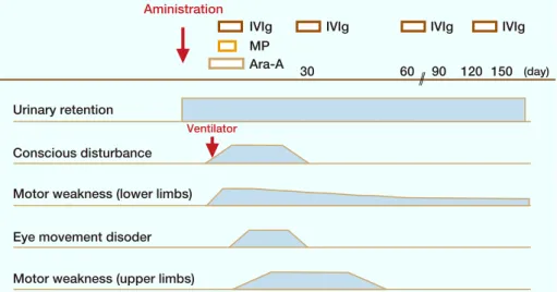
He was transferred to another hospital for further rehabilitation for 168 days after onset. Brain MRI showed high signal intensity in the pons, basal ganglia, cerebral white matter on T2 weighted imaging and fluid attenuated imaging 6 months after onset (Fig. 1). Cervical, thoracic and lumbar MRI were normal. Clinical course was shown in Fig. 2.
Fig. 2. The clinical course of
the present patient. Ara-A, vidarabine; IVIg, intravenous immunoglobulin; MP, methyl-prednisolone. MP (day) Ventilator Urinary retention Conscious disturbance Aministration
Motor weakness (lower limbs)
Motor weakness (upper limbs) Eye movement disoder
IVIg IVIg IVIg IVIg
30 60 90 120 150
Discussion
The patient presented urinary retention as the first symptom, followed by MER. There are some reports that urinary retention may precede menin-goencephalitis in GBS or meningo-encephalo-radiculitis as an initial symptom (Laarman et al., 1987; Tsugawa et al., 2004; Sasaki et al., 2006) (Table 1). Tsugawa et al. (2004) reported GBS with meningo-encephalitis in a patient present-ing urinary retention and muscle weakness as in initial symptoms. Laarman et al. (1987) reported a case of meningo-encephalitis and Guillain-Barré type ascending sensorimotor paralysis. In their case, headache and fever were shown as first symptoms, followed by difficult micturition 10 days after onset and consciousness disturbance 12 days after onset. The condition of our patient was similar to that described in these 2 reports; however, urinary retention as a first symptom was
Age (year) Sex Cerebrospinal fluid Precedent infection Cell (/mm3)/protein (mg/dL) Ganglioside antibody Oligoclonal band Myelin basic protein NCV and/or EMG EMG
Motor conduction velocity Sensory conduction velocity F wave
Brain MRI
High intensity area Spinal MRI
Enhanced
not emphasized in these previous reports. Sa-saki et al. (2006) reported 3 male patients with meningo-encephalo-radiculitis presenting with acute urinary retention. It was inferred that they may represent a variant of ADEM. The reports of Laarman et al. (1987) and Tsugawa et al. (2004) also showed peripheral disorder, suggesting the co-existence of GBS. However, demyelination of central nervous system by brain and spinal MRI was uncertain. On the other hand, in the report by Sasaki et al. (2006), central nervous system disorder was presented by MRI, but disorder of peripheral nervous system was uncertain. Our pa-tient presented muscle atrophy during the course of his condition, low amplitude of tibial, delay of right sural sensory nerve and tibial F-wave la-tency. Brain MRI showed high signal intensity in the pons, basal ganglia and cerebral white matter on T2 weighted imaging and fluid attenuated im-aging. Thus disorder of both the central nervous system and peripherall nervous system was
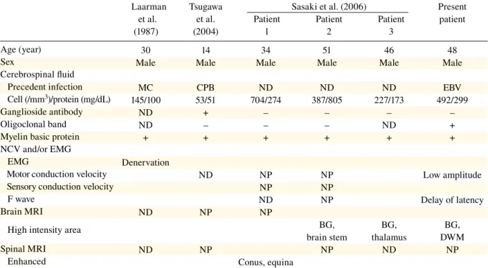
Laarman Tsugawa Sasaki et al. (2006) Present et al. et al. Patient Patient Patient patient (1987) (2004) 1 2 3
30 14 34 51 46 48 Male Male Male Male Male Male
MC CPB ND ND ND EBV 145/100 53/51 704/274 387/805 227/173 492/299 ND + – – – – ND – – – ND + + + + + + + Denervation ND NP NP Low amplitude NP NP ND NP Delay of latency ND NP NP BG, BG, BG, brain stem thalamus DWM ND NP NP ND NP
Conus, equina
Table 1. Clinical comparison among our patient and those reported in literature on urinary retention preceding meningoencephalitis with GBS or meningo-encephalo-radiculitis as the first symptom
BG, basal ganglia; CPB, campylobactor; DWM, deep white matter; EBV, Epstein-Barr virus; EMG, electromyography; GBS, Guillain-Barré syndrome; MC, mycoplasma; MRI, magnetic resonance imaging; NCV, nerve conduction velocity; ND, not detected; NP, not per-formed.
tainly shown in the present patient.
The etiology of urinary retention in our patient is thought to be a variant of the Elsberg syndrome that is generally regarded as a form of lumbosacral radiculopathy (Hemrika et al., 1985). The most common etiologies are viral infections, particularly genital herpes (HSV type 2) (Black et al., 1983; White et al., 1984; Hemrika et al., 1996; Lepori et al., 1992; Herbaut et al., 1990; Eber-hardt et al., 2004). EBV has also been reported as a cause of sacral myeloradiculitis (Sperber et al., 1973). Additionally, EBV has also been reported as a cause of myeloradiculitis and encephalomy-eloradiculitis (Merelli et al., 1997; Majid et al., 2002; Phowthongkum et al., 2007). Elevated CSF cell count is often observed in these condi-tions as in our case. To the best of our knowledge this is the first reported case of acute meningo-encephalo-radiculitis in EBV, in which urinary retention was an initial symptom. Although it remains to be determinated the pathomechanism of radiculitis and/or encephalitis in Epstein-Barr virus infection, our case report would serve as an aid to clarify this pathomechanism.
In the autonomic system examination, the sympathetic and parasympathetic nervous systems were both impaired. Postsynaptic impairment was suggested from the eye drops test. However, brain stem disorder also might affect urinary retention. It is important to keep in mind that urinary retention can be the initial sign of acute central or peripheral demyelination.
References
1 Amit R, Shapira Y, Blank A, Aker M. Acute, severe, central and peripheral nervous system combined de-myelination. Pediatr Neurol 1986;2:47–50.
2 Amit R, Glick B, Itzchak Y, Dgani Y, Meyeir S. Acute severe combined demyelination. Childs Nerv Syst 1992;8:354–356.
3 Baker AB. Guillain-Barre’s disease (encephalo-myelo-radiculitis): a review of 33 cases. Lancet 1943;63:384–398.
4 Black D, Stewart J, Melmed C. Sacral nerve dys-function plus generalized polyneuropathy in herpes
simplex genitalis. Ann Neurol 1983;14:692.
5 Blennow G, Gamstorp I, Rosenberg R. Encephalo-radiculo-neuropathy. Dev Med Child Neurol 1968;10:485–490.
6 Chikakiyo H, Kunishige M, Yoshino H, Asano A, Sumitomo Y, Endo I, et al. Delayed motor and sen-sory neuropathy in a patient with brainstem encepha-litis. J Neurol Sci 2005;234:105–108.
7 Eberhardt O, Kuler W, Dichgans J, Weller M. HSV-2 sacral radiculitis (Elsberg syndrome). Neurology 2004;63:758–759.
8 Gamstorp I. Encephalo-myelo-radiculo-neuropathy: involvement of the CNS in children with Guillain-Barré-Strohl syndrome. Dev Med Child Neurol 1974;16:654–658.
9 Guillain G. Radiculoneuritis with acellular hyper-albuminosis of the cerebrospinal fluid. Arch Neurol Psychiat 1936;36:975.
10 Hemrika DJ, Schutte MF, Bleker OP. Elsberg syn-drome: a neurologic basis for acute urinary reten-tion in patients with genital herpes. Obstet Gynecol 1986;68:37S–39S.
11 Herbaut AG, Nogueira MC, Wespes E. Urinary re-tention due to sacral myeloradiculitis: a clinical and neurophysiological study. J Urol 1990;144:1206– 1208.
12 Laarman GJ, Hoekstra JBL, Donker DNJ, Verbrugh HA, Hekker AC. Meningo-encephalitis and Guil-lain-Barré type ascending sensorimotor paralysis associated with mycoplasma pneumoniae infection. Neth J Med 1987;31:66–71.
13 Lepori P, Marcacci G, Gaglianone S. Elsberg syn-drome: radiculomyelopathy and acute urinary reten-tion in patient with genital herpes. Ital J Neurol Sci 1992;13:373–375.
14 Majid A, Galetta S, Sweeney C, Robinson C, Mahalingam R, Smith J, et al. Epstein-Barr virus myeloradiculitis and encephalomyeloradiculitis. Brain 2002;125:159–165.
15 Merelli E, Bedin R, Sola P, Gentilini M, Pietrosemoli P, Meacci M, et al. Encephalomyeloradiculopathy asso-ciated with Epstein-Barr virus: primary infection or reactivation? Acta Neurol Scand 1997;96:416–420. 16 Morley JB, Reynolds EH. Papilledema and
Landry-Guillain-Barré syndrome. Case report and review. Brain 1966; 89:205–222.
17 Okumura A, Ushida H, Maruyama K, Itomi K, Ishiguro Y, Takahashi M, et al. Guillain-Barré syndrome associated with central nervous system le-sions. Arch Dis Child 2002;86:304–306.
18 Pfausler B, Engelhardt K, Kampf l A, Spiss H, Taferner E, Schmutzhard E. Post-infectious central and peripheral nervous system diseases complicating Mycoplasma pneumoniae infection. Eur J Neurol 2002;9:93–96.
Suankratay C. Basal ganglia and brainstem enceph-alitis, optic neuritis, and radiculomyelitis in Epstein– Barr virus infection. J Infect 2007;54:141–144. 20 Sasa k i M, Oha ra S, Hayash i R, Iwa hash i T,
Tsuyuzaki J. Aseptic meningo-encephalitis pre-senting initially with urinary retention. J Neurol 2006;253:908–913.
21 Sperber A, Tessler AN, Berezeller P. Infectious mononucleosis with acute urinary retention. Urol-ogy 1973;2:456–457.
22 Tsugawa T, Nikaido K, Doi T, Koga M, Susuki K, Kubota T, et al. Guillain-Barré syndrome with
meningoencephalitis after Campylobacter jejuni in-fection. Ped Infect Dis J 2004;23:966–968.
23 White WB, Hanna M, Stewart JA. Systemic herpes simplex virus type 2 infection: proctitis, urinary re-tention, arthralgia, and meningitis in the absence of primary mucocutaneous lesions. Arch Intern Med 1984;144:826–827.
Received June 24, 2008; accepted August 22, 2008. Corresponding author: Yasumasa Asai, MD