タイトル
Title Distribution of diffuse plexiform neurofibroma on thebody surface in patients with neurofibromatosis 1
著者
Auther(s) Ehara, Yuko; Koga, Monji; Imafuku, Shinichi; Yamamoto,Osamu; Yoshida, Yuichi
掲載誌・巻号・ページ
Citation JOURNAL OF DERMATOLOGY , 47 (2) : 190 - 192
刊行日
Issue Date 2020-02
資源タイプ
Resource Type 学術雑誌論文 / Journal Article
版区分
Resource Version 著者版 / Author
権利
Rights (C) 2019 Japanese Dermatological Association
DOI 10.1111/1346-8138.15194
1
Title: Distribution of diffuse plexiform neurofibroma on the body surface in patients with neurofibromatosis 1.
Short title: Plexiform neurofibroma in NF1
The total number of words 1292, figures 2 and table 1 and references 15.
Yuko EHARA1, Monji KOGA2, Shinichi IMAFUKU2, Osamu YAMAMOTO 1, Yuichi
YOSHIDA 1
1Division of Dermatology, Department of Medicine of Sensory and Motor Organs,
Faculty of Medicine, Tottori University, Yonago, Japan
2Department of Dermatology, Fukuoka University School of Medicine, Fukuoka, Japan
Correspondence to: Y Yoshida, M.D.
Division of Dermatology, Department of Medicine of Sensory and Motor Organs, Faculty of Medicine, Tottori University, 86 Nishi-cho, Yonago-shi, Tottori 683-8503, Japan Tel: +81-859-38-6597 Fax: +81-859-38-6599 Email: yxyhifu1@med.tottori-u.ac.jp
2
ABSTRACT
Neurofibromatosis 1 is a genetic disease characterized by cutaneous, neurological and osseous abnormalities. About 20% of patients develop plexiform neurofibroma, resulting in impaired quality of life. To evaluate distribution of diffuse plexiform neurofibroma on the body surface, a retrospective study was conducted for 354 patients with neurofibromatosis 1 from 2007 to 2018 in Japan. We investigated a total of 40 patients with clinically apparent superficial diffuse plexiform neurofibroma. In the cases evaluated, 57.4% of the diffuse plexiform neurofibromas were located on the trunk, 19.2% were on the head and neck, 12.8% were on the lower limbs and 10.6% were on the upper limbs. Remarkably, 75.0% of the diffuse plexiform neurofibromas were located on the dorsal side. The frequency was significantly higher on the trunk than on the head and neck (p = 0.026). Our findings provide useful information for giving attention to the high possibility of diffuse plexiform neurofibroma on the dorsal side before the progression in childhood and for the future treatment in neurofibromatosis 1.
(Key words)
3 INTRODUCTION
Neurofibromatosis 1 (NF1) is an autosomal dominant genetic disease characterized by café-au-lait spots, neurofibromas, freckling, optic gliomas, Lisch nodules and bone deformity.1 It is caused by mutation of the NF1 gene on chromosome 17q11.2.2 The
worldwide prevalence of NF1 is approximately 1 in 3000-4000 individuals with nearly 100% penetrance.3 Diffuse plexiform neurofibroma (PN), known as elephantiasis
neurofibroma or patchydermatocele, is an overgrowth of cutaneous tissue associated with proliferation of neurofibromas.4 Complications of diffuse PN are limited limb movements,
neurological deficits, bleeding due to trauma, malignant transformation and disfigurement.5 It often causes cosmetic and functional disturbances, resulting in impaired
quality of life.6 To our knowledge, there have been a few reports on the assessment of
deeply located PN that was revealed by magnetic resonance imaging (MRI) .7-9 However,
the distribution of diffuse PN on the body surface has not been clarified. Therefore, we studied the features of superficial diffuse PN in patients with NF1.
PATIENTS AND METHODS
Study population (Patients)
A retrospective study was conducted for 354 NF1 patients referred to the Dermatology Department of Tottori University Hospital and the Dermatology Department of Fukuoka
4
University Hospital between 2007 and 2018. We investigated a total of 40 NF1 patients with clinically apparentdiffuse PN that could be recognized by physical examination (16
men and 24 women). All of the patients were evaluated by expert dermatologists and met the diagnostic criteria by National Institutes of Health for NF1.10 Patients with mosaic
NF1 (localized NF1)11 were excluded from our study. Demographic and clinical
information on the age and sex of patients and the size and number of diffuse PNs was obtained from medical records and photographic images. In this study, we included the hanging tumors (>5cm in size) with pigmented macule on the surface skin in most cases as diffuse plexiform neurofibroma. Subcutaneous nodular plexiform neurofibroma (solitary intraneural neurofibroma) was excluded from this study. For each patient, we evaluated the site of diffuse PN on the body surface based on the ratio of distribution of tumors and classified PNs into four sites: head and neck, trunk, upper limbs and lower limbs. Image examinations such as MRI were not necessarily performed. The study protocol was approved by the Ethics Committee of Tottori University Hospital (No. 1704A005) and the Ethics Committee of Fukuoka University Hospital (No. 17-4-03).
Statistical analysis
5
(ventral/dorsal side) between the head and the neck and trunk. P-values < 0.05 were considered statistically significant. All statistical analyses were conducted using EZR (Saitama Medical Center, Jichi Medical University, Saitama, Japan), which is a graphical user interface for R (The R Foundation for Statistical Computing, Vienna, Austria, version 2.13.0). More precisely, it is a modified version of R commander (version 1.32) designed to add statistical functions frequently used in biostatistics.
RESULTS
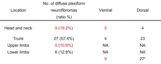
We evaluated 40 NF1 patients (16 men and 24 women [no pregnancy]; median age, 30 years, age range, 0-65 years) with diffuse PN on the body surface. There were 47 PNs in the 40 patients. A representative case (with 2 superficial diffuse PNs on the lower leg) is shown in Fig. 1. Among these cases, 57.4% of the diffuse PNs were located on the trunk, 19.2% were on the head and neck, 12.8% were on the lower limbs and 10.6% were on the upper limbs (TableⅠ). We mapped the distribution of diffuse PNs on the body surface per patient as shown in Fig. 2. and evaluated the predilection sites of the head and neck and the trunk in detail. In addition, we classified the location as the dorsal or ventral side based on the ratio of distribution of tumors on the head and neck and the trunk (Tumors on extremities were excluded from analysis). Interestingly, 75.0% (27/36) of the diffuse PNs were located on the dorsal side. The frequency was significantly higher on the trunk
6 than on the head and neck (p = 0.026).
DISCUSSION
NF1 has cutaneous, neurological and osseous abnormalities, and one of the major features of NF1 is PN (nodular and diffuse types).1 We previously reported that 21% of NF1
patients referred to our dermatology department had diffuse PNs.12 There have been some
studies that focused on deeply located PNs that were investigated by MRI.7-9 Mautner et
al. proposed 3 different growth patterns of PN (superficial, displacing and invasive growth patterns) and reported that the most frequent site of PNs was the head and neck region (36%) in256 tumors of 202 NF1 patients (mean age, 28.3 years).7 They found that
54% of the PNs located on the head and neck region showed an invasive growth pattern. On the other hand, Tucker et al. reported that the most frequent site of PNs was the extremities or the spine (30% each) in 44 tumors of 34 NF1 patients (median age, 10 years) .8 Nguyen et al. showed that the most frequent site was the trunk (38.4%) in 73
PNs of 37 NF1 patients (mean age, 10.5 years).9 In our study, 57.4% of the diffuse PNs
were found to be located on the trunk by evaluation of body surface PNs. Since we could not evaluate deeply located tumors such as tumors in the nearby spine, the relative ratio seems to be higher than that previously reported. In addition, the difference might be
7
related to referral facilities (i.e., department of maxillofacial surgery, department of medical genetics, MRI institute or department of dermatology). Some patients with severe facial diffuse PN might be referred to another department. In the present study, we focused on diffuse PNs that had developed on the surface of the body and found that the 75.0% of diffuse PNs were located on the dorsal side, especially on the dorsal side of the trunk. It is well known that diffuse plexiform neurofibroma is a congenital tumor. However, it is possible not to find the tumor early in childhood because only large pigmented macules were detectable in some patients. Since the tumor increase in size, especially in childhood, it is important for clinician to keep the high possibility of dorsal distribution of diffuse plexiform neurofibroma in mind before the progression in the follow-up period.
It is well known that Schwann cells play an important role in the pathogenesis of neurofibroma in NF1.13 It has been reported that complete loss of NF1 at the late
precursor-early immature Schwann cell stage causes dissociation of non-myelinating Schwann cells from Remak bundles, resulting in the development of PN.14 Schwann cells
are thought to originate from the neural crest. The neural crest is a transient embryonic state that arises between the newly formed ectoderm and the neural tube. While the neural tube gives rise to the central nervous system, embryonic neural crest-derived stem cells
8
migrate from the dorsal region to the ventral region and differentiate into cells of both mesodermal and ectodermal components. Therefore, we speculate that loss of function of the second NF1 allele in Schwann cells during migration is related to the frequent anatomical location of diffuse PN on the dorsal side of the trunk.
There is a number of limitations in our study. Since we investigated diffuse PN that developed on the body surface, image analysis was not necessarily performed. Therefore, we could not evaluate the volume of diffuse PN. In addition, the number of patients was relatively small because the survey was limited to the field of dermatology.
Recent clinical trials have shown promising success in treatment with the MAP kinase inhibitor selumetinib in children with PN.15 Our findings provide useful information for
giving attention to the high possibility of diffuse PN on the dorsal side in early childhood before progression and for treatment of NF1 in the future.
ACKNOWLEDGEMENTS:
This work was partly supported by Health Labour Sciences Research Grant (Y.Y and I.S) from the Ministry of Health, Labour and Welfare in Japan.
9 The authors have no conflict of interest to declare.
REFERENCES
1. Yoshida Y. 71 Neurofibromatosis. In: Kelly, A. P., Taylor, S. C., editors. Taylor and Kelly’s Dermatology for skin of Color 2nd eds. McGrawHill Press; 2016: p. 499-504.
2. Viskochil D, Buchberg AM, Xu G, et al. Deletions and a translocation interrupt a cloned gene at the neurofibromatosis type 1 locus. Cell 1990; 62: 187-192.
3. Lammert M, Friedman JM, Kluwe L, Mautner VF. Prevalence of neurofibromatosis 1 in German children at elementary school enrollment. Arch Dermatol 2005; 141: 71-74. 4. Korf BR. Plexiform neurofibromas. Am J Med Genet 1999; 89: 31-37.
5. Yoshida Y, Ehara Y, Koga M, Imafuku S, Yamamoto O. Epidemiological analysis of major complications requiring medical intervention in patients with neurofibromatosis 1. Acta Derm Venereol 2018; 98: 753-756.
6. Page PZ, Page GP, Ecosse E, Korf BR, Leplege A, Wolkenstein P. Impact of neurofibromatosis 1 on quality of life: a cross-sectional study of 176 American cases. Am J Med Genet A 2006; 140: 1893-1898.
7. Mautner VF, Hartmann M, Kluwe L, Friedrich RE, Funsterer C. MRI growth patterns of plexiform neurofibromas in patients with neurofibromatosis type 1. Neuroradiology 2006; 48: 160-165.
10
Longitudinal study of neurofibromatosis 1 associated plexiform neurofibromas. J Med Genet 2009; 46: 81-85.
9. Nguyen R, Kluwe L, Fuensterer C, Kentsch M, Friedrich RE, Mautner VF. Plexiform neurofibromas in children with neurofibromatosis type 1: frequency and associated clinical deficits. J Pediatr 2011; 159: 652-655.
10. Neurofibromatosis. Conference statement. National Institutes of Health Consensus Development Conference. Arch Neurol 1988; 45: 575-578.
11. Tanito K, Ota A, Kamide R, Nakagawa H, Niimura M. clinical features of 58 Japanese patients with mosaic neurofibromatosis 1. J Dermatol 2014; 41: 724-728.
12. Ehara Y, Yamamoto O, Kosaki K, Yoshida Y. Clinical severity in Japanese patients with neurofibromatosis 1 based on DNB classification. J Dermatol 2017; 44: 1262-1267. 13. Zhu Y, Ghosh P, Charnay P, Burns DK, Parada LF. Neurofibromas in NF1: Schwann cell origin and role of tumor environment. Science 2002; 296: 920-922
14. Parrinello S, Lloyd AC. Neurofibroma development in NF1-insights into tumour initiation. Trends cell Biol 2009; 19: 395-403.
15. Dombi E, Baldwin A, Marcus LJ, et al. Activity of Selumetinib in Neurofibromatosis Type 1-Related Plexiform Neurofibromas. N Engl J Med 2016; 375: 2550-2560.
11
Figure Legends
Fig. 1. A representative case of NF1. There were two superficial diffuse plexiform
neurofibromas on the lower limb (right thigh and knee).
12
Table 1. Clinical features of superficial diffuse plexiform neurofibromas in our study.
Location
No. of diffuse plexiform neurofibromas
(ratio %)
Ventral Dorsal
Head and neck 9 (19.2%) 5 4
Trunk 27(57.4%) 4 23
Upper limbs 5 (10.6%) NA NA
Lower limbs 6 (12.8%) NA NA
9 27*
N.A., not applicable. * p = 0.026