Effects of Testosterone Replacement on Renal Function and Apoptosis
on Mesangial and Renal Tubule Cells in Rats
Kuniyasu Muraoka
Department of Urology and First Department of Pathology, Tottori University Faculty of Medicine, Yonago 683-0826 Japan
The effect of testosterone replacement in middle-aged rats on renal function and apoptosis of mesangial and renal tubule cells was evaluated. Middle-aged (13-month-old) male Wistar rats were divided into 2 groups, one of which was given high-dose and the other low-dose testosterone replacement for 2 months. Experiments were performed with the testosterone replacement groups and a non-replacement group when they had reached the age of 15 months. Serum levels of total and free testosterone were measured, and renal function was evaluated by glomerular filtration rate (GFR), serum levels of creatinine, blood urea nitrogen (BUN), urinary protein and N-acetyl-β-D -glucosamini-dase (NAG). Apoptosis was evaluated by counting the number of terminal deoxynucleo-tidyl transferase-mediated deUTP-biotin nick end labeling (TUNEL)-positive cells in the mesangial and renal tubule cells. Correlations between total and free testosterone and each parameter were evaluated. Also, 12-week-old rats were evaluated in the same manner as the age control. Among 15-month-old rats, testosterone replacement did not affect serum creatinine, BUN or urinary protein, but the GFR was found to correlate negatively with the total and free testosterone level, and the urinary NAG level was found to correlate positively with the free testosterone level to a significant extent (P < 0.01). Percentages of apoptosis of mesangial and renal tubule cells were found to correlate positively with the total and free testosterone levels to a significant extent (P < 0.01). In conclusion, the results of this study suggested that testosterone replacement affected the deterioration of renal function in middle-aged rats.
Key words: apoptosis; renal function; testosterone
Abbreviations: BUN, blood urea nitrogen; dUTP, deoxyuridine triphosphate;GFR, glomerular filtration rate; NAG, N-acetyl-β-D-glucosaminidase; TUNEL, terminal deoxynucleotidyl transferase-mediated dUTP-biotin nick end labeling
It has been reported that aging females and cast-rated male rats excrete less protein in the urine than aging male rats (Gafter et al., 1990), and it has been concluded that androgens are a risk factor in the development of glomerulosclerotic injury (Mulronery et al., 1999; Reckelhoff et al., 1997; Neugarten et al., 1996).
In cultured rat mesangial cells, mesangial cells derived from aged rats show an increased rate of apoptosis when compared to those from young rats, and the rate of mesangial cell apo-ptosis increases as the concentration of testos-terone is increased (Singhal et al., 1997).
It is not known what kind of changes occur
in renal function and in mesangial and renal tubule cells when aging rats are subjected to tes-tosterone replacement.
In the present study, the effects of testos-terone replacement on renal function and on apoptosis of mesangial cells and renal tubule cells was evaluated in rats.
Materials and Methods
Male Wistar rats were obtained (Shimizu Ex-perimental Material Company, Kyoto, Japan). All rats were housed in a temperature-controlled
mean number of TUNEL-positive cells in 50 randomly selected fields in each kidney was calculated.
Urine level of creatinine, protein and NAG
Urinary creatinine was determined by the the alkaline picrate method, urinary protein was determined by the pyrogallolred method, and urinary NAG was determined by colorimetry by the Special Reference Laboratory (Hiroshima, Japan) .
Serum levels of total and free testoster-one, creatinine and BUN
Total and free testosterone levels were assessed by radioimmunoassay using a kit from Nihon DPC Corporation (Tokyo, Japan). The BUN level was assessed by the urease-UV method, and the serum creatinine level was determined by the alkaline picrate method.
Calculation of the glomerular filtration rate (GFR)
The GFR was determined by the creatinine clearance expressed per gram of kidney weight.
Statistical analysis
The data are presented as mean ± SD; Kruskal-Wallis one-way analysis of variance was perform-ed. When a difference found to be significant, room (22 ± 1˚C) with 14 h of illumination daily
(0600–2000) and given food and water. Experi-ments were performed when the rats had reach-ed the age of 12 weeks (Group A, n = 10) and 15 months. The 15-month-old rats were divided into 3 groups as in the following: rats with no testosterone replacement (Group B, n = 6), low-dose testosterone replacement (Group C, n = 8) and high-dose testosterone replacement (Group D, n = 8).
In the testosterone replacement groups (Groups C and D), rats were anesthetized for surgery by inhalation of diethyl ether. A silastic tube (Kaneka Medix Corp., Osaka, Japan) 3 cm in length and containing 40 mg of testosterone powder (Sigma, St. Louis, MO) was subcuta-neously implanted in the backs of the rats. Four silastic tubes (Group C) or 8 silastic tubes (Group D) were implanted in rats 13 months old. Tes-tosterone replacement was continued for 2 months.
In the experiment, 24-h urine collections were performed for fasting rats which were housed in individual metabolic cages, and urine levels of creatinine, protein and N-acetyl-β-D -glucosaminidase (NAG) were evaluated. After completion of urine volume for each day, rats were anesthetized with ether, and blood was withdrawn into heparinized syringes by vena cava puncture in order to determine the serum levels of total and free testosterone, creatinine and blood urea nitrogen (BUN), and then bi-lateral nephrectomy was performed, and the rats were killed. Each kidney was cut free of surrounding tissue, weighed on a Mettler Basbal scale (Delta Range, Tokyo, Japan), cut by a coronal section through the mid-portion of the kidney, and fixed in 10% buffered formalin. Several paraffin blocks were prepared from this section and several 3-µm sections were cut from each paraffin block and stained via termi-nal deoxynucleotidyl transferase-mediated dUTP-biotin nick end labeling (TUNEL). TUNEL staining was performed with an ApopTag in situ apoptosis detection kit (Oncor, Gaithersburg, MD). Semiquantitative analysis was performed by counting TUNEL-positive cells per field at × 400 magnification. The
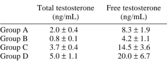
Table 1. Serum concentration of testos-terone
Total testosterone Free testosterone
(ng/mL) (ng/mL)
Group A 2.0 ± 0.4 8.3 ± 1.9 Group B 0.8 ± 0.1 4.2 ± 1.1
Group C 3.7 ± 0.4 14.5 ± 3.6
Group D 5.0 ± 1.1 20.0 ± 6.7 Data are expressed as mean ± SD.
Statistical analysis by Mann-Whitney U-test. Group A versus Group B, P < 0.05.
Group B versus Group C, P < 0.05. Group B versus Group D, P < 0.05.
the Mann-Whitney U-test for 2 independent sam-ples was used to compare each of the 2 groups. Statistical significance was defined as P < 0.05.
Results
Serum concentration of testosterone
The serum levels of total and free testosterone in Group B were significantly lower than those in Group A. Among groups of 15-month-old rats, the serum levels of total and free testos-terone in Groups C and D were significantly higher than those in Group B (Table 1).
Serum creatinin, BUN, urinary protein, urine NAG and GFR
The serum creatinin, BUN and urine NAG levels of Groups A and B were similar to that of the control group. The level of urinary protein in Group B was significantly higher than that of Group A, and GFR in Group B was significant-ly lower than that in Group A.
Among groups of 15-month-old rats, tes-tosterone replacement groups (Groups C and D) showed significantly higher levels of urinary protein and NAG, significantly lower values of GFR, but no significant difference in serum creatinin and BUN (Table 2).
Apoptosis of mesangial and renal tubule cells
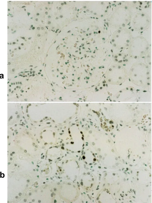
TUNEL-positive mesangial and renal tubule cells of high-dose testosterone replacement rats (Group D) are shown in Figs. 1a and b. The percentages of apoptotic mesangial and renal tubule cells were similar to those of Groups A and B (Table 3). Among groups of 15-month-old rats, mesangial and renal tubule cells of Groups C and D showed greater percentages of TUNEL-positive cells when compared to those of Group B.
Correlations between urinary protein, urinary NAG, GFR, apoptosis of mesan-gial and renal tubule cells and total and free testosterone
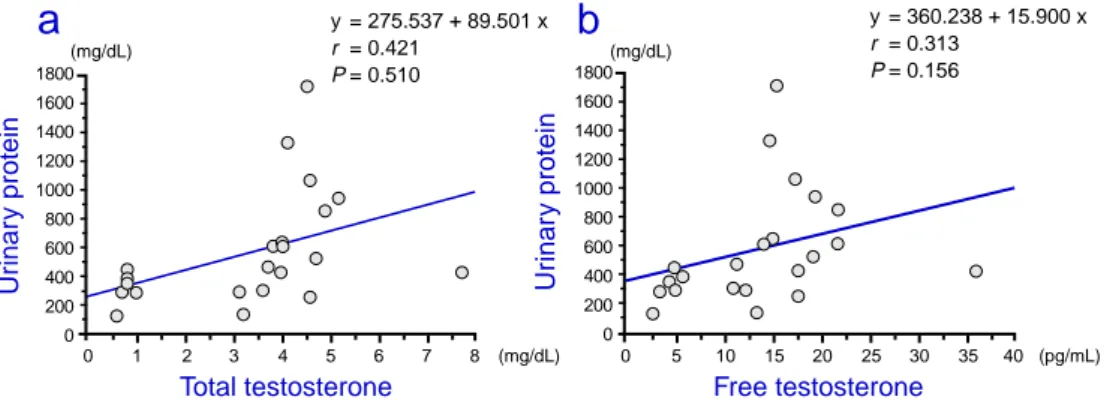
In the 15-month-old rats, the correlation be-tween the serum concentration of total and free testosterone and each parameter was examined using linear regression analysis. The urinary protein level was not found to correlate with the total and free testosterone levels (Figs. 2a and b). The urinary NAG level was found to cor-relate positively with the free testosterone level but not with the total testosterone levels (Figs. 3a and b). The GFR was found to correlate negatively with the total and free testosterone levels (Figs. 4a and b). Percentages of apopto-Table 2. Effects of testosterone replacement on renal function
Serum creatinine BUN Urinary protein Urinary NAG GFR
(mg/dL) (mg/dL) (mg/dL) (U/L) (mL/min/kw)
Group A 0.2 ± 0.1 15.6 ± 1.4 65.8 ± 27.3 5.9 ± 2.3 174.16 ± 37.90 Group B 0.3 ± 0.1 21.1 ± 1.6 327.2 ± 108.9 4.6 ± 7.3 124.40 ± 9.94 Group C 0.4 ± 0.1 20.4 ± 2.0 445.6 ± 181.4 14.0 ± 10.8 104.47 ± 8.22 Group D 0.3 ± 0.1 21.3 ± 2.3 899.1 ± 487.4 11.3 ± 11.3 98.51 ± 17.36 Data are expressed as mean ± SD.
BUN, blood urea nitrogen; GFR, glomerular filtration rate; kw, kidney weight (g); NAG, N-acetyl-β -D-glucosaminidase; NS, not significant.
Statistical analysis by Mann-Whitney U-test.
Group A versus Group B: serum creaninine, BUN and urine NAG = NS; urinary protein and GFR = P < 0.05. Group B versus Group C: serum creaninine, BUN and urinary protein = NS; urine NAG and GFR = P < 0.05. Group B versus Group D: serum creaninine and BUN = NS; urinary protein, urine NAG and GFR = P < 0.05.
a
b
Fig. 1. TUNEL-positive cells of a high-dose testosterone replacement rat at the age of 15 months. a: TUNEL-positive mesangial cells (original magnification × 400).
b: TUNEL-positive renal tubule cells (original magnification × 400).
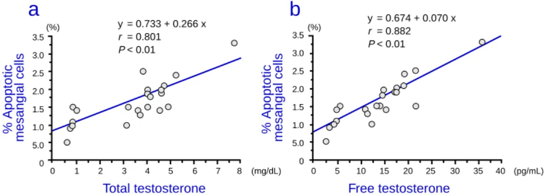
sis of mesangial and renal tubule cells were found to correlate positively with the total and free testosterone levels (Figs. 5a, 5b, 6a and 6b).
Discussion
In a previous study involving male Wistar rats, the GFR began diminishing at 16 months (Tanaka et al., 1995). In this previous study, 2 months of testosterone replacement at 13
Table 3. Effects of testosterone replacement on apoptosis of mesangial cells and renal tubules
% Apoptotic % Apoptotic mesangial cells renal tubule cells Group A 0.96 ± 0.25 0.46 ± 0.21 Group B 1.07 ± 0.36 0.87 ± 0.27 Group C 1.47 ± 0.40 1.23 ± 0.24 Group D 2.05 ± 0.60 1.60 ± 0.35 Data are expressed as mean ± SD.
Statistical analysis by Mann-Whitney U-test. Group A versus Group B, not significant. Group B versus Group C, P < 0.05. Group B versus Group D, P < 0.05.
Fig. 2. Correlation between urinary protein and total and free testosterone in 15-month-old rats.
0 1 2 3 4 5 6 7 8 (mg/dL) 1800 1600 1400 1200 1000 800 600 400 200 0 Total testosterone Ur inar y protein (mg/dL)
a
0 5 10 15 20 25 30 35 40 (pg/mL) 1800 1600 1400 1200 1000 800 600 400 200 0 Free testosterone Ur inar y protein (mg/dL)b
y = 360.238 + 15.900 x r = 0.313 P = 0.156 y = 275.537 + 89.501 x r = 0.421 P = 0.510 0 5 10 15 20 25 30 35 40 (pg/mL) 0 1 2 3 4 5 6 7 8 (mg/dL) 35 30 25 20 15 10 5 0 35 30 25 20 15 10 5 0a
b
Ur inar y NA G (U/L) Ur inar y NA G (U/L)Total testosterone Free testosterone
y = 2.577 + 2.331 x r = 0.416 P = 0.543 y = 2.051 + 0.631 x r = 0.458 P < 0.05
months old accelerated a reduction of the GFR. The direct effects of testosterone on cellular collagen synthesis have not been extensively studied; however, most available data suggests that it has a stimulatory effect (Fischer et al., 1985; Franchimont et al., 1991). For example, testosterone increases collagen synthesis by vascular smooth muscle cells in culture (Leitman et al., 1994), and administration of testosterone increases the accumulation of collagen and elastin in the aorta of normal and cholesterol-fed animals (Silbiger et al., 1995). It has been suggested that some changes related to collagen may occur along with a significant reduction in the GFR in rats that have under-gone testosterone replacement.
Rats that have undergone testosterone re-placement exhibited proteinuria. In this study, urine albumin was not detected, and it is not
clear what kind of protein was excreted, but it might be that proteinuria evolved from a leak in the basement membrane. Excretion of sex-dependent α2u-globulin (Roy et al., 1966;
Fig. 3. Correlation between urinary NAG and total and free testosterone in 15-month-old rats. NAG,
Neuhaus et al., 1975) can be abolished by cas-tration of male rats (Irwin et al., 1971). It would seem that sex-dependent globulin could only slightly contribute to proteinuria. It was found that urinary NAG increased and correlated positively with the free testosterone level but not with the total testosterone level. NAG is one of the glycoproteinases in lysosome, and proximal renal tubule epithelial cells contain a lot of NAG. Urinary NAG increases due to an injured proximal tubule. It is believed that free testosterone may injure proximal tubules.
In addition, it was demonstrated that testos-terone replacement increased the number of TUNEL-positive cells in the mesangial and renal tubule cells when compared to the control group in middle-aged rats. Cultured mesangial
cell apoptosis in male rats increases with the level of testosterone (Singhal et al., 1997). The present study demonstrated that this pheno-menon occurred similarly in vivo. In addition, renal tubule cell apoptosis in male rats also in-creased with the level of testosterone. It is known that cultured mesangial cells from male rats contain nuclear receptors for testosterone (Neugarten et al., 1994). In addition, it is be-lieved that renal tubule cells contain nuclear re-ceptors for testosterone (Davidoff et al., 1980). Free testosterone has true androgen activity, so it is believed that some changes in mesangial and renal tubule cells may occur with a sig-nificant increase in the number of TUNEL-positive cells by the direct effect of free testos-terone.
Fig. 4. Correlation between GFR and total and free testosterone in 15-month-old rats. GFR, glomerular
filtration rate; kw, kidney weight (g).
Fig. 5. Correlation between % apoptosis of mesangial cells and total and free testosterone in 15-month-old
rats. 0 5 10 15 20 25 30 35 40 (pg/mL) 140 130 120 110 100 90 80 70 140 130 120 110 100 90 80 70 0 1 2 3 4 5 6 7 8 (mg/dL) GFR (mL/min/kw)
a
GFR (mL/min/kw)b
Total testosterone Free testosterone
y = 130.371 – 6.693 x r = 0.770 P < 0.01 y = 130.296 – 1.641 x r = 0.792 P < 0.01 0 5 10 15 20 25 30 35 40 (pg/mL) 3.5 3.0 2.5 2.0 1.5 1.0 5.0 0 3.5 3.0 2.5 2.0 1.5 1.0 5.0 0 0 1 2 3 4 5 6 7 8 (mg/dL) % Apoptotic
mesangial cells % Apoptotic mesangial cells (%)
a
(%)
b
Total testosterone Free testosterone
y = 0.733 + 0.266 x r = 0.801 P < 0.01 y = 0.674 + 0.070 x r = 0.882 P < 0.01
On the other hand, it was surmised that tes-tosterone replacement caused a decrease of go-nadotrophic hormone by a feedback mecha-nism. However, the relation between the go-nadotrophic hormone and mesangial and renal tubule cells is not clear. Further, multiple apo-ptosis signaling pathways have been described, but further studies should be performed to de-termine the apoptosis pathway for mesangial and renal tubule cells treated with testosterone. The present study is the first report showing that testosterone replacement in middle-aged rats increased the rate of apoptosis of mesangial and renal tubule cells and the urine level of NAG and decreased the GFR. The concept that androgens are a risk factor for renal dysfunction is supported by the current findings, but further studies are needed to clarify their association with testosterone.
Acknowledgments: The author would like to thank Professor Ikuo Miyagawa of the Department of Urology, Tottori University Faculty of Medicine, for his kind advice and valuable suggestions concerning this study.
References
1 Davidoff M, Caffier H, Schiebler TH. Steroid hormone binding receptors in the rat kidney. Histochemistry 1980;69:39–48.
2 Franchimont P, Bassleer C. Effects of hormones and local growth factors on articular chondrocyte
metabolism. J Rheumatol 1991;18:68–70. 3 Fischer GM, Bashey RI, Rosenbaum H, Lyttle
CR. A possible mechanism in arterial wall for mediation of sex difference in atherosclerosis. Exp Mol Pathol 1985;43:288–296.
4 Gafter U, Ben Bassat M, Levi J. Castration inhibits glomerular hypertrophy and proteinuria in uninephrectomized male rats. Eur J Clin Invest 1990;4:360–365.
5 Irwin JF, Lane SE, Neuhaus OS. Synergistic effect of glucocorticoids and androgens on the biosynthesis of sex-dependent protein in the male rat. Biochim Biophys Acta 1971;252:328–334. 6 Leitman DC, Benson SC, Johnson LK.
Gluco-corticoids stimulate collagen and noncollagen protein synthesis in cultured vascular smooth muscle cells. J Cell Biol 1994;98:541–549. 7 Mulroney SE, Woda C, Johnson M, Pesce C.
Gender differences in renal growth and function after uninephrectomy in adult rats. Kidney Int 1999;56:944–953.
8 Neugarten J, Silbiger S. Nuclear receptor for estradiol and testosterone in mesangial cells. J Am Soc Nephrol 1994;5:951.
9 Neugarten J, Silbiger SR. Effects of sex hor-mones on mesangial cells. Am J Kidney Dis 1995;26:147–151.
10 Neuhaus OW, Flory W. The effect of dietary protein on the excretion of alpha2u, the sex-dependent protein of the adult male rat. Biochim Biophys Acta 1975;411:74–86.
11 Reckelhoff JF, Zhang H, Granger JP. Decline in renal hemodynamic function in aging SHR : role of androgens. Hypertension 1997;30(3 Pt 2): 677–681.
12 Roy AK, Neuhaus OW. Identification of rat uri-nary proteins by zone and immunoelectrophore-sis. Proc Soc Exp Biol Med 1966;121:894–899. 13 Silbiger S, Neugarten J. The impact of gender on
progressive renal functional impairment. Am J
Fig. 6. Correlation between % apoptosis of renal tubule cells and total and free testosterone in 15-month-old
rats. % Apoptotic renal tub ule cells 2.4 2.0 1.6 1.2 0.8 0.4 % Apoptotic renal tub ule cells 2.4 2.0 1.6 1.2 0.8 0.4 0 5 10 15 20 25 30 35 40 (pg/mL) 0 1 2 3 4 5 6 7 8 (mg/dL) (%)
a
(%)b
Total testosterone Free testosterone
y = 0.646 + 0.183 x r = 0.832 P < 0.01 y = 0.651 + 0.470 x r = 0.903 P < 0.01
Kidney Dis 1995;25:515–533.
14 Singhal PC, Reddy K, Franki N, Sanwal V, Kapasi A, Gibbons N, et al. Age and sex modulate renal expression SGP-2 and transglutaminase and apo-ptosis of splenocytes, thymocytes, and macro-phage. J Invest Med 1997;45: 567–575. 15 Tanaka A, Kyoukuwa M, Mori T, Kawashima S.
Acceleration of renal dysfunction with aging by the use of androgen in Wistar/Tw rats. In-vivo 1995;9:495–502.
Received December 15, 2000; accepted December 25, 2000 Corresponding author: Dr. Kuniyasu Muraoka