Analysis of the alveolar development based on
the mice with the combined HIF-3α knockout
and HIF-2α knockdown
著者(英)
ZULKIFLI AMIN FIRMAN
year
2019
その他のタイトル
HIF-3αノックアウトとHIF-2αノックダウンを組み
合わせたマウスに基づく肺胞発生解析
学位授与大学
筑波大学 (University of Tsukuba)
学位授与年度
2018
報告番号
12102甲第8882号
URL
http://doi.org/10.15068/00156452
筑
波 大 学
Analysis of the alveolar development based on
the mice with the combined HIF-3α knockout
and HIF-2α knockdown
(HIF-3αノックアウトと HIF-2αノックダウンを組
み合わせたマウスに基づく肺胞発生解析)
2018
筑波大学大学院博士課程人間総合科学研究科
Table of contents
Chapter I. General overview 1
1.1. Purpose 1
1.2. Material and method 1
1.3. Result 2
1.4. Conclusion 2
1.5. Abbreviation 3
Chapter II. Deterioration of alveolar development in mice with both
HIF-3α -/- and HIF-2α kd/kd 5
2.1. Introduction 5
2.1.1. The importance of studies on lung development 5
2.1.2. The process occurring early in lung development 5
2.1.3. Alveologenesis as a crucial phase in lung development
for gas exchange readiness 7
2.1.4. The role of the hypoxic environment in lung development 8 2.1.5. The regulating of hypoxia-inducible factors in lung development 10
2.2. Materials and methods 12
2.2.1. The generation of the double-mutant mice from HIF-3α -/- and
HIF-2α kd/+ mice 12
2.2.2. Genotyping for HIF-2α and HIF-3α gene of the double-mutant mice
from HIF-3α -/- and HIF-2α kd/+ mice 13
2.2.3. Isolation and culture of cells 14
2.2.4. Immunohistochemistry and section staining 15
2.2.5. Quantitative reverse transcription polymerase chain reaction (qPCR) 16
2.3. Results 17
2.3.1. No double-mutant mice died immediately after birth 17
2.3.2. The double-mutant mice had not died embryonically 18
2.3.3. The neonatal double-mutant mice showed the impaired alveolar sacs
and lung alveolar structure 19
2.3.4. The neonatal double-mutant mice showed the impaired expression of
HIF-1α, HIF-2α, VE-cadherin, and VCAM-1 in lung tissues 21
2.3.5. The neonatal double-mutant mouse lung cells exhibited suppressed
expressions of HIF-1α and HIF-2α 23
2.3.6. The neonatal double-mutant mouse lung cells exhibited the downregulation of HIF-target genes denoted by VE-cadherin,
VCAM-1, Ang-2, Tie-2, and VEGF 24
2.3.7. The neonatal double-mutant mice showed the decreased
endothelial cell numbers 26
2.4. Discussion 28
Chapter III. Conclusion and perspectives 37
3.1. Conclusion 37
3.2. Perspectives 37
References 39
Acknowledgments 47
1
Chapter I. General overview
1.1. Purpose
Studying the lung development to clarify the underlying mechanisms of diseases or the molecular identity of various cell types may provide further strategies to comply in models of human lung disease. In relation to this, previous studies using adult mice with single-mutation consisted of hypoxia-inducible factor 3α (HIF-3α) knockout (-/-) showed the impairments in lung remodeling and lung endothelial cells. Another previous research demonstrated that impaired expression of hypoxia-inducible factor 2α (HIF-2α) induced compensatory expression of hypoxia-hypoxia-inducible factor 1α (HIF-1α) in HIF-2α knockdown (HIF-2α kd/kd) mice. This recent study hence learns from those previous studies to uncover more insights by extending the investigation, utilizing neonatal mice with doublemutation consisted of both HIF3α -/- and HIF-2α kd/kd aiming to recover the impaired lung conditions due to the single-mutation.
1.2. Materials and methods
Firstly, the wild-type (WT) and mutant mouse lines were maintained on the C57BL/6J genetic background. The HIF-2α kd/kd mice and HIF-3α -/- (single-mutant) mice were each generated. Concerning this, the mating of single-mutant and 2α knockdown heterozygotes (2α kd/+) mice resulted in the combined HIF-3α -/- and HIF-2α kd/kd mice, which were called the double-mutant mice. Furthermore, the lungs from each of the WT and double-mutant mice were dissected at postnatal day 0 (P0) for hematoxylin and eosin staining and immunological staining with CD31, HIFs, VE-cadherin, and VCAM-1 antibodies. The lungs from each of the
2
WT, HIF-2α kd/kd, single-mutant, and double-mutant mice were dissected at postnatal 6-week-old for cell culture. Also, the gene expression of HIFs and their target genes were analyzed by quantitative reverse transcription polymerase chain reaction (qPCR).
1.3. Results
No mice with both HIF-3α -/- and HIF-2α kd/kd died immediately after birth. The double-mutant mice with both HIF-3α -/- and HIF-2α kd/kd exhibited impaired lung alveolar structure and HIFs expression. Transcriptional analysis of lung cells revealed the depressed expressions of HIF-1α, HIF-2α, VE-cadherin, VCAM-1, Tie-2, and Ang-2 in the double-mutant mice compared to that in the WT mice. The endothelial cell numbers were also decreased in the double-mutant mice as opposed to the WT mice.
1.4. Conclusion
The current study proposed the insightful ideas of the role of both HIF-3α and HIF-2α in lung development, which involved in the low number of endothelial cells and the impaired expression of angiogenic factors. Further studies are necessary to elucidate the underlying mechanism of how HIF-3α and HIF-2α regulate the lung endothelial cells as well as lung development. Nevertheless, it is apprehensible that studies of HIF-dependent responses on pulmonary vascular and airway epithelium are equally important. Thus, the future perspective on the present study will need to aim at elucidating the functional role of double-mutation consisted of both HIF-3α -/- and HIF-2α kd/kd in lung endothelial cells and alveolar epithelial cells of the double-mutant mice.
3
1.5. Abbreviation AT1: Alveolar type 1 AT2: Alveolar type 2 Ang-1: Angiopoietin 1 Ang-2: Angiopoietin 2
ARNT: Aryl hydrocarbon receptor nuclear translocator bHLH: Basic helix-loop-helix
CAD: C-terminus transactivation domain CBP: CREB-binding protein
cDNA: Complementary deoxyribonucleic acid CO2: Carbon dioxide
DMEM: Dulbecco’s modified Eagle’s medium FBS: Fetal bovine serum
FIH: Factor inhibiting hypoxia-inducible factor
HAVA: High glucose Dulbecco’s modified Eagle’s medium supplemented with 10% fetal bovine serum, 0.1 mmol/L nonessential amino acids, 2 mmol/L L-glutamine, penicillin-streptomycin, and 10-4mol/L β-mercaptoethanol
HIF: Hypoxia-inducible factors HIF-1α: Hypoxia-inducible factor 1α HIF-2α: Hypoxia-inducible factor 2α HIF-3α: Hypoxia-inducible factor 3α HRE: Hypoxia-responsive element kd/kd: Knockdown
kd/+: Knockdown heterozygotes mRNA: Messenger ribonucleic acid
4
n: Sample number
OCT: Optimum cutting temperature ODD: Oxygen-dependent degradation P: Postnatal day
PAS: PER-ARNT-SIM
PCR: Polymerase chain reaction PHD: Proline hydroxylase
pVHL: Von Hippel-Lindau tumor suppressor
qPCR: Quantitative real-time polymerase chain reaction RNA: Ribonucleic acid
VCAM-1: Vascular cell adhesion molecule 1 VE-cadherin: Vascular endothelial cadherin VEGF: Vascular endothelial growth factor WT: Wild-type -/-: Knockout +/+: Homozygotes +/-: Knockout heterozygotes µm: Micrometer °C: Celsius degree
5
Chapter II. Deterioration of alveolar development in mice with both HIF-3α -/- and HIF-2α kd/kd
2.1. Introduction
2.1.1. The importance of studies on lung development
Recent progress in regenerative medicine studies toward lung development has served many new advances about the background mechanisms involved. Nonetheless, there has still remained a gap of knowledge regarding the respiratory system ability in response to injury and disease.
The study of lung development is essential accordingly due to the fact that it can provide the underlying mechanisms to be taken care of for instance in the event of perinatal infection along with inflammation interfering with alveologenesis leading to bronchopulmonary dysplasia [1]. Another example of its importance can be in the form of knowing more detailed about molecular identity of various cell types that may provide further strategies to comply with the behavior of stem or progenitor cells in models of human lung disease. Therein, it is of necessity to depict of the mechanisms developing the respiratory system.
2.1.2. The process occurring early in lung development
Principally, the respiratory system of mammalian as in both of the adult human and mouse lung is similar. The lungs along with the trachea emerge from the anterior foregut endoderm. The latter is a tissue resulting in multiple organs, which involve the respiratory system, esophagus, thyroid, and liver. The specification of lung starts around embryonic day (E) 9.0 in the mouse marked by
6
the transcription factor Nkx2-1 that is expressed in endodermal cells on the ventral side of the anterior foregut. At E9.5, evagination of those epithelial cells produces in the formation of the trachea and two lung buds and the onset of the embryonic stage of lung development (E9.5 - E12.5). Through this phase, the trachea accomplishes its splitting from the esophagus. Furthermore, throughout the embryonic and pseudoglandular phases (E12.5 - E16.5), the two lung buds serve a highly arranged branching process named branching morphogenesis to yield a tree-like composition of airways accompanied by thousands of terminal branches [2–6].
Of the branching morphogenesis, two primary lung buds appearing around E9.5 in the mouse and 4-5 weeks gestation in the human provide the increment to the airway tree. The buds are arranged of a simple endodermal epithelium that the surrounding is mesoderm and a vascular plexus. The tissues are enwrapped in a thin layer of mesothelium leading a transient early role to mesenchymal lineages [7]. The buds elongate and diverge in a scheme that is at first really stereotypic but reduces further as development proceeds [4, 8, 9].
All lung endodermal cells in the beginning express Nkx2-1, by which this marker continues firmly into the adult. Nonetheless, along with the primary buds elongate and diverge, a distinct scheme of gene expression arises in the endoderm of the stalks compared to the buds. This proximal-distal difference is indicated by the expression domains of Sox2 and Sox9, which are two transcription factors needed for early lung development. Sox2 expression is limited to the proximal stalks. On the other hand, Sox9 is vibrantly expressed in the much higher proliferative cuboidal cells of the distal buds [10, 11].
7
2.1.3. Alveologenesis as a crucial phase in lung development for gas exchange readiness
In the mouse prenatal alveologenesis, at about E15, branching morphogenesis decreases and notable changes occur in the distal epithelium. The canalicular (E16.5 - E17.5) and saccular (E18.5 - postnatal day (P) 5) phases have followed on, by which those terminals branches narrow and make clusters of epithelial cell that will further develop into alveoli in the arrangement for respiration at birth. The tubes that have become canalicular have more tightly related to the surrounding vasculature. Cells located in the distal side of the tubules start to express genes trait of alveolar epithelial type 1 (AT1) and 2 (AT2) cells, which arrange for the mature alveoli. AT1 cells express Hopx, podoplanin (Pdpn, notable as T1alpha), and AGER. On the other side, AT2 cells express proteins correlated with high levels of surfactant production and secretion e.g. SftpA-C [12]. Furthermore, the tubules that get into the saccular stage implicate the budding of very small peripheral sacs outcast by primary septa. Postnatally, these sacs are then divided again by secondary septa.
In the mouse postnatal alveologenesis, the establishment of the alveoli does not finish at birth. The alveoli number and surface area rises significantly postnatally and keeps on for weeks in the mice and months in the humans with the development of new secondary septa. Ultimately, full maturation of the alveolus happens in the course of the alveolarization phase (P0 - P14). Through all phases of endodermal development, the lung mesoderm (or mesenchyme) expands and cooperates with the lung endoderm to promote branching and differentiation and to yield the various lineages within the lung, which involves airway and vascular smooth muscle and pericytes [2–6].
8
2.1.4. The role of the hypoxic environment in lung development
The respiratory process in lung takes place in the alveoli that consist of alveoli, alveolar duct, and alveolar sacs. Alveolar sacs are composed of AT1 and AT2 cell. Gas exchange occurring in the alveoli is mediated by AT1, by which oxygen from alveoli will enter the blood circulation through endothelial cells to go throughout the body and vice versa for the carbon dioxide. On the other side, AT2 cells will maintain alveolar fluid balance or fluid homeostasis; produce surfactant proteins [13, 14]; and are adult stem cells, competent of both self-renewal and differentiation into AT1 cells [15–18].
In utero, the fetal lung develops in a relatively hypoxic environment. Most
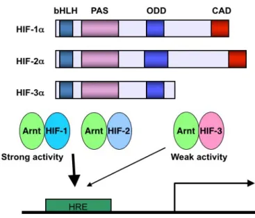
hypoxia-induced regulatory states are mediated by HIFs giving the contribution to normal lung development [19]. HIFs are heterodimers that consist of three oxygen-sensitive α-subunits (HIF-1α, HIF-2α, and HIF-3α) and a β-subunit, the aryl hydrocarbon receptor nuclear translocator (ARNT) [20, 21]. Under the normoxic condition, the proline residue on oxygen-dependent degradation (ODD) domain of HIF is hydroxylated by proline hydroxylase (PHD) using oxygen in the cytosol (Figure 1) [22, 23]. E3 ubiquitin ligase recognizes hydroxyl proline and ubiquitinates HIF protein. Ubiquitinated HIF protein is degraded immediately. On the other side, under the hypoxic condition, PHD cannot undergo hydroxylation because there is no substrate for hydroxylation. HIF protein is stabilized and dimerized with ARNT, then, bind to the hypoxia-responsive element (HRE) enhancer leading to activation of HIF-target genes [24, 25].
9
Figure 1. The regulation of HIFs during normoxic and hypoxic conditions. ARNT, aryl hydrocarbon receptor nuclear translocator; CBP, CREB-binding protein; FIH, factor inhibiting hypoxia-inducible factor; HIF, hypoxia-inducible factor; PHD, proline hydroxylase; pVHL, von Hippel-Lindau tumor suppressor.
Both HIF-1α and HIF-2α have strong transcriptional activities for binding to HRE. While, HIF-3α transcription activity is weaker compared to HIF-1α and HIF-2α because of no C-terminus transactivation domain (CAD) in the structure (Figure 2) [20, 26].
10
Figure 2. The role of CAD in the structure of HIFs. ARNT, aryl hydrocarbon receptor nuclear translocator; bHLH, basic helix-loop-helix; CAD, C-terminus transactivation domain; HIF, hypoxia-inducible factor; HRE, hypoxia-responsive element; ODD, oxygen-dependent degradation; PAS, PER-ARNT-SIM.
2.1.5. The regulating of hypoxia-inducible factors in lung development
The lung development is widely known to occur in a hypoxic environment. Therefore, HIFs have without any wonder become the substantive contributors to embryogenesis. The mRNA and protein levels of HIFs have sufficiently high in the fetal lung [27–29]. In human, the whole system of HIFs has started from 8 weeks of gestation [28]. Hypoxia generated the protein expression of HIF-1α in cultured cells that came from, showing control, all kind of cells known in the adult lung [30]. Meanwhile, the protein expression of HIF-2α limited only control to the vascular endothelium and AT2 cells [31].
The lung of the normal embryo is at the hypoxic stage throughout fetal development [32]. HIF-1α protein is at high levels, which primarily restricted in
11
branching epithelium based on analysis of the spatial pattern of expression in the lungs of human through the early development of the pulmonary vasculature. HIF-2α has as well presented in the epithelium, and also in mesenchymal structures that provide the increase to the vascular endothelium [28].
Many of HIF-target genes involved in endothelial cell proliferation and survival, for instance, angiopoietin 1 (Ang-1) and 2 (Ang-2) [24]. Nevertheless, all members of the HIF regulating pathway, in the human fetal lung, is also expressed in the epithelium [28, 33]. HIFs act as regulators of the molecular hypoxic response [20, 21]; in a study examining normal alveolarization in fostered newborn rats, HIFs promoted alveolar development and regeneration by preventing and repairing oxygen-induced alveolar damage [34]. Nonetheless, HIF-1α inhibition using antisense knockdown in vitro during early lung development decreased vascular development and epithelial branching morphogenesis in lung explants [35]. In contrast, the conditional overexpression of HIF-1α in embryonic lung epithelium also impaired branching morphogenesis and lung maturation and affected vascular lung abnormalities, including hemorrhages and increased lymphangiogenesis [36]. Collectively, these data suggest that interference in the alveolar epithelium by oxygen pressure changes, including hypoxia, can affect alveolar homeostasis, leading to epithelial injuries and diseases such as lung fibrosis [37–40].
A previous study that used adult single-mutant mice with HIF-3α -/- showed impaired lung remodeling exhibited by the walls of the secondary septa in subdivided alveoli, and immunostaining of alveolar endothelial cells presented an increase in defective space in the interalveolar septa and hyperplasia of endothelial cells during the maturation of alveolar formation in these knockout
12
mice [26]. Additionally, another study on the lung endothelial cells of adult single-mutant mice revealed that the defect of HIF-3α induced HIF-2α overexpression, and excess HIF-2α caused overproduction of VE-cadherin. Hence, the adult single-mutant mice showed impairments in lung endothelial cells presented by slow growth and a decreased number of tubes formed by endothelial cells [41]. Furthermore, a different but related study preceding the present study demonstrated that impaired expression of HIF-2α in HIF-2α kd/kd mice induced compensatory expression of HIF-1α [42]. The current study uncovers more insights by extending the investigation from those previously stated findings before the current study utilizing the double-mutant mice with both HIF-3α -/- and HIF-2α kd/kd. The purpose of the current study using mice with the double-mutation of both HIF-3α -/- and HIF-2α kd/kd is to recover the impaired lung conditions due to the single-mutation of HIF-3α -/-. For this reason, in this study, male and female HIF-3α -/- and HIF-2α kd/+ mice were interbred, resulting in the double-mutant mice previously mentioned.
2.2. Materials and methods
2.2.1. The generation of the double-mutant mice from HIF-3α -/- and HIF-2α kd/+ mice
All of the experiments performed were approved by the ethics committee of the University of Tsukuba. All WT and mutant mouse strain were of the C57BL/6J genetic background. Because the HIF-2α -/- causes the embryonic mortality, the HIF-2α kd/kd mice were selected for the present study. In order to obtain the HIF-2α kd/kd mice, firstly, the HIF-2α kd/+ mice, which are heterozygous, were generated as previously reported for the mutated HIF-2α gene
13
[43]. Briefly, a targeting vector was inserted a PGK promoter-neomycin gene cassette, which was a sandwiched between two loxP sequences into upstream exon 1 that encoded the 5’-UTR sequence by homologous recombination. This vector was utilized to disrupt the mouse HIF-2α gene. Next, the heterozygous embryonic stem (ES) cell line for the recombinant allele as for HIF-2α kd/+ was utilized to generate the chimeric mice by blastocyst injection. The resulting chimeric mice were crossed with WT mice to obtain the HIF-2α kd/+ mice. The resulting HIF-2α kd/+ mice were thereafter interbred with each other to yield the HIF-2α kd/kd mice. The generating mice were genotyped for HIF-2α gene.
HIF-3α -/- mice were obtained as previously published [41]. Briefly, a pBluescript SK(+) vector contained the chimeric DNA replacing part of the second exon. The following intron of the HIF-3α gene and a neomycin resistance gene were sandwiched between loxP sequences. Thereafter, the GFP gene was constructed. Then, the vector was transfected into ES cells resulting in recombinant ES cells that were hence injected into WT mice by blastocyst injection. The resulting chimeric mice were intercrossed with WT mice obtaining HIF-3α -/- mice that the generating mice were genotyped for HIF-3α gene.
In order to generate the double-mutant mice, the mating of 12 pairs of HIF-3α -/- and HIF-2α kd/+ mice for a breeding period of one year was performed. The double-mutant neonatal pups were confirmed by genotyping for 2α and HIF-3α.
2.2.2. Genotyping for HIF-2α and HIF-3α gene of the double-mutant mice from HIF-3α -/- and HIF-2α kd/+ mice
14
genotype of the mouse was determined by polymerase chain reaction (PCR). The sequences of primer for HIF-3α genotyping were 5’-primer
TGCCATTTCCTTCCCACAGGTCGAACACCG and 3’-primer
CTTGGATTTGGGTTCAATCTCCAGTATGGC, and for HIF-2α genotyping
were 5’-primer TCGGCAGTGTCCTGAGACTG and 3’-primer
AAGGGACCTGGAGTTCCGTT.
2.2.3. Isolation and culture of cells
In order to analyze the expression of HIFs and their target genes, and clarify the molecular mechanisms by which HIFs affect the lung development, the whole lung cells were isolated and cultured. The overdose treatment of anesthetic reagent (isoflurane; WAKO, Japan) was given to the WT, HIF-2α kd/kd, HIF-3α -/-, and double-mutant mice that were sacrificed, and lung tissues from these mice were harvested as previously mentioned [44]. Furthermore, lungs from the WT, HIF-2α kd/kd, HIF-3α -/-, and double-mutant mice were dissected at postnatal week 6 for cell culture. Collagenase digestion (Nitta Gelatin, Osaka, Japan) was used to prepare the lung cell suspensions. Afterward, these cells were cultured, in high glucose Dulbecco’s modified Eagle’s medium (DMEM; GIBCO) supplemented with 10% fetal bovine serum (FBS), 0.1 mmol/L nonessential amino acids, 2 mmol/L L-glutamine, penicillin-streptomycin, and 10-4 mol/L β-mercaptoethanol (HAVA medium) [40], and maintained at 33°C in 20% CO2
without any addition of growth supplements for a total of 7 days with no propagation.
15
2.2.4. Immunohistochemistry and section staining
The lungs dissected at P0 that obtained from three mice (n = 3) from each of the WT, single-mutant, and double-mutant mice for hematoxylin and eosin staining and at P2-P3 from each of the WT and double-mutant mice for immunological staining, which were fixed with 4% paraformaldehyde combined with phosphate-buffered saline at 4°C overnight and embedded in OCT compound (Sakura Finetek, Tokyo, Japan). Sections (10 µm) were then prepared for hematoxylin and eosin staining and immunological staining. Sections were washed with phosphate-buffered saline (PBS) for removing OCT compound and soaked into Mayer’s hematoxylin solution (Wako, Osaka, Japan) at 3 minutes. After washing with water, sections were soaked into eosin solution (Wako) at 30 seconds. Samples were dehydrated with ethanol series and penetrated with xylene. Section was mounted with Malinol (Muto pure chemical, Tokyo, Japan). Serial cryostat sections were incubated with anti CD31 (1:1000; clone: MEC 13.3; BD Biosciences, San Diego, CA, USA) antibody. The sections, after being washed, were incubated with an HRP-conjugated secondary antibody (1:2000; Vector Laboratories, Burlingame, CA, USA). Different sections were incubated with anti-HIF-1α, anti-HIF-2α (1:1000; Novus Biologicals, CO, USA), anti-vascular cell adhesion molecule 1 (VCAM-1), and anti-vascular endothelial cadherin (VE-cadherin) (1:200; Santa Cruz Biotechnology Inc. Texas, USA) antibodies for overnight at 4°C. After washing 3 times, sections were incubated with the biotin-conjugated secondary antibody (1:2000; Vector Laboratories). After washing, sections were treated with ABC labeling kit (Vector Laboratories), and the positive signal was stained with 3,3’-diaminobenzidine reagent (Dojindo, Japan). The sample sections were counterstained with hematoxylin (Wako). The
CD31-
16
positive cell number was counted by the observation using the microscope (Olympus, Tokyo, Japan). The number of the CD31 positive cell was shown as the value per field (200x higher magnification). For a sample, a total of nine fields from three slides of the sample was randomly selected out of one section and counted. The experiment was performed in triplicate.
2.2.5. Quantitative reverse transcription polymerase chain reaction (qPCR) Total RNA was obtained from three samples of three independent mice (n = 3) with the use of the extraction reagent (Sepasol-RNA I Super G; Nakalai Tesque, Kyoto, Japan). The cDNA was then synthesized by reverse transcription from one microgram of total RNA using a reverse transcriptase (RT)-polymerase chain reaction (PCR) kit (ReveTra Ace; TOYOBO, Osaka, Japan). The resulting cDNAs were analyzed using a GeneAmp PCR System (Life Technologies, CA) with 25-35 cycles of denaturation at 95oC for 5 seconds followed by annealing and extension at 68°C for 30 seconds then performed a fluorescence reading step. The reaction mixtures for the quantitative PCR were prepared by using SYBR-Green Real-Time PCR Mastermix (Life Technologies, Carlsbad, CA, USA) and analyzed by using a 7500 Fast Real-Time PCR machine (Applied Biosystems, Carlsbad, CA, USA) with SYBR-green. Experiments were performed in triplicate, and the resulting data were analyzed by the delta CT method. The sequences of primer used for this method were as follows: β-actin was 5’-primer
GTCGTACCACAGGCATTGTGATGGACT and 3’-primer
CACCAGAGAGCACTGTGTTGGCATAGA; HIF-1α was 5’-primer
GAAGCACCTTCCACGTTGCTGACTTGAT and 3’-primer
17
CTTTGTGTGTGTTGAGAGTTTTATTGT and 3’-primer
AAGGAAGTTAGGATTCACAAATGTAGT; VE-cadherin was 5’-primer CCGCCAGAATGCTAAGTATGTGC and 3’-primer TCCACAATGAGGGC
AGTAAGGAA; and VCAM-1 was 5’-primer
GAACACTTTTCCCAGACACTTTTCACGTGG and 3’-primer
TAAGGATCTGGGTTTTATAAATAATGTCTC.
2.2.6. Statistical analysis
The ANOVA.SPSS software (IBM Corp., NY, USA) was used to perform the statistical analyses. The p values of <0.05 are considered significance. The data are presented as the mean ± standard deviation (SD).
2.3. Results
2.3.1. No double-mutant mice died immediately after birth
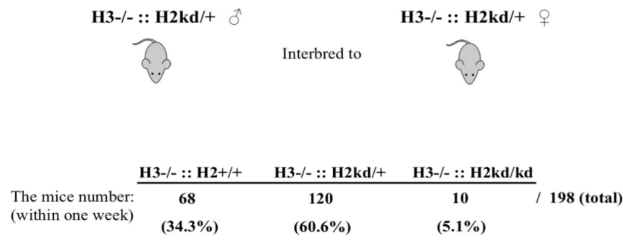
In order to study the role of both HIF-2α and HIF-3α in lung development, I performed the mating of single-mutant and HIF-2α kd/+ mice. Interbreeding between male and female single-mutant and HIF-2α kd/+ mice resulted in 198 offspring within one week (Figure 3). Of the total number of offspring, 5.1% were surprisingly the double-mutant mice, with very few pups still alive at birth, 34.3% were HIF-3α -/- and HIF-2α +/+, and 60.6% were HIF-3α -/- and HIF-2α kd/+. Almost none of the double-mutant mice were alive at birth. The majority of double mutant mice died within one week after birth due to respiratory failure resulting in the survived mice.
18
Figure 3. No double-mutant mice died immediately after birth. The offspring from the interbreeding of male and female single-mutant and HIF-2α kd/+ mice comprised HIF-3α -/- and HIF-2α +/+ mice, HIF-3α -/- and HIF-2α kd/+ mice, and double-mutant mice. H3, hypoxia-inducible factor 3α; H2, hypoxia-inducible factor 2α; ::, intercrossed with; -/-, knockout; kd/+, knockdown heterozygotes; +/+, homozygotes; ♂, male; ♀, female.
2.3.2. The double-mutant mice had not died embryonically
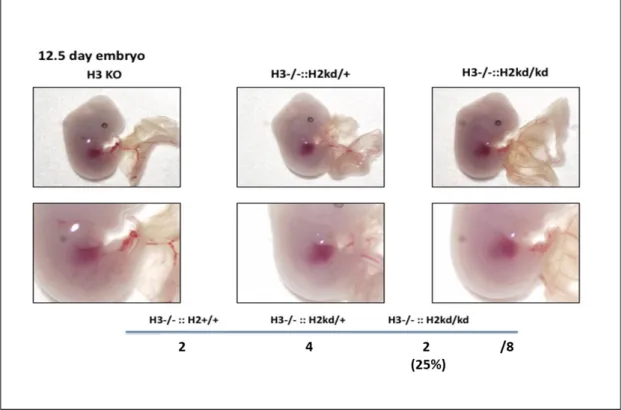
In E12.5, I checked the embryo development of double-mutant mice. The double-mutant mice had not died embryonically, and vasculogenesis on yolk sac and hematopoiesis (fetal liver) were nothing different in between the genotypes of HIF-3α -/- and HIF-2α +/+, HIF-3α -/- and HIF-2α kd/+, and double-mutant mice (Figure 4). These data clarified that the double-mutant mice were died later, even though have been shown not immediately, after birth.
19
Figure 4. The double-mutant mice had not died embryonically. At E12.5, the littermate mice were analyzed by using Stereomicroscope. The vascular on yolk sac and fetal liver of double-mutant mice were nothing different compared to other genotype mice. H3, hypoxia-inducible factor 3α; H2, hypoxia-inducible factor 2α; ::, intercrossed with; -/-, knockout; kd/+, knockdown heterozygotes; KO, knockout; +/+, homozygotes.
2.3.3. The neonatal double-mutant mice showed the impaired alveolar sacs and lung alveolar structure
The previous report from my laboratory described that adult single-mutant mice with HIF-3α -/- showed an enlargement of the right ventricular and impaired lung remodeling during development [26]. Therefore, in the present study, firstly I examined the influences of double-mutation on the lung of the mice. Hematoxylin and eosin staining of the lung in the neonatal WT, single-mutant,
20
and double-mutant mice was performed at P0 (Figure 5). In contrary to the expectation from the study purpose, I found that double-mutation caused the impaired structure of the lungs. In particular, alveolar sacs were found almost imperceptible in the lungs of neonatal double-mutant mice, and the appearance of the blood vessels in the lungs of these mice was different from that in the lungs of neonatal single-mutant and WT mice. The neonatal double-mutant mice had less immature alveoli with a lot of broader alveolar sacs as compared with the neonatal single-mutant mice. There is no phenotype data for neonatal HIF-2α kd/kd mice at present. All of these showed important roles of both HIF-3α and HIF-2α in lung development and may further suggest the possibility of the relationship among HIFs.
Figure 5. The neonatal double-mutant mice had the impaired alveolar sacs and lung alveolar structure. The lung of neonatal WT mice, neonatal single-mutant mice, and neonatal double-mutant mice at P0 was examined based on hematoxylin and eosin staining. H3, inducible factor 3α; H2,
hypoxia-
21
inducible factor 2α; ::, intercrossed with; -/-, knockout; kd/kd, knockdown; WT, wild-type.
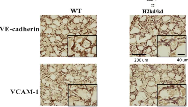
2.3.4. The neonatal double-mutant mice showed the impaired expression of HIF-1α, HIF-2α, VE-cadherin, and VCAM-1 in lung tissues
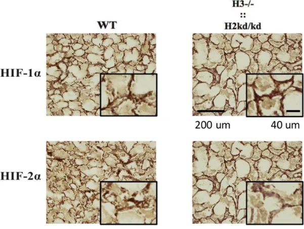
In order to study the relationship among HIFs in lung development, I thereafter examined the expression of HIF-1α and HIF-2α in lung tissues of the neonatal double-mutant mice by immunological staining. I found that the neonatal double-mutant mice showed the impaired expressions of both HIF-1α and HIF-2α in lung tissues (Figure 6).
Figure 6. The double-mutant mice showed the impaired expression of HIF-1α and HIF-2α in lung tissues. Immunohistochemistry analysis performed to sections from the neonatal WT and double-mutant mice at two to three days of age by
22
staining with HIF-1α and HIF-2α antibodies. H3, hypoxia-inducible factor 3α; H2, hypoxia-inducible factor 2α; ::, intercrossed with; -/-, knockout; kd/kd, knockdown; WT, wild-type; HIF-2α, hypoxia-inducible factor 2α; HIF-1α, hypoxia-inducible factor 1α; µm, micrometer.
Next, I examined the expression of HIF-target genes in lung tissues. I found that consistent with the impaired expression of HIF-1α and HIF-2α, the neonatal double mutant mice lung also showed the impaired expression of adhesion molecules denoted by VE-cadherin and VCAM-1 (Figure 7).
Figure 7. The double-mutant mice showed the impaired expression of VE-cadherin and VCAM-1 in lung tissues. Immunohistochemistry analysis performed to sections from the neonatal WT and double-mutant mice at two to three days of age by staining with VE-cadherin and VCAM-1 antibodies. H3, hypoxia-inducible factor 3α; H2, hypoxia-hypoxia-inducible factor 2α; ::, intercrossed with; -/-, knockout; kd/kd, knockdown; WT, wild-type; VE-cadherin, vascular endothelial cadherin; VCAM-1, vascular cell adhesion molecule 1; µm, micrometer.
23
2.3.5. The neonatal double-mutant mouse lung cells exhibited suppressed expressions of HIF-1α and HIF-2α
Kobayashi et al. reported the increased mRNA expression of HIF-2α, but not HIF-1α, by HIF-3α ablation in the pulmonary endothelial cells isolated from the adult single-mutant mice having such HIF-3α -/- compared to those from the adult WT mice, suggesting the role of HIF-3α in the regulation of HIF-2α [41]. Therefore, next, I examined the transcriptional relation of HIFs in the whole lung cells isolated from the neonatal WT, HIF-2α kd/kd, single-mutant, and double mutant mice. This analysis utilized the whole lung cells isolated at two to three days of age, and the relative mRNA expression levels were examined. Inconsistent with the finding utilizing the adult single-mutant mice showing no effects of HIF-3α on the expression of HIF-1α in pulmonary cells, I found that in whole lung cells of the neonatal single-mutant mice, the single mutation of HIF-3α -/- impaired the transcription of both HIF-1α and HIF-2α. Interestingly, the double-mutation even much further suppressed the expression of HIF-1α and HIF-2α genes in lung cells of the neonatal double-mutant mice (Figure 8).
24
Figure 8. The neonatal double-mutant mouse lung cells exhibited suppressed expressions of HIF-1α and HIF-2α. The utilizing qPCR performed the analysis of mRNA relative expressions for HIF-1α, HIF-2α. WT, wild-type, H3, hypoxia-inducible factor 3α; H2, hypoxia-hypoxia-inducible factor 2α; ::, intercrossed with; -/-, knockout; kd/kd, knockdown; HIF-1α, hypoxia-inducible factor 1α; HIF-2α, hypoxia-inducible factor 2α; mRNA, messenger ribonucleic acid. ****p<0.0001, ***p<0.001, **p<0.01, *p<0.05, ns: no significance.
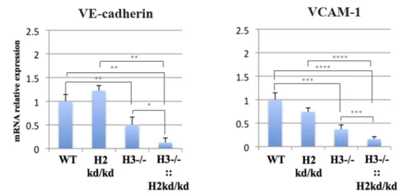
2.3.6. The neonatal double-mutant mouse lung cells exhibited the downregulation of HIF-target genes denoted by VE-cadherin, VCAM-1, Ang-2, Tie-2, and VEGF
Next, the relative mRNA expression levels of HIF-target genes denoted by VE-cadherin, VCAM-1, angiopoietin 1 (Ang-1) and 2 (Ang-2), Tie-2, vascular endothelial growth factor (VEGF), and Flk-1 were analyzed. The analysis utilized the whole lung tissue from the neonatal WT, HIF-2α kd/kd, single-mutant, and double-mutant mice at two to three days of age. In whole lung cells of the neonatal single-mutant mice, the single mutation of HIF-3α -/- reduced expression of VE-cadherin and VCAM-1. Furthermore, consistent with the impaired expression of VE-cadherin and VCAM-1 in lung tissues of the neonatal double-mutant mice, the whole lung cells from the neonatal double-mutant mice showed the downregulation of these adhesion molecules (Figure 9).
25
Figure 9. The neonatal double-mutant mouse lung cells showed the decreased expressions of adhesion molecules, VE-cadherin and VCAM-1. The utilizing qPCR performed the analysis of mRNA relative expressions for VE-cadherin and VCAM-1. WT, wild-type, H3, inducible factor 3α; H2, hypoxia-inducible factor 2α; ::, intercrossed with; -/-, knockout; kd/kd, knockdown; VCAM-1, vascular cell adhesion molecule 1; VE-cadherin, vascular endothelial cadherin; mRNA, messenger ribonucleic acid. ****p<0.0001, ***p<0.001, **p<0.01, *p<0.05.
In addition to the adhesion molecules, I found the impaired expression of other genes that related to the angiogenesis in whole lung cells of the neonatal single-mutant and double single-mutant mice. The Ang-1, Ang-2, and Tie-2 expressions were reduced in the lung of the neonatal single-mutant mice and Ang-2 and Tie-2, respectively, even more, depressed in the lung of the neonatal double-mutant mice (Figure 10). The result also exhibited that, unexpectedly, Ang-1 was expressed excessively in the lung of the neonatal double-mutant mice.
26
Figure 10. The double-mutant mouse lung cells exhibited depressed expressions of angiogenic factors denoted by Ang-2 and Tie-2. The utilizing qPCR performed the analysis of mRNA relative expressions for Ang-2, Tie-2, VGEF, Ang-1, and Flk-1 that normalized to β-actin. Ang-1, angiopoietin 1; Ang-2, angiopoietin 2; VEGF, vascular endothelial growth factor; WT, wild-type, H3, hypoxia-inducible factor 3α; H2, hypoxia-inducible factor 2α; ::, intercrossed with; -/-, knockout; kd/kd, knockdown; mRNA, messenger ribonucleic acid. ****p<0.0001, ***p<0.001, **p<0.01, *p<0.05, ns: no significance.
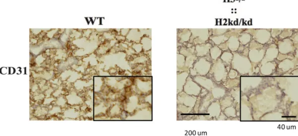
2.3.7. The neonatal double-mutant mice showed the decreased endothelial cell numbers
The previous study showed that the single-mutation of HIF-3α -/- impaired the proliferation and functions of pulmonary endothelial cells [41]. In the present study, I found that the double-mutation caused the abnormal appearance of blood
27
vessels in the lungs of the mice suggesting the involvement of endothelial cells in this phenomenon. Therefore, next, I examined the effect of the double-mutation on the endothelial cell numbers in the neonatal double-mutant mice lung. I performed the immunohistochemistry analysis of sections from the neonatal WT and double-mutant mice at two to three days of age (Figure 11) by CD31, a marker of endothelial cells. As expected, I found that consistent with the impaired lung vessel structure, the neonatal double-mutant mice showed the decreased pulmonary endothelial cell numbers. Particularly, the counting of the CD31 positive cell number per field in 200x higher magnification showed that the neonatal WT mice had 156.3 ± 11.1 and the neonatal double-mutant mice had much lower, 44.3 ± 6.03 (n=3; p=0.000104) (Figure 12).
Figure 11. Immunohistochemistry analysis performed to sections from the neonatal WT and double-mutant mice at two to three days of age by staining with the CD31 antibody. H3, hypoxia-inducible factor 3α; H2, hypoxia-inducible factor 2α; ::, intercrossed with; -/-, knockout; kd/kd, knockdown; WT, wild-type; µm, micrometer.
28
Figure 12. CD31-positive cells calculated from sections from the neonatal WT and double-mutant mice at two to three days of age. H3, hypoxia-inducible factor 3α; H2, hypoxia-inducible factor 2α; ::, intercrossed with; -/-, knockout; kd/kd, knockdown; WT, wild-type; µm, micrometer. ***p<0.001.
2.4. Discussion
Resulting from the diverse expression pattern yielded in the lung development, HIF-1α and HIF-2α may act dissimilar, and specific, roles in epithelial and vascular morphogenesis. The outcomes from various studies evaluating HIF-1α and HIF-2α loss-of-function models bolster this likelihood [19]. On the other hand, the study of the role of HIF-3α in lung development remains very limited; nevertheless, there is a previous study exhibited that adult single-mutant mice with HIF-3α -/- have impaired lung remodeling [26]. Another study on the lung endothelial cells of single-mutant mice with HIF-3α -/- further reported that the defect of HIF-3α induced HIF-2α overexpression resulting in the overproduction of VE-cadherin; hence, impaired the lung endothelial cells functions [41]. Regarding this, up to now, there is no report about the complete role of both HIF-3α and HIF-2α in lung development. In the current study, I hypothesized that inhibition of the overexpression of HIF-2α in the single-mutant mice with HIF-3α -/- may help to rescue the impaired lung effects due
0 20 40 60 80 100 120 140 160 180 WT H3-/-::H2kd/kd CD 3 1 -p osi ti ve c el ls ***
29
to the single-mutation of HIF-3α -/-. In order to examine my hypothesis, I performed the current study by utilizing the double-mutant mice possessing both HIF-3α -/- and HIF-2α kd/kd and analyzing whether the impaired lung conditions, which caused by the single-mutation of HIF-3α -/-, can be rescued.
This kind of interference to the distinct HIF genes exhibits the particular and critical role of the individual HIF isoforms (Table 1). Of the existed literature, the genetic ablation of HIF-1α or HIF-1β/ARNT produces in the fetal death at about E10, with advanced cardiovascular malformations [45–49]. Vice versa, loss of HIF-2α caused fetal death in around 50% of embryos, with the surviving offspring denoting impaired lung development, decreased the production of surfactant, postnatal respiratory distress, and neonatal lethality [45, 50]. The results that span from early embryonic lethality to adulthood in these HIF-2α -/- mice depend on the genetic background of the mice strain [51–54]. Even though homozygous ablation of HIF-1α or HIF-2α proven to be lethal, the heterozygous condition for either HIF surprisingly exhibits normal development, and survival until the adulthood on the animals [45, 50]. The architecture and pulmonary function appear visibly normal under normoxic conditions.
30
HIF-modified type Mice phenotype
HIF-1α -/- Fetal death with severe cardiovascular malformations [45–47, 49]
HIF-1β -/- or ARNT -/- Fetal death with severe cardiovascular malformations, defect on angiogenesis of the yolk sac and branchial arches, stunted development, and embryo wasting [48, 55] HIF-2α -/- Fetal death in around 50% of embryos; the
surviving offspring showed impaired lung development, decreased the production of surfactant, postnatal respiratory distress, and neonatal lethality [45, 50–54]
HIF-2α +/- Normal development with the architecture and pulmonary function appear visibly normal under normoxic conditions and survival until adulthood [45, 50]
HIF-2α kd/kd Grow well under normal conditions [43] HIF-3α -/- Alive at birth with impaired lung
remodeling at the late embryonic stage showed by the walls of the secondary septa in subdivided alveoli, increase in defective space in the interalveolar septa, and hyperplasia of endothelial cells during the maturation of alveolar formation resulting in right ventricular enlargement in the adult stage [26]
Table 1. The mice phenotype resulted from interference to the distinct HIF genes. ARNT, aryl hydrocarbon receptor nuclear translocator; HIF-1α, inducible factor 1α; HIF-1β, inducible factor 1β; HIF-2α, hypoxia-inducible factor 2α; HIF-3α, hypoxia-hypoxia-inducible factor 3α; -/-, knockout; +/-, knockout heterozygotes; kd/kd, knockdown.
Of the little, I thought HIF-3α may play a substantive role as well during lung development. Nonetheless, its exact role in the pulmonary formation remains not well understood. A previous study showed the HIF-3α positive cells in the epithelium of the developing lung from the end of E18.5 and AT2 cells from the lungs of the adult mice of 8 weeks after birth [56]. Utilizing transgenic mice with an inducible HIF-3α
31
gene, the developing lung had no visible defects early during lung development; nonetheless, exhibited a late, post-pseudoglandular, branching morphogenesis defect accompanied by a decreased number of alveolar spaces and explicitly of AT1 and AT2 cells. It is suggested the non-apparent defects due to putative-related factors of HIF-3α, as of HIF-1α and HIF-2α, at the early stages of lung development. In addition, the role of HIF-3α in balancing the function of HIF regulated genes was found by the binding to the Sox2 promoter. HIF-3α expressions, and also HIF-1α and HIF-2α, is strictly regulated to make sure balance among the total number of proximal and distal cells [56].
Of the first finding from my current study, I have elucidated the role of both HIF-3α and HIF-2α in lung development based on the result showed that no double-mutant mice died immediately after birth. Most of the double-double-mutant mice were later died within one week postnatal because of respiratory failure. In relation to this, evaluation to the embryo development of the double-mutant mice at E12.5 showing that the double-mutant mice had not died embryonically, and vasculogenesis on yolk sac and hematopoiesis (fetal liver) were nothing different to other genotypes generated from the interbreeding clarified that the double-mutant mice have died later, but shown not immediately, after birth. This new finding is slightly opposed to the common knowledge that HIF deficiency is immediately postnatally lethal. Such double-mutant mice having inactivation of HIF-3α resulting in no outwardly apparent defects during embryo development followed with lethal later after birth, but not immediately, showed comparable results to the study findings generated from ectopic activation of HIF-3α [56]. The neonatal double-mutant mice possessing as well the reduced HIF-2α that still survived, and eventually died due to respiratory failure demonstrated also comparable findings to the study results generated either from
32
inactivation or ectopic activation of HIF-2α on a mixed genetic background of mice strain [51, 57], which the survived neonates dealt with respiratory distress and the deficiency of surfactant, and later afterward died. A lead to understanding the current study findings emerges from existing works derived from inactivation and ectopic expression studies [51, 57], which have shown that HIF-3α, as well HIF-2α, positively expressed in AT2 cells as the same place, suggesting that AT2 cells may demand distinct levels of HIF-3α and HIF-2α at different phases of the maturation of AT2 cells. A further study needs to examine this hypothesis.
Furthermore, my present study has also become the first report showing that the alveolar sacs of the mice with the double-mutation consisted of both HIF-3α -/- and HIF-2α kd/kd are impaired. This result has unexpectedly opposed the initial purpose of this study due to the double-mutation has not rescued the impaired lung because of the single-mutation. This current study result showed comparable findings that revealed incomplete alveolar sacs, together with some other complete alveolar sacs occasionally seen; similar appearance yet broadened the understanding to the previous study result observed in the P15 and adult of single-mutant mice having HIF-3α -/- [26]. The explanation to the present findings, utilizing the double-mutant mice also having the reduced HIF-2α, may come from the defined role of HIF-2α as the key regulator in the formation of mature alveoli and AT2 cell differentiation [57] leading to the present result partly showed of alveolar maturation. The previous finding from Yamashita et al. utilizing the single-mutant mice with HIF-3α -/- [26] that exhibited incomplete alveolar spaces occasionally despite with the full presence of HIF-2α may show the importance of the defined HIF-3α presence in AT2 cells [56] to some extent in the alveolar maturation apart from the HIF-2α role. Further works are necessary to test this hypothesis.
33
Previous works had shown that HIF-1α and HIF-2α protein expressions in the embryonic lung of primates and sheep elevated in the third trimester; then, rapidly reduced at the time when delivery that leads to severe impacts on lung development: vascular and alveolar hypoplasia, bronchopulmonary dysplasia, and neonatal respiratory distress [27, 29]. In addition, the use of PHD inhibitors in vivo increasing HIF protein expressions rectify lung growth and function of premature baboon model [58, 59]; induction of HIF-1 protein in vitro in fetal lung buds is adequate to generate lung development including in non-hypoxic conditions [28]; and the antisense oligonucleotides declining HIF-1α lower lung branching morphogenesis and vascularization [35]. In my current study, utilizing mice without HIF-3α as in the single- and double-mutant mice resulting in the impaired expressions of both HIF-1α and HIF-2α in lung tissues based on immunological staining suggested the interaction among HIFs in lung development. There is the possibility that the expression of HIF-3α was gradually reduced during development, which results in a reduced level of HIF-1α and HIF-2α. Further analysis of the expression of HIF-3α during development is necessary to clarify this issue. In the molecular level, I found the impairment of HIF-1α and HIF-2α in the neonatal double-mutant mice lung suggesting that the decreased of HIF-1α and HIF-2α expressions causing of lacking to overcome the happening of oxygen homeostasis disruption also deteriorated such lung alveolar structure. The neonatal double mutant mice lung showing also the impaired expression of HIF-target genes denoted by VE-cadherin and VCAM-1 are hence in parallel with the impaired expression of HIF-1α and HIF-2α. Such pathological features may provide further insight into the molecular mechanism of alveolar development especially for further investigation at the embryonic stage.
34
The effect of HIF-3α on HIF-2α expression has been reported previously. The increased HIF-3α expression resulted in a reduced level of HIF-2α, but not HIF-1α, in HIF-3α-transgenic mice [56]. In addition, the upregulation of HIF-2α was reported in the HIF-3α -/- lung endothelial cells [41]. Nevertheless, in my current study, I found the downregulation of both HIF-1α and HIF-2α in the single- and double-mutant mice. A previous study utilizing human umbilical venous endothelial cells showed that overexpression of IκBαm, an irreversible inhibitor of NF-κB, produces a very high inhibition to the upregulation of HIF-3α under hypoxia, but no changes under normoxia, and contributes to the mRNA and protein levels of HIF-1α and HIF-2α tend to decrease; hence, suggesting the regulation between HIFs by NF-κB [60]. On the other side, the combined siHIF-1α and siHIF-2α, but not each alone, produces a slight yet significant reduction of HIF-3α levels under hypoxia [60]. Therefore, I hypothesized the role of NF-κB in the downregulation of HIF-1α and HIF-2α in the single- and double-mutant mice.
The current results on the gene expressions in the lung of the neonatal single-mutant mice are acknowledged to be partly in line as well as in contrary to the results showed on the former study utilizing the adult single-mutant mice lung endothelial cells [41]. The contrary results in the current study are mostly correlated to the angiogenic gene regulations due to the depressed mRNA levels of HIF-1α, HIF-2α, VCAM-1, VE-cadherin, Ang-1, Ang-2, and Tie-2 in the lung of the neonatal single-mutant mice. The mRNA levels of HIF-1α, HIF-2α, VCAM-1, VE-cadherin, Ang-2, and Tie-2 are even more depressed in the lung of the neonatal double-mutant mice. The contrast of the downregulation of both HIF-1α and HIF-2α on the current study findings with vice versa on the previous study by Kobayashi et al. despite with the same condition consisting of HIF-3α -/- suggests for the cell type-specific pattern of
35
HIF-3α function, as Augstein et al. had also proposed [60]. The understanding of those new findings may also apprehend in light of knowledge that HIF-2α has also presented in mesenchymal structures that provide the increase to the vascular endothelium, besides as well presented in the epithelium [28], and the angiopoietin/Tie-2 signaling pathways are vital for the maintenance of endothelial cell homeostasis [61–63], which can add to explain impaired alveolar sacs and lung alveolar structure conditions. Ang-1 that is interestingly highly expressed even though HIF-1α is expressed inversely in the lung of the neonatal double-mutant mice, which against the previous report concluding HIF-1α targets explicitly Ang-1 [64] may indicate other regulations involved.
Hereof, it is understandable that studies of HIF-dependent responses on pulmonary vascular and airway epithelium are equally important. Kobayashi et al. reported that the pulmonary endothelial cells isolated from adult single-mutant mice exhibited impaired proliferation, migration ability, and angiogenic functions [41]. The present immunohistochemistry results from the lung tissues of the neonatal double-mutant mice have further widened the comprehension showing the decreased of endothelial cell numbers. The phenomena of the decreased CD31 positive cell number of the neonatal double-mutant mice may implicate the suppression of the angiogenic factors. The immunohistochemistry data cannot clearly show the decreased VCAM-1 and VE-cadherin expressions per one endothelial cell. Meanwhile, the depressed of mRNA expressions correlated with the angiogenic gene regulations occurred because already only slight endothelial cells persisted in the whole lung. Aside from the low pulmonary endothelial cell numbers, the impaired proliferative and angiogenic activities of these cells appeared to contribute on the structure impairment of lung alveolar of the neonatal double-mutant mice. Further experiments related to the
36
functions of pulmonary endothelial cells isolated from the double-mutant mice are required to reveal this hypothesis.
37
Chapter III. Conclusion and perspectives
3.1. Conclusion
In the present study, I demonstrated that the double-mutant mice having double-mutation, which resulted from interbreeding between male and female HIF-3α -/- and HIF-2α kd/+ mice showed no immediate death at birth. Of note, both HIF-3α and HIF-2α exhibited their roles in lung development of mice. The double mutant mice revealed the abnormal structure of lung alveolar and appearance of blood vessels.
In addition, involving the impaired alveolar sacs of the double-mutant mice, I found the impaired expression of HIF-1α and HIF-2α in lung tissues of the neonatal double-mutant mice suggesting the lacking to overcome the oxygen homeostasis disruption, which deteriorated the lung alveolar structure. Lastly, there was the downregulation of angiogenic genes denoted by VCAM-1, VE-cadherin, Ang-2, and Tie-2 in lung cells isolated, and the decreased numbers of endothelial cells in lung tissues, from the neonatal double-mutant mice, which may be involved in the impaired vascularization leading subsequently to the abnormal appearance of blood vessels.
3.2. Perspectives
Although the common knowledge shows that HIF deficiency is immediately postnatally lethal, interestingly, my present study revealed even not all of the double-mutant mice bringing the double-mutation condition died immediately after birth. Therefore, in perspective, the current study described quite appealing results and proposed insightful ideas of the role of both HIF-3α and HIF-2α in the survival and
38
lung development of mice. Nonetheless, there are still obstacles from the limited number of double-mutant mice that survive postnatally, which may hinder the acquisition of sufficient samples for the isolation and identification.
Overall, the findings need to be further investigated to elucidate the impaired development occurred in the lung endothelial cells as well as alveolar epithelial cells, in particular for AT2 cells. The future plan for the current study finding will by then aim to know the functional role of double-mutation consisted of both HIF-3α -/- and HIF-2α kd/kd in lung endothelial cells and AT2 cells of the double-mutant mice.
39 References
[1]. Blackwell TS, Hipps AN, Yamamoto Y, Han W, Barham WJ, Ostrowski MC, et al. NF-κB signaling in fetal lung macrophages disrupts airway morphogenesis. J Immunol. 2011;187:2740-7.
[2]. Cardoso WV, Whitsett JA. Resident cellular components of the lung: developmental aspects. Proc Am Thorac Soc. 2008;5:767-71.
[3]. Herriges M, Morrisey EE. Lung development: orchestrating the generation and regeneration of a complex organ. Development. 2014;141:502-13.
[4]. Hogan BL, Barkauskas CE, Chapman HA, Epstein JA, Jain R, Hsia CC, et al. Repair and regeneration of the respiratory system: complexity, plasticity, and mechanisms of lung stem cell function. Cell Stem Cell. 2014;15:123-38.
[5]. Ornitz DM, Yin Y. Signaling networks regulating development of the lower respiratory tract. Cold Spring Harb Perspect Biol. 2012;4
[6]. Shi W, Chen F, Cardoso WV. Mechanisms of lung development: contribution to adult lung disease and relevance to chronic obstructive pulmonary disease. Proc Am Thorac Soc. 2009;6:558-63.
[7]. Dixit R, Ai X, Fine A. Derivation of lung mesenchymal lineages from the fetal mesothelium requires hedgehog signaling for mesothelial cell entry. Development. 2013;140:4398-406.
[8]. Metzger RJ, Klein OD, Martin GR, Krasnow MA. The branching programme of mouse lung development. Nature. 2008;453:745-50.
[9]. Short K, Hodson M, Smyth I. Spatial mapping and quantification of developmental branching morphogenesis. Development. 2013;140:471-8.
[10]. Chang DR, Martinez Alanis D, Miller RK, Ji H, Akiyama H, McCrea PD, et al. Lung epithelial branching program antagonizes alveolar differentiation. Proc
40
Natl Acad Sci U S A. 2013;110:18042-51.
[11]. Rockich BE, Hrycaj SM, Shih HP, Nagy MS, Ferguson MA, Kopp JL, et al. Sox9 plays multiple roles in the lung epithelium during branching morphogenesis. Proc Natl Acad Sci U S A. 2013;110:E4456-64.
[12]. Treutlein B, Brownfield DG, Wu AR, Neff NF, Mantalas GL, Espinoza FH, et al. Reconstructing lineage hierarchies of the distal lung epithelium using single-cell RNA-seq. Nature. 2014;509:371-5.
[13]. Fehrenbach H. Alveolar epithelial type II cell: defender of the alveolus revisited. Respir Res. 2001;2:33-46.
[14]. McCormack FX, Whitsett JA. The pulmonary collectins, SP-A and SP-D, orchestrate innate immunity in the lung. J Clin Invest. 2002;109:707-12.
[15]. Barkauskas CE, Cronce MJ, Rackley CR, Bowie EJ, Keene DR, Stripp BR, et al. Type 2 alveolar cells are stem cells in adult lung. J Clin Invest. 2013;123:3025-36.
[16]. Crapo JD, Barry BE, Foscue HA, Shelburne J. Structural and biochemical changes in rat lungs occurring during exposures to lethal and adaptive doses of oxygen. Am Rev Respir Dis. 1980;122:123-43.
[17]. Evans MJ, Cabral LJ, Stephens RJ, Freeman G. Transformation of alveolar type 2 cells to type 1 cells following exposure to NO2. Exp Mol Pathol. 1975;22:142-50.
[18]. Kapanci Y, Weibel ER, Kaplan HP, Robinson FR. Pathogenesis and reversibility of the pulmonary lesions of oxygen toxicity in monkeys. II. Ultrastructural and morphometric studies. Lab Invest. 1969;20:101-18.
[19]. Shimoda LA, Semenza GL. HIF and the lung: role of hypoxia-inducible factors in pulmonary development and disease. Am J Respir Crit Care Med.
41 2011;183:152-6.
[20]. Gu YZ, Moran SM, Hogenesch JB, Wartman L, Bradfield CA. Molecular characterization and chromosomal localization of a third alpha-class hypoxia inducible factor subunit, HIF3alpha. Gene Expr. 1998;7:205-13.
[21]. Jiang BH, Rue E, Wang GL, Roe R, Semenza GL. Dimerization, DNA binding, and transactivation properties of hypoxia-inducible factor 1. J Biol Chem. 1996;271:17771-8.
[22]. Epstein AC, Gleadle JM, McNeill LA, Hewitson KS, O’Rourke J, Mole DR, et al. C. elegans EGL-9 and mammalian homologs define a family of dioxygenases that regulate HIF by prolyl hydroxylation. Cell. 2001;107:43-54.
[23]. Huang LE, Gu J, Schau M, Bunn HF. Regulation of hypoxia-inducible factor 1alpha is mediated by an O2-dependent degradation domain via the ubiquitin-proteasome pathway. Proc Natl Acad Sci U S A. 1998;95:7987-92.
[24]. Manalo DJ, Rowan A, Lavoie T, Natarajan L, Kelly BD, Ye SQ, et al. Transcriptional regulation of vascular endothelial cell responses to hypoxia by HIF-1. Blood. 2005;105:659-69.
[25]. Wenger RH, Stiehl DP, Camenisch G. Integration of oxygen signaling at the consensus HRE. Sci STKE. 2005;2005:re12.
[26]. Yamashita T, Ohneda O, Nagano M, Iemitsu M, Makino Y, Tanaka H, et al. Abnormal heart development and lung remodeling in mice lacking the hypoxia-inducible factor-related basic helix-loop-helix PAS protein NEPAS. Mol Cell Biol. 2008;28:1285-97.
[27]. Asikainen TM, Ahmad A, Schneider BK, White CW. Effect of preterm birth on hypoxia-inducible factors and vascular endothelial growth factor in primate lungs. Pediatr Pulmonol. 2005;40:538-46.
42
[28]. Groenman F, Rutter M, Caniggia I, Tibboel D, Post M. Hypoxia-inducible factors in the first trimester human lung. J Histochem Cytochem. 2007;55:355-63.
[29]. Grover TR, Asikainen TM, Kinsella JP, Abman SH, White CW. Hypoxia-inducible factors HIF-1alpha and HIF-2alpha are decreased in an experimental model of severe respiratory distress syndrome in preterm lambs. Am J Physiol Lung Cell Mol Physiol. 2007;292:L1345-51.
[30]. Yu AY, Frid MG, Shimoda LA, Wiener CM, Stenmark K, Semenza GL. Temporal, spatial, and oxygen-regulated expression of hypoxia-inducible factor-1 in the lung. Am J Physiol. factor-1998;275:L8factor-18-26.
[31]. Wiesener MS, Jürgensen JS, Rosenberger C, Scholze CK, Hörstrup JH, Warnecke C, et al. Widespread hypoxia-inducible expression of HIF-2alpha in distinct cell populations of different organs. FASEB J. 2003;17:271-3.
[32]. Lee YM, Jeong CH, Koo SY, Son MJ, Song HS, Bae SK, et al. Determination of hypoxic region by hypoxia marker in developing mouse embryos in vivo: a possible signal for vessel development. Dev Dyn. 2001;220:175-86.
[33]. Rajatapiti P, van der Horst IW, de Rooij JD, Tran MG, Maxwell PH, Tibboel D, et al. Expression of hypoxia-inducible factors in normal human lung development. Pediatr Dev Pathol. 2008;11:193-9.
[34]. Vadivel A, Alphonse RS, Etches N, van Haaften T, Collins JJ, O’Reilly M, et al. Hypoxia-inducible factors promote alveolar development and regeneration. Am J Respir Cell Mol Biol. 2014;50:96-105.
[35]. van Tuyl M, Liu J, Wang J, Kuliszewski M, Tibboel D, Post M. Role of oxygen and vascular development in epithelial branching morphogenesis of the developing mouse lung. Am J Physiol Lung Cell Mol Physiol.
2005;288:L167- 43 78.
[36]. Bridges JP, Lin S, Ikegami M, Shannon JM. Conditional hypoxia inducible factor-1α induction in embryonic pulmonary epithelium impairs maturation and augments lymphangiogenesis. Dev Biol. 2012;362:24-41.
[37]. Adamson IY, Young L, Bowden DH. Relationship of alveolar epithelial injury and repair to the induction of pulmonary fibrosis. Am J Pathol. 1988;130:377-83.
[38]. Fine A, Janssen-Heininger Y, Soultanakis RP, Swisher SG, Uhal BD. Apoptosis in lung pathophysiology. Am J Physiol Lung Cell Mol Physiol. 2000;279:L423-7.
[39]. Kuwano K, Hagimoto N, Maeyama T, Fujita M, Yoshimi M, Inoshima I, et al. Mitochondria-mediated apoptosis of lung epithelial cells in idiopathic interstitial pneumonias. Lab Invest. 2002;82:1695-706.
[40]. Uhal BD. Apoptosis in lung fibrosis and repair. Chest. 2002;122:293S-8S. [41]. Kobayashi S, Yamashita T, Ohneda K, Nagano M, Kimura K, Nakai H, et al.
Hypoxia-inducible factor-3α promotes angiogenic activity of pulmonary endothelial cells by repressing the expression of the VE-cadherin gene. Genes Cells. 2015;20:224-41.
[42]. Tsuboi I, Yamashita T, Nagano M, Kimura K, To’a Salazar G, Ohneda O. Impaired expression of HIF-2α induces compensatory expression of HIF-1α for the recovery from anemia. J Cell Physiol. 2015;230:1534-48.
[43]. Morita M, Ohneda O, Yamashita T, Takahashi S, Suzuki N, Nakajima O, et al. HLF/HIF-2alpha is a key factor in retinopathy of prematurity in association with erythropoietin. EMBO J. 2003;22:1134-46.
44
transgenic mice harboring temperature-sensitive simian virus 40 large T-antigen gene. Exp Cell Res. 1991;197:50-6.
[45]. Compernolle V, Brusselmans K, Franco D, Moorman A, Dewerchin M, Collen D, et al. Cardia bifida, defective heart development and abnormal neural crest migration in embryos lacking hypoxia-inducible factor-1alpha. Cardiovasc Res. 2003;60:569-79.
[46]. Iyer NV, Kotch LE, Agani F, Leung SW, Laughner E, Wenger RH, et al. Cellular and developmental control of O2 homeostasis by hypoxia-inducible factor 1 alpha. Genes Dev. 1998;12:149-62.
[47]. Kotch LE, Iyer NV, Laughner E, Semenza GL. Defective vascularization of HIF-1alpha-null embryos is not associated with VEGF deficiency but with mesenchymal cell death. Dev Biol. 1999;209:254-67.
[48]. Maltepe E, Schmidt JV, Baunoch D, Bradfield CA, Simon MC. Abnormal angiogenesis and responses to glucose and oxygen deprivation in mice lacking the protein ARNT. Nature. 1997;386:403-7.
[49]. Ryan HE, Lo J, Johnson RS. HIF-1 alpha is required for solid tumor formation and embryonic vascularization. EMBO J. 1998;17:3005-15.
[50]. Brusselmans K, Compernolle V, Tjwa M, Wiesener MS, Maxwell PH, Collen D, et al. Heterozygous deficiency of hypoxia-inducible factor-2alpha protects mice against pulmonary hypertension and right ventricular dysfunction during prolonged hypoxia. J Clin Invest. 2003;111:1519-27.
[51]. Compernolle V, Brusselmans K, Acker T, Hoet P, Tjwa M, Beck H, et al. Loss of HIF-2alpha and inhibition of VEGF impair fetal lung maturation, whereas treatment with VEGF prevents fatal respiratory distress in premature mice. Nat Med. 2002;8:702-10.