INTRODUCTION
The cloning of vertebrates is carried out by transferring a somatic cell nucleus into an enu-cleated oocyte. Since 1996, when Dolly, the first cloned vertebrate, was born1), a generation of cloned animals has been reported in several ver-tebral species including mice, rabbits, horses,
donkeys, pigs, cows, goats, deer, ferrets, cattle and dogs2). Among them, mice have been wide-ly used as laboratory animals for various biologi-cal and genetic studies because their generation cycle is relatively short and a large space is not required for breeding. However, there are a few laboratories where the somatic cell nuclear transfer (SCNT) technique and animal cloning are applied to experiments using mice, because the success rate of the SCNT and cloning is still very low in mice although a decade has passed since production of the first cloned mouse, Cumulina, was reported3,4).
Recently, the treatment of reconstructed
Effects of Sirtinol on Early Development of the Cloned Murine Embryos
Shuji H
IRATA1), Hiroko F
UKASAWA1), Hikaru T
AGAYA1), Tomoko S
HODA1),
Teruhiko W
AKAYAMA2)and Kazuhiko H
OSHI3)1)Department of Obstetrics and Gynecology, Interdisciplinary Graduate School of Medicine and Engineering,
University of Yamanashi, Shimokato 1110, Chuo, Yamanashi 409-3898
2)Laboratory for Genomic Reprogramming, Center for Developmental Biology, RIKEN Kobe,
Minatojima-Minamimachi 2-2-3, Chuo, Kobe, Hyogo 650-0047 and 3)Director, University of Yamanashi Hospital, University of Yamanashi,
Shimokato 1110, Chuo, Yamanashi 409-3898, Japan.
Abstract: Since 1996, when Dolly was born, cloned vertebrates have been produced in many species. However, the success rate of animal cloning has remained very low especially in mice. This low rate is a severe hindrance in the application of animal cloning on various fields of studies. Recently, the treatment of reconstructed embryos with trichostatin A (TSA), one of the inhibitors for classes I and II histone deacetylases (HDACs), has been reported to significantly improve the development of cloned mice. In the present study, we have investigated the effect of sirtinol, an inhibitor for class III HDAC, on the early development of a cloned murine embryos. The embryos, reconstructed with an enucleated oocyte and cumulus cell nucleus, were treated with sirtinol during activation and/or for 12 hr after activation. The rate of blastocyst formation was significantly improved by treatment with 100µM sirtinol for 12 hr after activation. Moreover, six live offspring were born after transfer of the cloned embryos, which were treated with TSA and sirtinol, into surrogate mothers. These results indicate that sirtinol improves the early development of cloned murine embryos without adverse effects on subsequent development.
Key words: SCNT (somatic cell nuclear transfer), cloned mouse, early development, HDACi (his-tone deacetylase inhibitor), sirtinol
Correspondence to: Dr. Shuji Hirata, Department of Obstetrics and Gynecology, Interdisciplinary Graduate School of Medicine and Engineering, University of Yamanashi, Shimokato 1110, Chuo, Yamanashi, 409-3898, Japan.
Received June 2, 2008 Accepted July 3, 2008
embryos with trichostatin A (TSA), a histone deacetylase inhibitor (HDACi), has been report-ed to drastically improve the development of the murine cloned embryos5,6). HDAC removes acetyl groups from histone tails, causing the DNA to be wrapped more tightly around the histones and interfering with the transcription of genes by blocking access by transcription fac-tors7,8). HDAC also deacetylates the non-histone regulatory proteins involved in various cellular functions7,8). HDACi inhibits this enzymatic activity resulting in up-regulation of the tran-scription and/or in modification of function of the regulatory proteins. The improvement of mouse cloning by TSA indicates that the nuclear reprogramming is supported by the acetylated proteins induced by TSA. At least 12 HDACs have been identified and classified into 3 groups: classes I, II and III, depending on the sequence similarity and domain structure7) (Some researchers define HDAC 11 as class IV because the enzyme has homology to both class I and class II8)). Moreover, various inhibitors for these HDACs have been reported; inhibitors for classes I and II HDACs include TSA, butyrate and suberic bishydroxamate, and inhibitors for class III HDAC include sirtinol and splitomicin9- 11). Although little information has been available on differing roles and functions between classes I/II HDACs and class III HDAC in the murine cloned embryo so far, it is possible that the effect of the inhibitor for class III HDAC on the development of murine clone embryos might be different from that of the inhibitor for class-es I and II HDACs.
In this context, we studied the effects of the treatment of the reconstructed embryos with sirtinol11), an inhibitor for class III HDAC, on the early development of murine cloned embryos generated with the enucleated oocyte and cumulus cell nucleus. The additive effect of
sirtinol on the TSA-treated cloned embryos has been also studied. Furthermore, in order to investigate the effect of the simultaneous treat-ment of the reconstructed embryos with both compounds on fetal and postnatal develop-ment, embryos that were treated by sirtinol and TSA were transferred into pseudopregnant sur-rogate mothers, and their live offspring were examined.
MATERIALS ANDMETHODS
Animals
Mice were purchased from CLEA Japan Inc. (Higashiyama, Tokyo, Japan). Six-week-old female B6D2F1 mice (C57BL/6 × DBA/2, reg-istered as “Jcl:BDF1”), 4-week-old male C57BL/6 mice (registered as “C57BL/6JJcl”), and 6-week-old female and male ICR mice (reg-istered as “Jcl:ICR”) were obtained. The mice were handled according to the Guidelines of the Center for Life Science Research, University of Yamanashi, and the experimental protocol of this study was approved by the Animal Care and Use Committee, University of Yamanashi. The mice were fed food and water ad libitum and maintained under 12 hr: 12 hr light-dark cycle.
Chemicals and laboratory equipments
All reagents used in these studies were of the highest grade available. Water was double dis-tilled and autoclaved before use. Sirtinol (Cat. #566320) and TSA (Cat. #647925) were obtained from Calbiochem (San Diego, CA, U.S.A.). Polyvinylpyrolidone (PVP, 10%) was obtained from IrvineScientific (Cat. #99311, Santa Ana, CA, U.S.A.). Mineral oil was pur-chased from Purdure Products (Cat. #MIN016, Stamford, CT, U.S.A.). Pregnant mere serum gonadotropin (PMSG, SenotropinR) and human chorionic gonadotropin (hCG,
Gonat-ropinR) were obtained from Asuka Pharmaceu-tics (Shibaura, Tokyo, Japan). BSA (Cat. #A-6003), cytochlasin B (Cat. #C-6762) and the other reagents were purchased from Sigma-Aldrich (St. Louis, MO, U.S.A.).
SCNT was carried out using an inverted microscope IX-70 (Olympus, Shinjuku, Tokyo, Japan) and a MO-202ND micromanipulator sys-tem (Narishige Minami-Karasuyama, Tokyo, Japan) equipped with a PIEZO drive (Prime Tech Japan, Tsuchiura, Ibaraki, Japan). A glass pipette for enucleation and nuclear transfer was made using a Sutter P97 micropipette puller (Sutter Instruments, Novato, CA, U.S.A.) and a microforge (MF-900, Narishige).
Preparation of oocytes
8-10-week-old female B6D2F1 mice were superovulated with subcutaneous administra-tion of 5 IU PMSG followed by 5 IU hCG at 48 hr after the PMSG administration. Oocytes collected from oviducts after 14 hr of hCG administration were treated with 0.05% hyaluronidase in modified CZB (Chatot-Tasca-Ziomek) medium (Hepes-CZB, H-CZB)12). Denuded oocytes were transferred into a CZB medium13) and stored at 37C under a humidi-fied condition of 5% CO2-95% air.
SCNT procedure
The SCNT was carried out according to the method described in Ref.14 with some modifica-tions14). The oocyte at metaphase II (MII) was treated with 5µg/ml cytochlasin B (CB)-1.0% dimethylsulfoxide (DMSO, used as a solvent of CB and HDACis) in H-CZB. The MII chromo-some-spindle complex was drawn out using a micropipette (a procedure called “enucle-ation”). The cumulus cells surrounding the oocytes before hyaluronidase treatment were used as donor cells of the nucleus. The nucleus,
prepared by breaking the plasma membrane of the donor cell in the injection pipette using PIEZO pulses, was injected into the enucleated oocyte to generate the reconstructed embryos. The reconstructed embryos were activated in the activation medium (calcium-free CZB with 10 mM SrCl2-5µg/ml cytochlasin B-1.0% DMSO) for 6 hr. After activation, the embryos were transferred into the CZB medium and incubated.
HDACi treatment (Experiments 1-3)
In experiment 1, to know the optimal con-centration of sirtinol, reconstructed embryos were incubated for 12 hr after activation in CZB containing 100µM sirtinol-0.4% DMSO (termed the “S100” group), 10µM sirtinol-0.4% DMSO (the “S10” group), or 0.4 % DMSO (the “S0” group) (Fig. 1A).
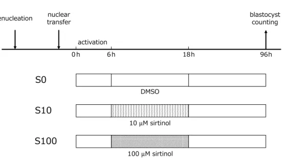
In experiment 2, to know the effective ment period of sirtinol, the embryos were treat-ed with 100µM sirtinol during activation (6 hr) and/or after activation (12 hr). In the “S-S” group, the embryos were activated in the activa-tion medium containing 100µM sirtinol-1.4% DMSO and incubated for 12 hr after activation with CZB containing 100µM sirtinol-0.4% DMSO. In the “S-N” and “N-S” groups, the embryos were treated with sirtinol only during and only after activation, respectively. In the “N-N” group, only DMSO was added in activation buffer and CZB using in incubation of 12 hr after activation (Fig. 2A).
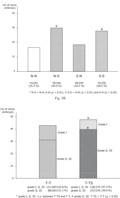
In experiment 3, the embryos were activated with the activation medium containing 5 nM TSA-1.5% DMSO, followed by 12 hr of incuba-tion in the CZB containing 5 nM TSA-0.9% DMSO (the “T-T” group) or 5 nM TSA-100µM sirtinol-0.9% DMSO (the “T-TS” group) (Fig. 3A).
Fig. 1. A: The reconstructed embryos were incubated for 12 hr after activation in CZB containing 100µM sirtinol-0.4% DMSO (“S100” group), 10 µM sirtinol-0.4% DMSO (“S10” group), or 0.4% DMSO (“S0” group). B: The rate of formation of the total blastocyst (grades I-III) of the “S100” group was significantly higher than that of the “S10” group and “S0 group”.
Fig. 2. A: In “S-S” group, the reconstructed embryos were activated in the activation medium containing 100µM sirtinol-1.4% DMSO and incubated for 12 hr after activation with CZB containing 100 µM sirtinol-0.4% DMSO. In “S-N” and “N-S” groups, the embryos were treated with sirtinol only during and only after activation, respectively. In N-N” group, only DMSO was activated in the activation medium containing 1.4% DMSO and incubated for 12 hr after activation with CZB containing 0.4% DMSO. B: The rate of formation of the total blastocysts (grades I-III) of “N-S” group was sig-nificantly higher than that of “N-N” group and “S-N” group. The rate of “S-S” group was signifi-cantly higher than that of “N-N” group and “S-N” group.
Evaluation of early embryonal development
We used a grading system for the murine blas-tocyst defined by Chen T-C et al. in this study; in brief, grade I , II and III blastocysts correspond-ed to non-expandcorrespond-ed, and expandcorrespond-ed, and hatched blastocysts, respectively15). In experi-ments 1 and 2, the number of total blastocysts (grades I-III) was counted. In experiment 3 the number of blastocysts of grades II and III as well as total blastocysts (grades I-III) were counted.
Generation of offspring derived from the “T-TS” treat-ed embryos
A female ICR mouse, used as a surrogate mother, was mated with a vasectomized male ICR mouse. The “T-TS” treated embr yos obtained by the method indicated in experi-ment 3 were transferred into a surrogate moth-er. The 2-cell stage and blastocyst stage embryos were transferred into the oviduct and uterus of the mother, respectively. The offspring were delivered by cesarean section after 18.5 days of SCNT, and the body weight and placental
weight of the live offspring and were recorded. The newborn cloned mice were transferred to a foster mother of B6D2F1 strain. Moreover, in order to analyze the fertility of the “T-TS” treat-ed embryo-derivtreat-ed clontreat-ed mice, two clontreat-ed mice that reached adulthood (the cloned mouse No. 3 named “Chuo”, and No. 6 named “Kai”) were mated with C57BL/6 male mice of the same age, respectively, and the pups of the cloned mice were observed.
Statistics
The data were compared by chi-square test. A value of p < 0.05 or 0.01 was judged as statistical-ly significant.
RESULTS
Experiment 1
In experiment 1, the effect of the treatment of the reconstructed embryos with sirtinol for 12 hr after activation on the early development of the cloned embryos was examined. The rate Fig. 3. A: The reconstructed embryos were activated with the activation medium containing 5 nM
TSA-1.5% DMSO, followed by 12 hr of incubation in the CZB containing 5 nM TSA-0.9% DMSO (“T-T” group) or 5 nM TSA-100µM sirtinol-0.9% DMSO (“T-TS” group). B: The rate of formation of grades I-III blastocysts of “T-TS” group was not statistically different from that of the “T-T” group. However, the rate of formation of the grades II and III blastocysts of the “T-TS” group was signifi-cantly higher than that of the “T-T” group.
of formation of the total blastocysts (grades I-III) of the “S100” group (31.4%, n=204) was sig-nificantly higher than that of the “S10” group (20.3%, n=207) (p < 0.01) and the control “S0 group” (19.1%, n=199) (p < 0.01) (Fig. 1B).
Experiment 2
The effect of activation with sirtinol and/or incubation with sirtinol for 12 hr after activation on blastocyst formation of the cloned embryos was examined in experiment 2. The rate of for-mation of the total blastocysts (grades I-III) of the “N-S” group (30.0%, n=260) was significant-ly higher than that of the “N-N” group (16.4%, n=261) (p < 0.01) and the “S-N” group (18.5%, n=259) (p < 0.01). The rate of the “S-S” group (28.0%, n=250) was significantly higher than that of the “N-N” group (p < 0.01) and the “S-N” group (p < 0.05). No significant differences were observed between the “N-S” group and the “S-S” group, or between the “N-N” group and the “S-N” group (Fig. 2B).
Experiment 3
In experiment 3, the additive effect of the treatment of the reconstructed embryos with sirtinol for 12 hr after activation on the early development of the TSA-treated cloned embryos was examined. The rate of formation of grades I-III blastocysts of the “T-TS” group (47.4%, n=270) was not statistically different from that of the “T-T” group (42.8%, n=283). However, when the rate of formation of the grades II and III blastocysts was compared, the rate of the “T-TS” group (39.6%) was signifi-cantly higher than that of the “T-T” group (31.1%) (p < 0 .05) (Fig. 3B).
Offspring of the cloned mouse derived from the “T-TS” treated embryos
For generation of offspring derived from the “T-TS” treated embryos, SCNT experiments were carried out 26 times between 06/27/2007 and 09/16/2007. We obtained 2405 recon-structed embryos. 1338 embryos at 2 cell stage and 142 embryos at blastocyst stage were trans-ferred into 40 and 8 surrogate mothers,
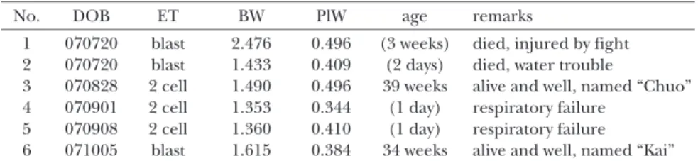
tively. Six live born offspring, weighing 1.621 +/– 0.430 g (mean +/– SD), were delivered (Table 1). The weights of the placenta were 0.423 +/– 0.061 g. Of the live born offspring, 2 cloned mice reached adulthood and were named “Chuo” and “Kai”. “Chuo” and “Kai”
(cloned mice No. 3 and 6 in Table 1), mated with C57BL/6 male mice, became pregnant and delivered their pups, and could nurse their pups. The first eight pups of “Chuo” (born on 2007/10/29 weighing 1.333 +/– 0.105 g) at 15 days of age were shown in Fig.4.
Fig. 3B Fig. 2B
DISCUSSION
Sirtuins are a family of homologues of Sir-2
(Silent mating type Information Regulation-2) that has been identified in yeast. Sir-2 is known as a “lifespan-extending gene” because overex-Table 1. The live born offsprings derived from the “T-TS” treated embryo
No. DOB ET BW PlW age remarks
1 070720 blast 2.476 0.496 (3 weeks) died, injured by fight 2 070720 blast 1.433 0.409 (2 days) died, water trouble
3 070828 2 cell 1.490 0.496 39 weeks alive and well, named “Chuo” 4 070901 2 cell 1.353 0.344 (1 day) respiratory failure
5 070908 2 cell 1.360 0.410 (1 day) respiratory failure 6 071005 blast 1.615 0.384 34 weeks alive and well, named “Kai” “T-TS” treatment: the embryos were treated with TSA and sirtinol as indicated in methods of the experiment 3.
DOB: date of birth
ET: type of embryonal transfer, 2 cell: the embryos were transferred at the 2 cell stage into the oviduct of the surrogate mother, blast: the embryos were transferred at the blastocyst stage into the uterus of the surrogate mother.
BW: body weight of new born offspring, 1.621 +/– 0.430 g (mean +/– SD). PlW: placental weight, 0.423 +/- 0.061 g (mean +/– SD).
age: the age on 6/1/2008. The days or weeks franked by the parenthesis indicated the length of life.
Fig. 4. Two of six live born offspring, which were derived from the “T-TS” treated embryo, named “Chuo” and “Kai” were mated with C57BL/6 male mice. They became pregnant and delivered pups, and could nurse their pups. The photo indicates “Chuo” and her first 8 pups born on 10/29/2007 (The photo was taken on 11/14/2007).
pression of the gene induces deacetylation of histones and results in about a 30% extension of lifespan in yeast16). Sirtuins remove acetyl groups from histones and other non-histone proteins in the presence of NAD+, and are classi-fied as a class III HDAC. Sirtuins have also been identified in mammals including mice. Sirtinol and splitomicin are general inhibitors for sirtu-ins, class III HDACs, in mammalian cells10,11).
Recent studies revealed that treatment of the reconstructed embryos with TSA, an inhibitor for classes I and II HDACs, significantly improves the development of the cloned mice5). Because the substrate proteins of class III HDACs are reported to be different from those of classes I and II HDACs7,8), the effects of sirti-nol on murine reconstructed embryos are also considered different from that of TSA. There-fore, we hypothesized that the simultaneous treatment of the murine cloned embryos with TSA and sirtinol might induce additional improvement of embryonal and fetal develop-ment. In order to verify this hypothesis, the pre-sent study was performed.
In experiment 1, we examined the effect of the treatment of reconstructed embryos with 100, 10 or 0 (control)µM sirtinol for 12 hr after activation on early embryonal development (Fig. 1B). The results indicate that sirtinol sig-nificantly improves the development of the cloned embryos to the blastocyst stage at the concentration of 100µM but not at that of 10µM, indicating that acetylation of the sub-strate proteins of class III HDACs during 12 hr after activation are involved in the reprogram-ming of the somatic nucleus. We could not examine the effect of sirtinol at a higher con-centration, because it was difficult to dissolve this compound in CZB at 200µM or higher.
In experiment 2, we studied the effect of the treatment with 100µM sirtinol during activation
and/or for 12 hr after activation on the early development of a cloned embryos. The results show that the treatment during activation has little or no benefit in contrast to the treatment for 12 hr after activation (Fig. 2B). Therefore, the acetylation of substrate proteins of class III HDACs during activation might not induce nuclear reprogramming. Interestingly, it has been reported that the embryonal development is improved by the treatment with TSA only dur-ing activation as well as only durdur-ing 14 hr after activation5), suggesting that the acetylation of the substrate proteins of classes I and II HDACs during activation as well as during 14 hr after activation induce reprogramming. Taken togeth-er, it is considered that the acetylated proteins responsible for nuclear reprogramming in the cloned embryos by sirtinol are not the same as those by TSA. Thus, we considered that the treatment with sirtinol after activation could additively improve the development of the TSA-treated cloned embryos. To further investigate this issue, we carried out experiment 3.
In experiment 3, treatment with 100µM sirti-nol for 12 hr after activation in addition to the TSA-treatment (“T-TS” group) did not improve the rate of formation of the total blastocysts (grades I, II and III blastocysts) compared to the TSA-only treated embryos (“T-T” group). However, the rate of formation of the expanded (grade II) blastocysts and the hatched (grade III) blastocysts of the “T-TS” group was signifi-cantly higher than that of the “T-T” group (Fig.3B). These results imply that the substrates, which were responsible for nuclear reprogram-ming, modified after treatment with sirtinol are different from those modified after treatment with TSA, and that the simultaneous treatment of the inhibitor for classes I/II and class III HDAC, TSA and sirtinol, could further improve the success rate of the murine SCNT
experi-ments.
Although the results from experiments 1-3 indicate that sirtinol could improve the early development of murine cloned embryos, it was possible that sirtinol, a general inhibitor for sir-tuins, might interfere with the development after the blastocyst stage, because, as mentioned above, sirtuins was thought to have a “lifespan-extending” effect16). In order to investigate this point, we generated offspring derived from the “T-TS” treated embryos. As shown in Table 1, six live offspring were obtained from the embryos, and two of the clones reached adulthood. These two cloned mice, “Chuo” and “Kai”, delivered their pups and could nurse them. These find-ings indicate that the treatment with sirtinol for 12 hr after activation does not affect embryonal, fetal or postnatal development.
In conclusion, the treatment of the recon-structed embryos with sirtinol for 12 hr after activation improves early embryonal develop-ment without adverse effects on subsequent development. This treatment also improves early embryonal development of the TSA-treat-ed clonTSA-treat-ed embryos, implying that the proteins, which were responsible for nuclear reprogram-ming, acetylated after the treatment with the inhibitor for class III HDAC would be different from those acetylated after the treatment with the inhibitor for classes I and II in the cloned murine embryo. At the present time, the target substrate proteins of the respective class of the HDACs in the cloned embryo have not been clarified. Since HDACis induce gene transcrip-tion9), it is considered that the genes responsi-ble for nuclear reprogramming might be up-regulated by the treatment of HDACis in the reconstructed embryo. However, it is also possi-ble that the HDACis might interact with the non-histone regulatory proteins involved in nuclear reprogramming in the cloned
embryo9,10). Further studies are necessary to clarify the mechanism by which the HDACis improve the development of the cloned murine embr yo. Based on such studies, the more improved the murine SCNT technique could be, the more widely the technique would be applied to the various fields of studies in labora-tories using mouse as a laboratory animal.
ACKNOWLEDGEMENTS
The authors gratefully acknowledge the tech-nical assistance of Ms. Michiko Yoneyama.
REFERENCES
1) Wilmut I, Schnieke AE, McWhir J, Kind AJ, Campbell KH. Viable offspring derived from fetal and adult mammalian cells. Nature 385: 810–813, 1997.
2) Keefer GL. Lessons learned from nuclear trans-fer (cloning). Theriogenology 69: 48–54, 2008. 3) Wakayama T, Perry AC, Zuccotti M, Johnson KR,
Yanagimachi R. Full-term development of mice from enucleated oocytes injected with cumulus cell nuclei. Nature 394: 369–374, 1998.
4) Wakayama T. Production of cloned mice and ES cells from adult somatic cells by nuclear transfer: how to improve cloning efficiency. J Reprod Dev 53: 13-26, 2007.
5) Kishigami S, Mizutani E, Ohta H, Hikichi T, Thuan NV, et al.: Significant improvement of mouse cloning technique by treatment with tri-chostatin A after somatic nuclear transfer. Biochem Biophys Res Commun 340: 183–189, 2006.
6) Kishigami S, Bui HT, Wakayama S, Tokunaga K, Van Thuan N, et al. Successful mouse cloning of an outbred strain by trichostatin A treatment after somatic nuclear transfer. J Reprod Dev 53: 165–170, 2007.
7) Marks PA, Miller T, Richon V. Histone deacety-lases. Curr Opin Pharmacol 3: 344–351, 2003. 8) Gallinari P, Di Marco S, Jones P, Pallaoro M,
Steinkühler C. HDACs, histone deacetylation and gene transcription: from molecular biology to cancer therapy. Cell Res 17: 195–211, 2007. 9) Xu WS, Parmigiani RB, Marks PA. Histone
action. Oncogene 26: 5541–5552, 2007.
10) Denu JM. The Sir2 family of protein deacety-lases. Curr Opin Chem Biol 9: 431–440, 2005. 11) Gronzinger CM, Chao ED, Blackwell HE,
Moazed D, Schreiber SL. Identification of a class of small molecule inhibitors of the sirtuin family of NAD-dependent deacetylases by phenotype screening. J Biol Chem 276: 38837–38843, 2001. 12) Kimura Y, Yanagimachi R. Intracytoplasmic
sperm injection in the mouse. Biol Reprod 52: 709–720, 1995.
13) Chatot CL, Lewis JL, Torres I, Ziomek CA. Devel-opment of 1-cell embryos from different strains of mice in CZB medium. Biol Reprod 42: 432–440, 1990.
14) Kishigami S, Wakayama S, Thuan NV, Ohta H, Mizutani E, et al. Production of cloned mice by somatic cell nuclear transfer. Nat Protoc 1: 125-138, 2006.
15) Cheng TC, Huang CC, Huang LS, Chen CI, Lee MS, et al. Evaluation of mouse blastocyst implan-tation rate by morphology grading. Chin J Physi-ol 47: 43–47, 2004.
16) Lin SJ, Defossez PA, Guarente L. Requirement of NAD and SIR2 for life-span extension by calorie restriction in Saccharomyces cerevisiae. Science 289: 2126–2128, 2000.