INTRODUCTION
The Mr 190,000 multidrug resistance protein (MRP)1 is an identified member of the ATP-binding cassette (ABC) transmembrane transport proteins (1). In ad-dition to the glioma cell lines (T98G and Gli36) from which these proteins were isolated, overexpression of MRP1 has been detected in many multidrug resis-tant tumor cell lines derived from various tissues and selected in different natural product type chemothera-peutic agents (2).
The efficacy of topoisomerase Ⅱ inhibitors (e.g., epipodophyllotoxins, aminoacridine, and
mitoxan-trone) is limited by the occurrence of drug resistance in the tumor cell population. Cellular insensitivity to drugs that stabilize the cleavable complex is frequently expressed as multidrug resistance (MDR)(2). In some cell lines, overexpression of MDR-1/P-glycoprotein or the multidrug resistance associated protein (MRP) 1-5, has been demonstrated and implicated as the mecha-nism of resistance(2, 3). These proteins are known to confer multidrug resistant phenotypes to the cancer cells. Although the primary structures of P-glycoprotein and MRP share only 15% amino acid homology (1), both P-glycoprotein and MRP are involved in the drug re-sistance to a similar profile of chemotherapeutic agents that include anthracyclines, vinka alkaloid, and epipo-dophyllotoxins (3). Another transporter, termed breast cancer-resistance protein (BCRP), also known as Mitoxantrone-resistance protein and placenta ABC protein, was described in breast and colon carcinoma
ORIGINAL
Reduction of expression of the multidrug resistance protein
(MRP)1 in glioma cells by antisense phosphorothioate
oligonucleotides
Yoshihito Matsumoto
1, Keisuke Miyake
1,Katsuzou Kunishio
1, Takashi Tamiya
1,
Seigo Nagao
1 1Department of Neurological Surgery, Kagawa University School of Medicine, Kagawa, Japan
Abstract : The tumor cells’ acquisition of resistance to multiple drugs due to overexpression of the multidrug resistance protein (MRP) 1 gene is one of major obstacles in cancer chemotherapy. We have attempted to reverse the multidrug resistance (MDR) phenotype by treating etoposide resistant glioma cell lines (T98G-VP and Gli36-VP) with MRP1 antisense oligonucleotides. 20-mer phosphorothioate oligodeoxynucleotide (0.3µM), complementary to the coding region in the MRP cDNA sequence, could significantly inhibit the growth of multidrug resistant cell lines, T98G-VP and Gli36-VP, cultured in etoposide containing medium. No such effect was observed for the parental T98G and Gli36 cell lines. Further investigations by the reverse transcription-polymerase chain reaction and immunoblotting revealed that antisense oligomer could result in a reduction in the level of MRP1 mRNA, probably through hindering MRP1 gene transcription. This study demonstrates that the antisense oligonucleotides can increase the sensitivity of the tumor cells to the anticancer drug by decreasing the expression of the MRP gene. This strategy may be applicable to cure cancer patients with MRP mediated MDR phenotype. J. Med. Invest. 51 : 194-201, August, 2004
Keywords : antisense, multidrug resistance, gene therapy, MRP, glioma
Received for publication February 18, 2004; accepted July 22, 2004. Address correspondence and reprint requests to Yoshito Matsumoto, M.D., Department of Neurological Surgery, Kagawa University School of Medicine, Miki-cho, Kita-Gun 761-0793, Kagawa, Japan and Fax : +81-87-891-2208.
The Journal of Medical Investigation Vol. 51 2004 194
cell lines selected for higher-level resistance to the an-tineoplastic drug, Mitoxantrone (3).
The cells that overexpress P-glycoprotein may be sensitized to the cytotoxic effects of chemotherapeutic agents by the co-administration of a wide variety of the so-called “reversing agents”(4). Two of the most extensively studied reversing agents both in vitro and
in vivoare verapamil, a calcium channel blocker, and cyclosporin A, an immunosuppressive cyclic peptide. However, both verapamil and cyclosporin A are relatively ineffective and/or non-specific chemosensitizers in MRP expressing cell lines, promoting a search for alterna-tive ways to reverse the MRP1-mediated resistance (4). One particularly attractive approach is to use antisense oligonucleotides, because of the potentially high degree of specificity of these reagents. Rather than inhibiting the protein function, the antisense oligonucleotides decrease mRNA and protein synthesis by binding to its complementary nucleic acid target in a sequence specific manner (5-7).
This study demonstrates that the antisense oligonu-cleotides can increase the sensitivity of the tumor cells to the anticancer drugs by decreasing the expression of the MRP1 gene. This strategy may be applicable to cure the cancer patients with MRP1 mediated multidrug resistance phenotype.
MATERIALS AND METHODS
Oligonucleotides
The oligonucleotides used in this study was described previously (7). The oligonucleotides was 20 bases in length and was phosphorothioate oligodeoxynucleo-tides. The sequences of antisense oligonucleotides for the MRP1 was as follows :2107
5’-TGCTGTTCGTGCC CCCGCCG-3’
Cell culture and treatment
The T98G-VP and Gli36-VP are multidrug resistant human glioma cell lines overexpressing the MRP, which were derived from the parental T98G and Gli36 cells by culture in etoposide (VP-16)(8).
These cells were grown in 75cm2
flasks until 70-80% confluent and then were washed twice with serum-free MEM medium before the addition of lipofectin/oli-gonucleotide complexes. Lipofectin (GIBCO/BRL life technologies, Burlington, Ontario, Canada)(10µg/ml) and the oligonucleotides were allowed to form complexes in serum-free MEM medium at room temperature for 15 min after gentle mixing. The cells were incubated with lipofectin/oligonucleotide at 37℃ for 4hr, washed
once with MEM/10% fetal bovine serum, and then incubated in fresh MEM/10% fetal bovine serum until harvested.
Reverse transcription and polymerase chain reaction (PCR) analysis for the expression of the MRP 1
Total RNA was isolated from cells by IsogenR
(Nippon gene) according to the manufacture’s instructions. RT-PCR was performed using First-Strand cDNA Syn-thesis KitR
(Amersham pharmacia biotech). 1µg of total RNA was added to 14µl of RT-mixture. After mix-ing, the samples were incubated at 37℃ for 45 min, 95℃ for 5 min and 4℃ for at least 5min. 35µl of PCR-mixture containing 10nM primers and Taq DNA po-lymerase (Amersham pharmacia biotech) was added to RT-products. Initial denaturation for 2 min at 94℃ was followed by 30 cycles of 1 min at 94℃, 1 min at 55℃ and 2 min at 72℃ and a final extension for 10 min at 72℃. The PCR-products were separated on 2% aga-rose gels, and stained with ethidium (9).
The primer sequences for MRP1 were as follows 5’primer :733
5’-CGGAAACCATCCACGACCCTAATCC-3’ 3’primer :1027
5’-ACCTCCTCATTCGCATCCACCTTGG-3’
Immunoblotting
Immunoblotting for the the MRP1 was performed using detergent-solubilized membrane proteins resolved on a 6% SDS-polyacrylamide. Immunoblotting was performed with a1 : 2000 dilution of a monoclonal MRP1 antibody, kindly provided by Dr. Rik Scheper (Free University Hospital, Netherlands). After three washes in TBS-T, the membranes were incubated with HRP-conjugated anti-mouse secondary antibody at a dilution of 1:1000 for 1 hr. Five washes were then performed and immunolabeled protein was detected by chemilumi-nescence(2).
Cellular Accumulation for[3
H]-VP-16
3.5×10⁵
cells were plated in each well of a six-well plate and incubated for 24 hr at 37℃. Growth medium was then replaced with serum-free MEM, and the cells were incubated with 2.6µM [3
H]-VP 16 for various times up to 90 min. at 37℃. At the completion of the incuba-tion period, cells were quickly washed three times with ice-cold PBS, lysed with 2ml of 0.05% SDS, and mixed with 3ml of Scintisol (2, 10). Data derived from the drug accumulation analysis was compared using ANOVA. A probability value of less than 0.05 was accepted as statistically significant (2).
Cloning of topoisomerase IIα, MRP1-5, and MDR-1 cDNA by Reverse Transcription-PCR(2)
Synthetic oligonucleotides corresponding to the published cDNA sequence of human MRP1-5were used to isolate by RT-PCR specific products for direct cloning into pGEM-3 z vectors (Promega, Madison, WI). The identity of the cDNA clones was confirmed by direct sequence analysis prior to their use as probes for northern analysis. The sequences of the oligonucleotides used in the RT-PCR were as follows: topoisomerase IIα: -91 5‘TGTGGAGAAGCGGCTTGGTC 3 ’ and 449 5‘TAGTTACTAGAAGTTAGGAGCT 3’ ; MRP 1 : 733 5 ‘ CGGAAACCATCCACGACCCTAATCC 3’ and 1027 5 ‘ACCTCCTCATTCGCATCCACCTTGG 3’; MRP 2 : 4136 5‘CTGCCTCTTCAGAATCTTAG’ and 4376 5‘CCCAAGTTGCAGGCTGGCC’ ; MRP 3 : 5
‘GATACGCTCGCCACAGTCC3’ and 5‘CAGTTGG
CCGTGATGTggCTG 3’ ; MRP 4 : 5‘CCATTGAAG ATCTTCCTGG3’ and 5‘GGTGTTCAATCTGTGTGC 3’ ; MRP 5 : 5‘GGATAACTTCTCAGTGGG 3’and 5
‘GGAATGGCAATGCTCTAAAG 3’ ; MDR-1 : 352
5‘CAGTGTTTGCCATAGTATTTTCAAGGATTG 3’ and 741 5‘CCCTTTAACACTAGAAGCATCAC 3’.
Northern Blotting
Total RNA(20µg/lane) was separated on a 6% for-maldehyde gel and transferred to Hybond N+with 10 X SSC. The membranes were hybridized at 42℃ overnight with the radiolabeled probe in Hybrisol I (Oncor, Gaithersburg, MD)2
. We used the RNA of ZR-VP 13that overexpressed all drug resistant genes (MDR-1, MRP 1-5, topoisomerase Ⅱα) as positive control (2).
Cytotoxicity Assays
Cytotoxicity assays were performed as previously described (11). Briefly, cells were plated at 10000 per well in 96-well microtiter plates with various concen-trations of the anticancer drugs〔VP-16, adriamycin, vin-cristine, cisplatin, and1-(4-amino-2-methyl-5-pyrimidinyl) methyl-3-(2-chloroethyl)-3-nitrosourea (ACNU)〕and were cultured for 2 days. Then the cells were incu-bated with 0.2 mg/ml of MTT in MEM for 4 hr, and the formazan product was dissolved in dimethyl sulfoxide and its absorbance at 540 nm was measured. The cy-totoxic effects of the drugs were expressed quantitatively as the dose required to kill 50% of the cells (IC50).
RESULTS
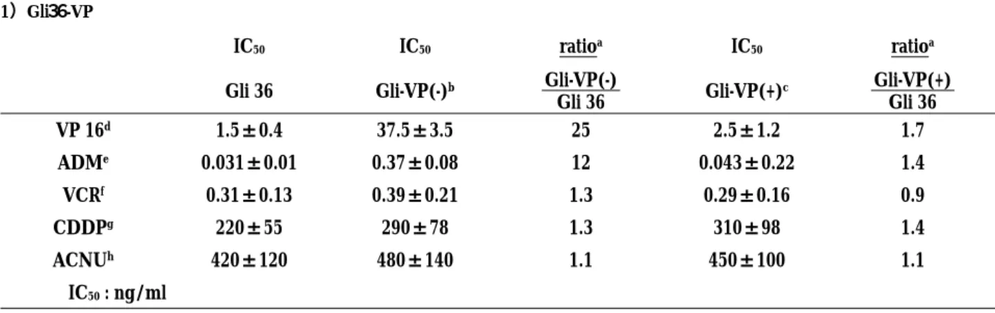
Table1 depicts the cross-resistance profile of the
two sublines to various chemotherapeutic agents. Cross-resistance to two topoisomerase Ⅱ poisons (VP-16, adriamycin) was observed in these drug resistant cells with little cross resistant to non-topoisomerase Ⅱ poisons, Vincristine, cisplatin, and ACNU. Resistant sublines treated with the MRP1 antisense oligonucleo-tide displayed an enhanced sensitivity to topoisomerase Ⅱ inhibitors, VP-16 and Adriamycin.
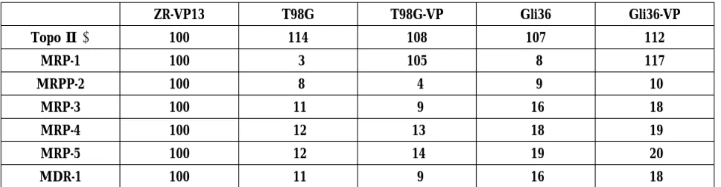
When expressions of topoisomerase IIα, MRP1-5, and MDR-1 were examined, the results shown in Fig.1 and Table2 were obtained. Northern analysis showed that these resistant cells the overexpress MRP1, but do not overexpress MDR-1 or MRP1-5 except 1. Moreover, in the results of northern blot analysis of mRNA for topoisomerase IIαand topoisomerase Ⅱ activity in decatanation assay using kinetoplast DNA (data not shown), identical results are observed in parental cell lines and their resistant cell lines, suggesting that alterations in topoisomerase II do not account for the resistance in these cells.
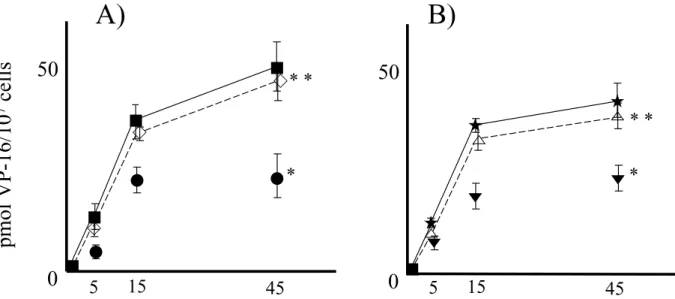
The VP-16 accumulation study was carried out in the parental cell lines and their drug resistant sublines (Fig.2). Figure 2 demonstrates the3
H-VP-16 accu-mulation in the parental cells and the drug resistant sublines. Accumulation over 45 min. period is displayed as p mol of VP-16/107
cells.3
H-VP-16 accumulations for the resistant sublines relative to parental cells were T 98 G-VP, 48% and gli 36-VP, 58%. Increased accumu-lation was observed in the two resistant sublines treated with the MRP1 antisense oligonucleotide, with partial restoration of levels to that of the parental cells (T98 G-VP with MRP1 antisense treatment, 96% of parental T 98 G ; and Gli36-VP with the MRP1 antisense treat-ment, 94% of parental Gli 36).
Figure 3 depicts concentration-dependent reduction of MRP1 mRNA and protein in VP-16 resistant cell lines by a treatment with the MRP1 antisense oligonucleotide. The T98G-VP and Gli-VP cells were exposed for 4 hr to selected concentrations of the MRP1 antisense oli-gonucleotide. The MRP 1 mRNA was isolated 24 hr after treatment, and RT-PCR and immunoblotting were performed. A concentration-dependent decrease in the MRP1 mRNA was observed after a single treatment of the T98G-VP with the MRP antisense oligonucleotide. A significant decrease of the MRP mRNA and protein were observed at 0.1µM, and virtually no MRP mRNA was detectable at oligonucleotide concentration of 0.3µM (upper panel). Similar results were found in the experiment using the Gli36-VP cell line. Significant reduced MRP mRNA and protein were detectable at oligonucleotide concentration of 0.3µM (lower panel). Y. Matsumoto et al. Reduction of MRP 1 expression by antisense
Figure 1:Northern blot analysis of topoisomerase IIα(4.5 KB), MRP 1-5(4.5 KB, 4.4 KB, 4.7 KB, 4.3 KB, 6.5 KB), and MDR-1 (4.5 KB) expression in drug resistant sublines and their parental cell lines. The RNA of ZR-VP13 that overexpressed all drug resistant genes (MDR-1, MRP 1-5, topoisomerase Ⅱα) was used as positive control.β-actin was used as internal control.
Figure 2: VP-16 accumulation study was carried out in parental cell lines and their drug resistant sublines (A; T98G, B; Gli36). * significantly different (p<0.05) between resistant cell lines and their parental cell lines(incubation time at 45 min). * * significantly different (p<0.05) between resistant cell lines and their own transfected MRP 2 antisense cDNA (incubation time at 45 min)
left panel : ■ ; T98G, ● ; T98G-VP, ◇ ; T98G-VP with MRP 1 antisense treatment right panel : ★ ; Gli 36,▼ ; Gli 36-VP, △ ; Gli 36-VP with MRP 1 antisense treatment
Figure 3: Concentration-dependent reduction of MRP1 mRNA and protein in etoposide resistant cell lines by a treatment with the MRP1 antisense oligonucleotide. Upper panel;a concentration-dependent decrease in the MRP1 mRNA and protein were observed after a single treatment of T98G-VP and Gli 36-VP with MRP antisense oligonucleotides.
Table 1. Cross resistace profile of the drug resistant cell lines 1)Gli36-VP
IC50 IC50 ratioa IC50 ratioa
Gli 36 Gli-VP(-)b Gli-VP(-)
Gli 36 Gli-VP(+) c Gli-VP(+) Gli 36 VP 16d 1.5±0.4 37.5±3.5 25 2.5±1.2 1.7 ADMe 0.031±0.01 0.37±0.08 12 0.043±0.22 1.4 VCRf 0.31±0.13 0.39±0.21 1.3 0.29±0.16 0.9 CDDPg 220±55 290±78 1.3 310±98 1.4 ACNUh 420±120 480±140 1.1 450±100 1.1 IC50: ng/ml 2)T98G-VP IC50 IC50 ratioa IC50 ratioa T98G T98G-VP(-)b T98G-VP(-) T98G T98G-VP(+)c T98G-VP(+) T98G VP 16d 7.5±2.4 137.5±33.5 18 8.5±5.2 1.1 ADMe 0.081±0.01 0.47±0.08 6 0.096±0.02 1.2 VCRf 1.31±0.33 1.39±0.51 1.1 1.39±0.36 1.1 CDDPg 420±155 490±178 1.2 510±198 1.2 ACNUh 390±220 480±140 1.2 450±178 1.2 IC50: ng/ml
a : ratio of the individual cell lines
b : without MRP antisense oligonucleotides c : with MRP antisense oligonucleotides
d : etoposide, e : adriamycin, f : vincristine, g : cispatin, h : 1-(4-aminio-2-methyl-5-pyrimidinyl) methyl-3-(2-choloroethyl)-3-nitrosourea
Y. Matsumoto et al. Reduction of MRP 1 expression by antisense 198
DISCUSSION
Since the development of MDR by cancer cells still represents one of the major reasons for anticancer chemotherapy failure (12-18), the aim of the present study was to attempt modulation and eventually reversal of MDR in the glioma cell lines in vitro, using antisense oligonucleotides to targeted to the MRP1 mRNA. In a previous study, the 16 oligonucleotides were screened initially for their ability to decrease the MRP1 protein and mRNA levels in MRP1-overexpressing H69AR cells (7). One of the oligonucleotides, which we used in this study, was complementary to nucleotide 2107-2126 of MRP1 mRNA and reproducibly the most ef-fective in lowering the MRP1 protein and mRNA levels. It was also demonstrated that the decrease in MRP1 mRNA results from site specific cleavage, consistent with an RNAseH-dependent antisense mechanism (10, 19-21).
The ability of MRP1 antisense oligonucleotide to increase VP-16 cytotoxicity in the T98G-VP and Gli36-VP cells and thus to reverse the MDR was assessed in combination experiments in which the T98G-VP and Gli36-VP cells were first exposed to MRP antisense oligonucleotide and then to 16. Treatment with VP-16 alone had modest effects on the T98G-VP and Gli 36-VP cell proliferation. The in vitro restoration of VP-16 sensitivity in T98 G-VP and Gli36-VP was confirmed by the VP-16 IC50value for each treatment. A strong
decrease of the IC50value was observed in the
T98G-VP and Gli36-T98G-VP cells treated with combination of MRP antisense oligonucleotide plus VP-16. Noteworthy, the IC50
value of the resistant cells exposed to antisense oli-gonucleotide plus VP-16 was essentially identical to that of the T98G and Gli 36 parental cells exposed to the drug alone, consistent with a complete restoration of the VP-16 accumulation in T98G-VP and Gli36-VP cells. Transfection experiments with MRP expression vectors
have shown that MRP can confer resistance to adriamycin and VP-16(22). It has been shown that MRP gene encodes glutathione S-conjugate export carrier, and that the molecules which are transported by MRP include adria-mycin glutathione conjugates and glucuronosyl VP-16. These reports are consistent with our present study using the MRP antisense oligonucleotide.
MRP expression has also been observed in other clinical cancers, including colorectal carcinoma, lung cancer, lung cancer, breast carcinoma, retinoblastoma, and acute leukemia. Regarding the nervous system, it has been reported that MRP mRNA and protein are not detectable in the brain, and glioma cell lines with higher MRP mRNA levels show increased drug resis-tance. Since all the present clinical specimens examined were primary tumors, we considered that the MRP expression was concerned with intrinsic drug resis-tance as well as acquired drug resisresis-tance (19, 22). In an attempt to correlate the anti-proliferative effect of the MRP1 antisense oligonucleotide with the levels of MRP1 mRNA, we evaluated the MRP1 mRNA and protein expression by RT-PCR and immunoblotting in the T98G-VP and Gli36-VP cells immediately after treatment with MRP1 antisense oligonucleotide plus VP-16. In comparison to the untreated cells, the cells treated with the MRP1 antisense oligonucleotide ex-hibited a marked reduction of the MRP1mRNA and protein. The level ofβ-actin mRNA were essentially unchanged in the cells receiving different treatments, confirming the specificity of the effects induced by the MRP1 antisense oligonucleotide.
In conclusion, our studies raise the possibility that antisense oligonucleotide targeted to MRP1 mRNA might be useful in the attempt to reverse the MDR in the cancer patients.
Table 2. Northern blot analysis of topoisomerase IIα, MRP1-5, and MDR-1 expression in drug resistant sublines and their parental cell lines ZR-VP13 T98G T98G-VP Gli36 Gli36-VP Topo Ⅱα 100 114 108 107 112 MRP-1 100 3 105 8 117 MRPP-2 100 8 4 9 10 MRP-3 100 11 9 16 18 MRP-4 100 12 13 18 19 MRP-5 100 12 14 19 20 MDR-1 100 11 9 16 18
Densitometric quantitations of drug resistance gene expressions were performed by comparing with those in ZR-VP13 cells
REFERENCES
1. Cole SPC, Bhardwaj G, Gerlach JH, Mackie JE, Grant CE, Almquist KC, Stewart AJ, Kurz EU, Duncan AMV, Deeley RG : Overexpression of a transporter gene in a multidrug resistant human lung cancer cell line. Science 258 : 1650-1654, 1992
2. Matsumoto Y, Takano M, Fojo T:Cellular adap-tation to drug exposure : Evolution of the Drug-resistant phenotype. Cancer Res 57 : 5086-5092, 1997
3. Miyake K, Mickley L, Fojo T, Bates SE : Molecular cloning of cDNAs which are highly overexpressed in Mitoxantrone-resistant cells. Cancer Res 59 : 8-13, 1999
4. Alvarez M, Robey R, Sandor V, Nishiyama K, Matsumoto Y, Paull K, Bates S, Fojo T : Using the National Cancer Institutes anticancer drug screen to assess the effect of MRP expression on drug sensitivity profiles. Molecular Pharmacology 54 : 802-814, 1998
5. Cuddo C, Calabretta B : In vitro and in vivo rever-sal of multidrug resistance in a human leukemia-resistant cell line by mdr 1 antisense oligonu-cleotides. Cancer Res 56 : 4332-4337, 1996 6. Li Y, Wang Y : Inhibition growth of multidrug
resistant KBV200cells by MDR1 antisense RNA.
Biochem Biophysical Research Communication 239 : 345-348, 1997
7. Stewart AJ, Canitrot Y, Baracchini E, Dean N M, Deeley R, G, Cole SPC : Reduction of expression of the multidrug resistance protein (MRP) in human tumor cells by antisense phosphorothioate oligonucleotides. Biochemical Pharmacology 51 : 461-469, 1996
8. Matsumoto Y, Matsumoto M, Minemura M, Takano H, Nagao S, Iglesias A, Fojo T : Expression of ATP binding cassette superfamily (multidrug resistance-1, multidrug resistance-associated pro-tein, human canalicular multispecific organ anion transporter) mRNA in etoposide and m-AMSA resistant cell lines. Jpn J Cancer Chemother 24: 1941-1946, 1997
9. Paulusma CC, Bosma PJ, Zaman GJ, Bakker CT, Otter M, Scheffer GL, Scheper RJ, Borst P : Congenital jaundice in rats with a mutation in a multidrug resistance associated protein gene. Science 271 : 1126-1127, 1996
10. Matsumoto Y, Takano H, Kunishio K, Nagao S, Fojo T: Hypophosphorylation of topoisomerase IIαin Etoposide(VP-16)-resistant human carcinoma
cell lines associated with carboxy-terminal trun-cation. Jpn J Cancer Res 92 : 799-805, 2001 11. Matsumoto Y, Sasaoka N, Tsuchida T, Fujiwara T,
Nagao S, Ohmoto T : Fluorescent dye Rhodamine 6 G as a molecular probe to study drug resistance of C6 rat glioma cells. Journal of Neurooncology 13 : 217-222, 1992
12. Cohen D, Higman S M, Hsu S, Horwitz S B : The involvement of a LINE-1 element in a DNA rearrangement upstream of the mdr 1 a gene in a Taxol multidrug-resistant murine cell line. J Biol Chem 267 : 20248-20254, 1992
13. Cole SPC, Sparks KE, Fraser K, Loe DW, Grant CE, Wilson GM : Pharmacological characterization of multidrug resistant MRP-transfected human tumor cells. Cancer Res 54 : 5902-5910, 1994 14. Fojo AT, Ueda K, Slamon DJ, Poplack DG,
Got-tesman MM, Pastan I : Expression of a multidrug resistance gene in human tumors and tissues. Proc Natl Acad Sci USA 84 : 265-269, 1987 15. Grant CE, Vadimarsson G, Hipfner DR, Almquist
KC, Cole SPC, Deeley RG : Overexpression of multidrug resistance associated protein increases resistance to natural product drugs. Cancer Res 54 : 357-361, 1994
16. Lee JS, Scala S, Matsumoto Y, Dickstein B, Robey R, Zhan Z, Altenberg G, Bates SE : Reduced drug accumulation and multidrug resistance in human breast cancer cells without associated P-glycoprotein or MRP overexpression. J Cell Biochem 65 : 513-526, 1997
17. Matsumoto Y, Fujiwara T, Nagao S : Determinants of drug response in camptothecin-11-resistant glioma cell lines. J Neurooncol 23 : 1-8, 1995 18. Taniguchi K, Wada M, Kohno K, Nakamura T,
Kawabe T, Kawakami M, Kagotani K, Okumura K, Akiyama S, Kuwano M : A Human canalicular multispecific organic anion transporter gene is overexpression in cisplatin resistant human cancer cell lines wit decreased drug accumulation. Cancer Res 56 : 4124-4129, 1996
19. Matsumoto Y, Takano H, Kunishio K, Nagao S, Fojo T: Expression of drug resistance genes in VP-16 and mAMSA-selected human carcinoma cells. Jpn J Cancer Res 92 : 778 -784, 2001 20. Matsumoto Y, Takano H, Kunishio K, Nagao S,
Fojo T. Incidence of Mutation and Deletion in Topoisomerase IIα mRNA of Etoposide and mAMSA Resistant Cell Lines. Jpn J Cancer Res 92 : 1133-1137, 2001
21. Matsumoto Y, Takano H, Nagao S, Fojo T : Al-tered topoisomerase IIαand multidrug resistance-Y. Matsumoto et al. Reduction of MRP 1 expression by antisense
associated protein levels during drug selection : Adaptation to increasing drug. Jpn J Cancer Res 92 : 968-974, 2001
22. Mohri M, Nitta H, Yamashita J : Expression of multidrug resistance-associated protein (MRP) in human gliomas. J Neurooncology 49 : 105-115, 2000