INTRODUCTION
Hyperkalemia can have multimodal effect on myo-cardial protection during ischemia/reperfusion. Ex-tracellular myocardial K+concentration ([K+]
o) in
ischemic regions increase to 10-20 mM after 3-10
min of ischemia (1, 2). Hyperkalemia may preserve adenosine triphosphate (ATP) by reducing excitabil-ity of cardiac cells in energy-limited state. Hyperka-lemic cardiac arrest is used as a protective interven-tion during cardiac surgery (3). In addiinterven-tion, Na+/
K+-ATPase activation induced by hyperkalemia may
exert a cardioprotective effect (4). However, mem-brane depolarization induced by hyperkalemia may cause detrimental effects in myocardial cells by inducing activation of a reversal mode of the Na+/
Ca2+ exchanger and/or slow Ca2+ channels (5).
Conversely, decreased myocardial Na+/K+-ATPase
ORIGINAL
Cardioprotective effects of hyperkalemia during simulated
ischemia/reperfusion in neonatal rat cardiomyocytes
-Preservation of Na
+/K
+-ATPase
activity-Kaori Takata
1, Yoshinobu Tomiyama
2, Katsuya Tanaka
1, and Shuzo Oshita
1 1Department of Anesthesiology, Institute of Health Biosciences, the University of Tokushima Graduate School,2
Division of Surgical Center, Tokushima University Hospital, the University of Tokushima
Abstract : Background : Hyperkalemia has multimodal effects on myocardial protection during ischemia/reperfusion. The preservation of Na+/K+-ATPase activity induced by
hy-perkalemia may have critical impact on myocardial protection. Methods : To elucidate the roles of hyperkalemia (16 mM) and Na+/K+-ATPase inhibition (100μM ouabain) in
myo-cardial protection during simulated ischemia (5 mM NaCN and 5.5 mM 2-deoxyglucose)/ reperfusion, we measured loss of membrane integrity and bleb formation using a vital dye calcein AM in cultured neonatal rat cardiomyocytes. The control perfusate was switched to treatment solution for 15 min, followed by reperfusion for 30 min. In a second set of experiments, myocardial excitability and diastolic intracellular calcium ion concentra-tion ([Ca2+]
i) were measured during a 45-min treatment using a calcium-sensitive
fluo-rescent dye fluo-4 AM. Results : Simulated ischemia/reperfusion under ouabain treatment induced loss of membrane integrity, which was suppressed by hyperkalemia. Simulated ischemia/reperfusion induced bleb formation, which was accelerated by ouabain. Hy-perkalemia delayed and inhibited the increase in diastolic [Ca2+]
i induced by simulated
ischemia. Furthermore, hyperkalemia almost completely inhibited the effects of ouabain on the diastolic [Ca2+]
i during ischemia. Conclusions : These results suggest that
hyperka-lemia during ischemia is cardioprotective against ischemia/reperfusion insults and that hyperkalemia inhibits the effects of ouabain during ischemia. J. Med. Invest. 60 : 66-76, February, 2013
Keywords : myocardial protection, hyperkalemia, ischemia/reperfusion, cardiotonic steroids
Received for publication October 30, 2012 ; accepted November 29, 2012.
Address correspondence and reprint requests to Dr Yoshinobu Tomiyama, Division of Surgical Center, Tokushima University Hospital, 2 - 50 - 1 Kuramoto, Tokushima, 770 - 8503, Japan and Fax : + 81 - 88 - 633 - 9191.
activity is associated with decreased post-ischemic recovery (6, 7). The Na+/K+-ATPase activity is
inhib-ited by various factors including the therapeutic use of cardiotonic steroids such as digoxin (8, 9), en-dogenous cardiotonic steroids (10-12), hypothermia (4), and ischemia (13, 14). Cardiotonic steroids may abolish the protective effects of hyperkalemia. In contrast, [K+]
oelevation decreases the affinity of
Na+/K+-ATPase for cardiotonic steroids (15-17).
Hyperkalemia may counteract the inhibition of Na+/
K+-ATPase induced by cardiotonic steroids during
ischemia. However, the effect has not yet been in-vestigated in ischemia/reperfusion studies.
We hypothesized that hyperkalemia during ische-mia has a cardioprotective effect during ischeische-mia/ reperfusion and that the effects are partly mediated by the preservation of Na+/K+-ATPase activity. To
test this hypothesis, we investigated whether hy-perkalemia and ouabain, a synthetic cardiotonic ster-oid, modulate the loss of membrane integrity and bleb formation during simulated ischemia/reperfu-sion in cultured neonatal rat cardiomyocytes. The suggested results were confirmed in a second set of experiments measuring diastolic [Ca2+]
iand
myo-cardial excitability during ischemia
MATERIALS AND METHODS
All experimental procedures and protocols used in this study were reviewed and approved by the Animal Investigation Committee of Tokushima Uni-versity and followed the guidelines of the American Physiological Society (Bethesda, Maryland) for the use of animals in research.
Cell culture
Primary cultures of neonatal rats were prepared from the hearts of 2-day-old Sprague-Dawley rats with a Neonatal Cardiomyocyte Isolation System (Worthington Biochemical Corp., Lakewood, NJ). This system, which utilizes purified enzyme prepa-rations, provides a reliable and consistent cell iso-lation method (18). Neonatal rats were anesthetized with sevoflurane and their hearts were quickly re-moved. Isolation was performed according to the manufacturer’s recommendations (18). The cells were maintained in M199 culture medium supple-mented with 10% fetal bovine serum, gentamycin (50μg"ml- 1), and amphotericin B (2.5 μg"ml- 1) in
35-mm glass-bottomed culture dishes (MacTek Corp, Gates Mills, OH). Experiments were initiated
after 4 days of incubation for calcein studies and 3 days of incubation for fluo-4 studies at 37!!in a 5% CO2 environment using monolayers of
spontane-ously beating myocytes.
Measurement of loss of membrane integrity and bleb formation during simulated ischemia/reperfusion
Simulated ischemia using NaCN, an inhibitor of mitochondrial cytochrome oxidase, and 2-deoxyglu-cose, an inhibitor of glycolysis, has been used in ischemia/reperfusion or ischemia studies (5, 19). The vital dye calcein-AM was used to assess mem-brane integrity (20-22). Necrotic blebs form in cells exposed to intense noxious stimuli such as hypoxia/ reoxygenation and appear prior to cell permeabili-zation (20, 23). We selected an ouabain concentra-tion (100μM) that resulted in 50% inhibition of Na+/
K+-ATPase but produces no loss of viability in
neo-natal rat cardiomyocytes (24).
Loading with the vital dye calcein AM (Molecular Probes, Eugene, OR) was accomplished by incuba-tion with calcein AM (200 nM in phosphate- buff-ered saline) for 20 min. The cells were maintained at 37!!for another 20 min to ensure complete hy-drolysis after washing with phosphate-buffered saline.
A cell perfusion system was designed to meas-ure cell fluorescence in response to rapid solution changes. The system used an inverted epifluores-cence microscope (Eclipse TS100, Nikon, Tokyo, Japan). A culture dish incubation system (Harvard Apparatus, DH-35i, Holliston, MA) was placed on the microscope stage. In addition, 35-mm glass-bottomed culture dishes and a chamber insert closed for 35-mm dishes (Harvard Apparatus, RC-37FC) were mounted on the incubation system. Cell perfusion solutions were infused using an in-fusion pump at a rate of 2 ml"min- 1. Solutions were
maintained at a constant temperature by a 35-mm tissue culture dish heater (Harvard Apparatus, DH-35), solution in-line heater (Harvard Apparatus, SH-27B), and chamber system heater controller (Harvard Apparatus, TC-344B).
Excitation of calcein was obtained using a Xenon lamp (50 W ; Nikon) filtered at 450-490 nm and re-flected to the microscope objective (
!
20, CFI Plan Fluor ELWD 20!
C ; Nikon, Tokyo, Japan) by a di-chroic mirror centered at 505 nm. Cell fluorescence was collected by the objective, passed through a 520-nm-long path filter, and directed to a cooled digital B/W CCD camera (ORCA, Hamamatsu Photonics, Hamamatsu, Japan).Relative changes in the calcein fluorescence in-tensity in each region of interest (ROI) relative to the baseline (%) were used as an index of mem-brane integrity. The times until bleb appearance in the ROIs were also recorded. The ROI included al-most all cells in a single field of view of the micro-scope, which included more than fifty cells. Fluo-rescence images with an exposure time of 555 msec were taken every 30 seconds using a computer-con-trolled shutter. The cells were initially perfused with “normal” perfusate i.e., Earle’s balanced salt solution (EBSS), containing (in mM) 116.4 NaCl, 5.4 KCl, 1.8 CaCl2, 0.8 MgSO4, 26.2 NaHCO3, 1 NaH2PO4,
and 5.6 D-glucose, which was titrated to pH 7.40. After equilibration (approximately 10 min), the per-fusate was randomly switched to one of treatment solutions : EBSS (CON ; n=6), EBSS containing 100 μM ouabain (OUA ; n=5), modified EBSS contain-ing NaCN and 2-deoxyglucose (ISC ; n=6), ISC+ HIK (n=6), ISC+OUA (n=6) or ISC+HIK+OUA (n=6), and measurements were performed for 15 min. The perfusate was then switched back to nor-mal perfusate to measure reperfusion injury for an additional 30 min at 37!!.
Measurement of myocardial excitability and diastolic [Ca2+]
iduring ischemia
Cultured neonatal rat cardiomyocytes have been used to study spontaneous beating (18, 25). A pha-sic rise and fall in fluorescence is well-established as representing the systolic rise and diastolic fall of [Ca2+]
iupon which contraction and relaxation
de-pend (25, 26). Therefore, we used the rhythmical
fluorescence changes as an index of myocardial ex-citability in the fluorescence images. Loading with the Ca2+indicator, fluo-4 AM (Molecular Probes,
Eugene, OR), was accomplished by incubation with fluo-4 AM (5μM in phosphate-buffered saline) for 20 min at room temperature ; the cells were main-tained at 37!!for another 20 min to ensure com-plete hydrolysis after washing with phosphate-buff-ered saline. We used the same cell perfusion and epifluorescence system as the calcein study. Fluo-rescence images with an exposure time of 334 msec were taken for 15 sec at 2 frames!s- 1at 5 minute
intervals using a computer-controlled shutter and an image processing system.
The cells were initially superfused with control perfusate. After equilibration (approximately 10 min), the perfusate was randomly switched to one of treatment solutions : CON (n=5), HIK (n=5), OUA (n=5), ISC (n=5), ISC+HIK (n=5), ISC+OUA (n=5) or I SC+HIK+OUA (n=5). Measurements were taken for 45 min at 37!!.
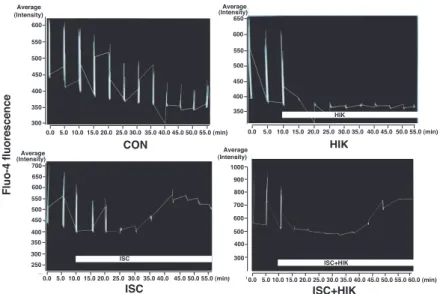
Data analysis was performed as follows. We chose 3-7 ROIs, which contained a cell or small clusters of cells and met the criteria for selection, at ran-dom from a single field of view of the microscope. The criteria for selection were that the fluorescences of ROI changed rhythmically and that the diastolic fluorescence did not increase until the intervention. Representative traces of fluo-4 fluorescence value are presented in Figure 1. Background values (mini-mum value in the windows of the ROIs that in-cluded the area outside of the cells) were always subtracted. Diastolic fluo-4 fluorescence values in
Figure 1. Graph showing representative traces of fluo - 4 fluorescence in the CON, HIK, ISC, and ISC + HIK groups. After equili-bration with “normal” perfusate (Earle’s balanced salt solution EBSS) for approximately 10 min, the perfusate was switched to either EBSS (CON), modified EBSS containing 16 mM KCl (HIK), modified EBSS containing 5 mM NaCN and 5.5 mM 2 - deoxyglucose (ISC), or modified EBSS containing 5 mM NaCN, 5.5 mM 2 - deoxyglucose, and 16 mM KCl (ISC + HIK).
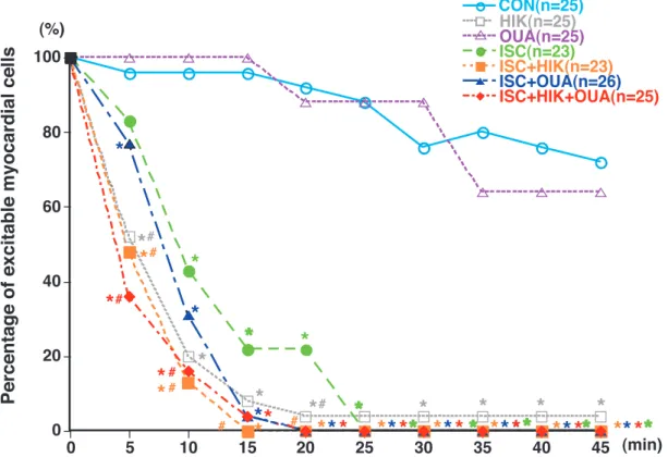
the ROIs were evaluated by calculating the mean value of the bottom five average pixel intensities in the 30 fluorescence images. Fluo-4 fluorescence was expressed relative to the baseline value (%). If at least one rhythmical increase in fluorescence was observed over 15 seconds, we considered the cell or cell mass to have retained excitability. Changes in excitability of cardiomyocytes were expressed as percentages of excitable myocardial cells (Figure 2). Statistical analysis
Data are expressed as the mean!SD. Compari-son of several means of diastolic intracellular [Ca2+]
i,
calcein fluorescence intensities, and the times un-til bleb appearance were performed using a two-way analysis of variance followed by a Student-Newman - Keuls post - hoc test for multiple com-parisons. All P-values are two-tailed. Comparisons of myocardial excitability were performed using Fisher’s exact test. A P-value of less than 0.05 was considered significant. Statistical analysis of the data was performed using the commercially available software program StatView 5.0 (SAS Institute Inc., Cary, NC).
RESULTS
Effects of hyperkalemia in the presence or absence of ouabain on membrane integrity and bleb formation during simulated ischemia/reperfusion
Three patterns were observed in the loss of cal-cein fluorescence : gradual loss, abrupt loss with bleb formation, and abrupt loss without bleb forma-tion (Figure 3). The latter two patterns were ob-served to have similar timings in this study to those reported in another study (22). The times until bleb appearance in the CON, OUA ISC, ISC+HIK, ISC+OUA ISC+OUA+HIK group were 35!12 min, 29!14 min, 20!13 min, 27!11 min, 13!4 min*, and 24!12 min, respectively (*P!0.05 vs. CON). During control perfusion at 37"!, gradual decreases in calcein fluorescence were observed (Figure 4, 5). However, appearance of bleb delayed until 35 min. Thus, although some cells in the CON group suf-fered a loss of membrane integrity, the majority of the decrease in calcein fluorescence was likely in-duced by dye leakage or photobleaching. Ouabain itself did not produce a significant difference from control. Abrupt losses of calcein fluorescence were
Figure 2. Time course of myocardial excitability in neonatal rat cardiomyocytes during treatment : control (CON), 16 mM KCl (HIK), 100μM ouabain (OUA), simulated ischemia (ISC), simulated ischemia plus 16 mM KCl (ISC+HIK), simulated ischemia plus 100μM ouabain (ISC+OUA), and simulated ischemia plus 16 mM KCl plus 100 μM ouabain (ISC+HIK+OUA). Simulated ischemia was induced by 5 mM NaCN and 5.5 mM 2 - deoxyglucose. If at least one rhythmical increase in fluorescence was observed over 15 seconds, we considered the cell or the cell mass to have retained excitability. Myocardial excitability was expressed as the percent-age of excitable cells in each group at every measured point (%). *P!0.05 vs. CON. #P!0.05 vs. ISC.
Figure 3. Figure showing representative changes in calcein fluorescence in the CON and ISC + OUA group. After equilibration with “normal” perfusate (Earle’s balanced salt solution EBSS) for approximately 10 min, the perfusate was switched to either EBSS (CON) or modified EBSS containing 5 mM NaCN, 5.5 mM 2 - deoxyglucose, and 100μM ouabain (ISC+OUA). Figures were ob-tained at baseline, 15, 30, and 45 min. Arrows indicate blebs.
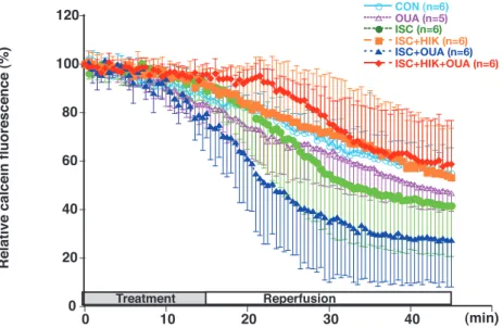
Figure 4. Time course of calcein fluorescence during and after treatment in time control (CON), 100μM ouabain (OUA), simu-lated ischemia (ISC), simusimu-lated ischemia plus 16 mM KCl (ISC + HIK), simusimu-lated ischemia plus 100μM ouabain (ISC+OUA), or simulated ischemia plus 16 mM KCl plus 100μM ouabain (ISC+HIK+OUA) groups. Simulated ischemia was induced by 5 mM NaCN and 5.5 mM 2 - deoxyglucose. Calcein fluorescence was measured relative to baseline (%). Data are represented as the mean!SD.
Figure 5. Effects of treatment/reperfusion on calcein fluorescence in cultured neonatal rat cardiomyocytes : control (CON), 100 μM ouabain (OUA), simulated ischemia (ISC), simulated ischemia plus 16 mM KCl (ISC+HIK), simulated ischemia plus 100 μM ouabain (ISC + OUA), and simulated ischemia plus 16 mM KCl plus 100μM ouabain (ISC+HIK+OUA). Simulated ischemia was in-duced by 5 mM NaCN and 5.5 mM 2 - deoxyglucose. Evaluation was performed at reperfusion, 15 min after reperfusion, and 30 min after reperfusion. Values are expressed as the mean!SD. *P!0.05 vs. CON. #P!0.05 vs. ISC+OUA.
observed after reperfusion in many cells in the ISC group (Figure 4). The timing of appearance of the bleb is consistent with the abrupt loss of calcein fluorescence, suggesting reperfusion injury.
Perfusion with ouabain during simulated ische-mia induced bleb formation in few cells (Figure 3, 4). Abrupt losses of calcein fluorescence were ob-served in most cells after reperfusion (Figure 4), although significant differences were not observed between the ISC and ISC+OUA groups. Difference with the CON group became significant in the times until bleb appearance. Thus, ouabain may acceler-ate and potentiacceler-ate the membrane injury induced by simulated ischemia/reperfusion. Perfusion with hy-perkalemia during simulated ischemia/reperfusion did not induce any difference in calcein fluorescence relative to the CON group, suggesting protective effects (Figure 5).
Hyperkalemia during simulated ischemia/reperfu-sion inhibits the decrease in calcein fluorescence in-duced by ischemia/reperfusion even in the presence of ouabain (Figure 4). Significant differences were observed in the calcein fluorescence between the ISC+OUA and the ISC+OUA+HIK group (Figure
5). Thus, the preventive effect became obvious in the presence of ouabain. Hyperkalemia nearly com-pletely inhibited the effects of ouabain during ische-mia/reperfusion.
Effects of hyperkalemia on myocardial excitability and diastolic [Ca2+
]iduring simulated ischemia in
the presence or absence of ouabain
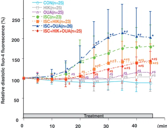
The change in myocardial excitability and diastolic [Ca2+]
iin the ouabain group occurred over a
simi-lar time course as that of the control (Figure 6). Hyperkalemia induced diastolic arrest within ap-proximately 5 min (Figure 6). Simulated ischemia in the absence and presence of ouabain also in-duced diastolic arrest within approximately 10 min and 8 min, respectively (Figure 6). Significant dif-ferences were observed between hyperkalemia groups and ischemia group at 5 min.
Hyperkalemia and ouabain did not significantly increase diastolic [Ca2+]
iin the absence of ischemia
(Figure 6, 7). Simulated ischemia with and without hyperkalemia induced significant increases in dia-stolic [Ca2+]
iafter 25 min and 15 min, respectively.
Significant differences between the ISC+HIK and
Figure 6. Time course of diastolic fluo - 4 fluorescence in neonatal rat cardiomyocytes during treatment : control (CON), 16 mM KCl (HIK), 100μM ouabain (OUA), simulated ischemia (ISC), simulated ischemia plus 16 mM KCl (ISC+HIK), simulated ischemia plus 100μM ouabain (ISC+OUA), and simulated ischemia plus 16 mM KCl plus 100 μM ouabain (ISC+HIK+OUA). Simulated ische-mia was induced by 5 mM NaCN and 5.5 mM 2 - deoxyglucose. Fluo - 4 fluorescence was measured relative to baseline (%). Data are presented as the mean!SD. *P!0.05 vs. CON. #P!0.05 vs. ISC. $P!0.05 vs. ISC+OUA
ISC group and between the ISC+HIK and HIK group were observed after 20 min and 30 min, re-spectively. Thus, hyperkalemia delayed and inhib-ited the increase in diastolic [Ca2+]
iinduced by
simu-lated ischemia, suggesting cardioprotective effects, although hyperkalemia did not fully abolished the effects of ischemia (Figure 6, 7).
Simulated ischemia with ouabain induced signifi-cant increases in diastolic [Ca2+]
iafter 10 min.
Sig-nificant differences between the ISC+OUA and ISC group were observed between 25 min and 40 min. Thus, ouabain accelerated and potentiated the in-crease in diastolic [Ca2+]
i induced by simulated
ischemia (Figure 6, 7).
Simulated ischemia under hyperkalemia condi-tions with and without ouabain induced significant increases in diastolic [Ca2+]
iafter 35 min and 25 min,
respectively. Thus, hyperkalemia almost completely inhibited the effects of ouabain during simulated ischemia (Figure 6, 7).
DISCUSSION
Cardiotonic steroids and effects of hyperkalemia on myocardial protection during ischemia/reperfusion Our results suggest that hyperkalemia during ischemia is cardioprotective against ischemia/reper-fusion insult. One of possible mechanisms is the antagonistic effect of hyperkalemia on cardiotonic steroids. Ouabain itself did not alter membrane in-tegrity, bleb formation, myocardial excitability, or
diastolic [Ca2+]
iin this study. Although nontoxic
con-centrations of ouabain (up to 100μM) cause a less than twofold increase in diastolic [Ca2+]
i(27), it does
not cause arrhythmic contraction and have no ef-fect on cell viability in neonatal rat cardiomyocytes (24, 27). Our results suggest that ouabain acceler-ated and potentiacceler-ated the increase in diastolic [Ca2+] i
during ischemia and possibly the membrane injury during ischemia/reperfusion. These data suggest that myocardial tolerance to ischemia is reduced during partial inhibition of Na+/K+-ATPase.
Hyperkalemia almost completely inhibited the ef-fects of ouabain during ischemia/reperfusion in this study. These results confirmed by the increase in diastolic [Ca2+]
iduring ischemia. Extracellular K+
is known to counteract the inhibition of Na+/K+
-ATPase by cardiotonic steroids (16, 17), including a neonatal rat study (15). Hermans et al. reported a possible mechanism of the antagonism (16). K+
and ouabain bind to different conformational states of the Na+/K+-ATPase (E
2-P and E2-P Na2,
respec-tively), which are transiently exposed to the exter-nal side of the sarcolemma. Upon K+ binding the
state (E2-P K2) is removed from the extracellular
side. If the K+concentration is increased, more E 2
-P K2is formed and, consequently, active Na+-K+
transport is enhanced. At the same time, less E2-P
Na2is available for ouabain binding (16). Many
stud-ies have demonstrated that serum (11) and myo-cardial (12, 28) cardiotonic steroid activity levels are significantly increased during acute myocardial ischemia and that anti-digoxin antiserum has a
Figure 7. Effects of treatment on diastolic fluo - 4 fluorescence in cultured neonatal rat cardiomyocytes : control (CON), 16 mM KCl (HIK), 100μM ouabain (OUA), simulated ischemia (ISC), simulated ischemia plus 16 mM KCl (ISC+HIK), simulated ischemia plus 100μM ouabain (ISC+OUA), and simulated ischemia plus 16 mM KCl plus 100 μM ouabain (ISC+HIK+OUA). Simulated ische-mia was induced by 5 mM NaCN and 5.5 mM 2 - deoxyglucose. Evaluation was performed at 20 min and 35 min. Values are expressed as mean!SD. *P!0.05 vs. CON. #P!0.05 vs. ISC. $P!0.05 vs. ISC+OUA
protective effect against myocardial ischemia-reper-fusion injury (11, 12). We did not provide direct evi-dence that simulated ischemia induces the release of cardiotonic steroids or hyperkalemia enhance myocardial membrane Na+/K+-ATPase activity in
the present study. In addition, released cardiotonic steroids could have been washout because myocar-dial cells were continuously superfused in this study. Therefore, whether this mechanism was responsi-ble for the results of the ISC group is not clear. However, it is clear that hyperkalemia strongly in-hibited the effects of ouabain during ischemia. Myo-cardial cells may be exposed to the condition such as that of the ISC+HIK+OUA group at the site of myocardial ischemia. Therefore, we suppose that the cardioprotective effects of hyperkalemia may be induced by the inhibition of cardiotonic steroids. The effects of activation of Na+/K+-ATPase induced
by hyperkalemia on myocardial protection during ischemia/reperfusion
Activation of Na+/K+-ATPase is another possible
mechanism of hyperkalemia-induced myocardial protection (4). Although Na+/K+-ATPase is activated
to a considerable extent under normal [K+] o
condi-tions (29), Terkildsen et al. suggested that Na+/K+
pump amplitude declines rapidly during phase one of simulated ischemia to a level!65% of normoxic pump current and the increases in [K+]
oare
sug-gested to stimulate pump activity during phase one and phase two (the plateau phase) (30). Ouabain did not abolish the protective effects of hyperkalemia in this study. Indeed, dihydro-ouabain did not affect the activation of Na+/K+ pump current on [K+]
o in
cardiac ventricular myocytes, although Na+/K+pump
current decreased in the presence of dihydro-ouabain (16). Yamamoto et al. reported that meta-bolic inhibition abolished the hyperkalemia-induced cardioprotective effects induced by Na+/K+-ATPase
activation during normothermic reperfusion (4). Therefore, activation of Na+/K+-ATPase may not be
the main mechanism of the cardioprotective effects of hyperkalemia during ischemia, although we can-not deny the mechanism.
Membrane potential and the effects of hyperkalemia on myocardial protection during ischemia/reper-fusion
Diastolic arrest may have beneficial effects in myocardial cells under an energy-limited condition and could be another possible mechanism. A pre-vious report suggested that 15 mM hyperkalemic
solutions induce a depolarization of the resting mem-brane potential (Vm) to -66 mV from a resting Vm
of -87 mV in adult rat cardiomyocytes (5). At this depolarized potential, the fast Na+channels are
in-activated because the threshold is -70 to -65 mV, resulting in diastolic arrest (3). Thus, high [K+]
o
may preserve ATP by reducing excitability of car-diac cells. However, ischemia also reduces the ex-citability of cardiac cells, although differences of sev-eral minutes were observed. The impact of the en-ergy saving effects on the results is unclear. Baczko
et al. reported that increased [K+]
oduring ischemia
and reperfusion can lead to increased Ca2+overload
and cell hypercontracture/death, which increases in parallel with depolarization (5). However, in their study, hyperkalemic conditions were maintained at the time of reperfusion, in contrast with our study. Reversal of increased [K+]
oafter reperfusion could
decrease membrane potential, which was suggested in our preliminary study using a voltage-sensitive dye bis - (1,3 - dibutylbarbituric acid) trimethine oxonol bis-oxonol (31) (Data not shown).
Experimental model and limitations of this study Modeling ischemia/reperfusion is difficult be-cause of complicated and interdependent processes (32). We determined minimum exposure time for the induction of reperfusion injury using bis-oxonol in a preliminary study (data not shown). And, reper-fusion-induced membrane injury and reperfusion-induced membrane depolarization observed in this study and in preliminary studies using bis-oxonol (data not shown) suggest successful simulation of reperfusion injury. Although relatively large dish-to-dish or cell-to-cell variation was observed, het-erogeneity of the myocardial cell response to ische-mia is well documented (32-34). To simplify the model, we investigated ischemia only and selected relatively intact cells in the second series of study. Ca2+overload increased prior to reperfusion and the
determinants of Na+overload are important
deter-minants of Ca2+overload in reperfused myocytes
(14). Calpain activation, which occurs when reper-fusion is induced by elevated diastolic [Ca2+]
i,
in-duces inactivation of Na+/K+-ATPase and further
calpain activation, resulting in sarcolemmal rup-ture and cell death (14). In fact, we observed that hyperkalemia applied only during the ischemic pe-riod affected membrane injury and bleb formation after reperfusion.
In our study, we used cultured neonatal rat cardio-myocytes as a model. Several studies have suggested
that immature myocytes have poorly developed sarcoplasmic reticulum and lack fully developed Ca2+channels (35). There is a slight developmental
change in a2and a3isoforms 14-21 days after birth
(36). However, neonatal and adult cells have com-parable time courses of the rise in [Ca2+]
i,
develop-ment of sarcolemmal blebs and cumulative enzyme release during energy depletion. (19). Although we must explore this result to adult heart carefully, myocardial protection of juvenile heart is itself clini-cally important (37). Eliminations of spontaneous activity were observed in some cells during fluo-4 loading and control perfusion in this study, suggest-ing damage caused by these procedures (38).
Although hyperkalemic cardioplegic solutions have a number of problems in cardiac surgery (3), hyperkalemia may counteract the effects of car-diotonic steroids if applied under normothermia.
In conclusion, the data obtained in the present study suggest three important findings. First, 15 mM hyperkalemia during ischemia may be cardio-protective against ischemia/reperfusion insult in neonatal cardiomyocytes if applied to intact cells un-der normothermia. Second, inhibition of Na+/K+
-ATPase activity during ischemia accelerates and potentiates myocardial injury induced by ischemia/ reperfusion. Third, hyperkalemia during ischemia almost completely inhibits the effects of cardiotonic steroids during ischemia/reperfusion. We propose that preservation of Na+/K+-ATPase activity induced
by the antagonistic effect of [K+]
oon endogenous
or therapeutic cardiotonic steroids may play a role in the cardioprotective effects of hyperkalemia.
ACKNOWLEDGEMENTS
This study was supported in part by a Grant-in Aid (#19591801) for Scientific Research (C) from the Japan Society for the Promotion of Science, Tokyo, Japan.
This study was presented at the Annual Meeting of the American Society of Anesthesiologists, New Orleans LA, October 19, 2009 and San Diego CA, October 16, 2010.
REFERENCES
1. Carmeliet E : Cardiac ionic currents and acute ischemia : from channels to arrhythmias. Physiol
Rev 79 : 917-1017, 1999
2. Hill J, Gettes L : Effect of acute coronary ar-tery occlusion on local myocardial extracellular K+activity in swine. Circulation 61 : 768-778,
1980
3. Chambers DJ, Hearse DJ : Developments in cardioprotection : “polarized” arrest as an alter-native to “depolarized” arrest. Ann Thorac Surg 68 : 1960-1966, 1999
4. Yamamoto H, Magishi K, Goh K, Sasajima T, Yamamoto F : Cardioprotective effects of nor-mothermic reperfusion with oxygenated potas-sium cardioplegia : a possible mechanism. In-teract CardioVasc Thorac Surg 9 : 598-604, 2009
5. Baczko I, Giles WR, Light PE : Resting mem-brane potential regulates Na+-Ca2+
exchange-mediated Ca2+overload during hypoxia -
reoxy-genation in rat ventricular myocytes. J Physiol 550 : 889-898, 2003
6. Lundmark JL, Ramasamy R, Vulliet PR, Schaefer S : Chelerythrine increases Na-K-ATPase activity and limits ischemic injury in isolated rat hearts. Am J Physiol 277 : H999-H1006, 1999
7. Imahashi K, Nishimura T, Yoshioka J, Kusuoka H : Role of intracellular Na+kinetics in
precondi-tioned rat heart. Circ Res 88 : 1176-1182, 2001 8. Hunt S, Abraham W, Chin M, Feldman A, Francis G, Ganiats T, Jessup M, Konstam M, Mancini D, Michl K, Oates J, Rahko P, Silver M, Stevenson L, Yancy C : 2009 focused update incorporated into the ACC/AHA 2005 Guide-lines for the Diagnosis and Management of Heart Failure in Adults : a report of the Ameri-can College of Cardiology Foundation/Ameri-can Heart Association Task Force on Practice Guidelines : developed in collaboration with the International Society for Heart and Lung Trans-plantation. Circulation 119 : e391-479, 2009 9. Friberg L, Hammar N, Rosenqvist M : Digoxin
in atrial fibrillation : report from the Stockholm Cohort study of Atrial Fibrillation (SCAF). Heart 96 : 275-280, 2010
10. Schoner W, Scheiner-Bobis G : Endogenous and exogenous cardiac glycosides : their roles in hypertension, salt metabolism, and cell growth. Am J Physiol 293 : C509-C536, 2007 11. Bagrov AY, Shapiro JI, Fedorova OV :
Endoge-nous cardiotonic steroids : physiology, pharma-cology, and novel therapeutic targets. Pharma-col Rev 61 : 9-38, 2009
12. Ke Y, Liu Z, Wang D, Wang H : Effects of anti-digoxin antiserum on endoxin levels, apotosis and the expression of bax and BCL-2 protein in ischemia-reperfusion myocardium. Clin Exp Pharmacol Physiol 31 : 691-695, 2004
13. Chen W-J, Lin-Shiau S-Y, Huang H-C, Lee Y-T : Ischemia-induced alternation of myocardial Na+-K+-ATPase activity and ouabain binding
sites in hypercholesterolemic rabbits. Athero-sclerosis 127 : 59-64, 1996
14. Inserte J, Garcia-Dorado D, Hernando V, Soler-Soler J : Calpain-mediated impairment of Na+/
K+-ATPase activity during early reperfusion
contributes to cell death after myocardial ische-mia. Circ Res 97 : 465-473, 2005
15. Meldgaard L, Steiness E, Waldorff S : Time course of ouabain uptake in isolated myocar-dial cells : dependence on extracellular potas-sium and calcium concentration. Br J Pharma-col 73 : 341-345, 1981
16. Hermans AN, Glitsch HG, Verdonck F : The an-tagonistic effect of K+
oand dihydro-ouabain on
the Na+pump current of single rat and
guinea-pig cardiac cells. J Physiol 484 : 617-628, 1995 17. Akimova O, Tremblay J, Hamet P, Orlov SN :
The Na+/K+-ATPase as [K+]
osensor : Role in
cardiovascular disease pathogenesis and aug-mented production of endogenous cardiotonic steroids. Pathophysiology 13 : 209-216, 2006 18. Kang JX, Leaf A : Effects of long-chain
polyun-saturated fatty acids on the contraction of neo-natal rat cardiac myocytes. Proc Natl Acad Sci USA 91 : 9886-9890, 1994
19. Atsma DE, Lars Bastiaanse EM, Jerzewski A, Van der Valk LJM, Van der Laarse A : Role of calcium-activated neutral protease (Calpain) in cell death in cultured neonatal rat cardiomyo-cytes during metabolic inhibition. Circ Res 76 : 1071-1078, 1995
20. Nishimura Y, Lemasters J : Glycine blocks opening of a death channel in cultured hepatic sinusoidal endothelial cells during chemical hy-poxia. Cell Death Differ 8 : 850-858, 2001 21. Neumar R, Xu Y, Gada H, Guttmann R, Siman
R : Cross-talk between calpain and caspase pro-teolytic systems during neuronal apoptosis. J Biol Chem 278 : 14162-14167, 2003
22. Sardão VA, Oliveira PJ, Holy J, Oliveira CR, Wallace KB : Vital imaging of H9c2 myoblasts exposed to tert-butylhydroperoxide- characteri-zation of morphological features of cell death. BMC Cell Biol 8 : 11, 2007
23. Barros L, Kanaseki T, Sabirov R, Morishima S, Castro J, Bittner C, Maeno E, Ando-Akatsuka Y, Okada Y : Apoptotic and necrotic blebs in epithelial cells display similar neck diameters but different kinase dependency. Cell Death Differ 10 : 687-697, 2003
24. Peng M, Huang L, Xie Z, Huang W, Askari A : Partial inhibition of Na+/K+-ATPase by ouabain
induces the Ca2+-dependent expressions of
early-response genes in cardiac myocytes. J Biol Chem 271 : 10372-10378, 1996
25. McCaslin PP, Butterworth J : Bupivacaine sup-presses [Ca2+]
ioscillations in neonatal rat
car-diomyocytes with increased extracellular K+and
is reversed with increased extracellular Mg2+.
Anesth Analg 91 : 82-88, 2000
26. Northover BJ : Continuous fluorimetric assess-ment of the changes in cytoplasmic calcium concentration during exposure of rat isolated myocardium to conditions of simulated ischae-mia. Br J Pharmacol 100 : 477-482, 1990 27. Tian J, Gong X, Xie Z : Signal-transducing
func-tion of Na+-K+
-ATPase is essential for ouabain’s effect on [Ca2+]
iin rat cardiac myocytes. Am J
Physiol 281 : H1899-1907, 2001
28. D’Urso G, Frascarelli S, Balzan S, Zucchi R, Montali U : Production of ouabain-like factor in normal and ischemic rat heart. J Cardiovasc Pharmacol 43 : 657-662, 2004
29. Glitsch HG : Electrophysiology of the sodium-potassium ATPase in cardiac cells. Physiol Rev 81 : 1791-1826, 2001
30. Terkildsen JR, Crampin EJ, Smith NP : The bal-ance between inactivation and activation of the Na+-K+pump underlies the triphasic
accumula-tion of extracellular K+during myocardial
ische-mia. Am J Physiol 293 : H3036-H3045, 2007 31. Baczko I, Giles WR, Light PE :
Pharmacologi-cal activation of plasma-membrane KATP
chan-nels reduces reoxygenation-induced Ca2+
over-load in cardiac myocytes via modulation of the diastolic membrane potential. Br J Pharmacol 141 : 1059-1067, 2004
32. Li QA, Altschuld RA, Stokes BT : Myocyte deenergization and intracellular free calcium dynamics. Am J Physiol 255 : C162-168, 1988 33. Gomez LA, Alekseev AE, Aleksandrova LA,
Brady PA, Terzic A : Use of the MTT assay in adult ventricular cardiomyocytes to assess vi-ability : effects of adenosine and potassium on cellular survival. J Mol Cell Cardiol 29 : 1255-1266, 1997
34. de Groot JR, Schumacher CA, Verkerk AO, Baartscheer A, Fiolet JWT, Coronel R : Intrin-sic heterogeneity in repolarization is increased in isolated failing rabbit cardiomyocytes during simulated ischemia. Cardiovasc Res 59 : 705-714, 2003
35. Ichiba T, Matsuda N, Takemoto N, Ishiguro S, Kuroda H, Mori T : Regulation of intracellu-lar calcium concentrations by calcium and mag-nesium in cardioplegic solutions protects rat neonatal myocytes from simulated ischemia. J Mol Cell Cardiol 30 : 1105-1114, 1998
36. Yalcin Y, Carman D, Shao V, Ismail-Beigi F, Klein IL, Ojamaa K : Regulation of Na/K-ATPase gene expression by thyroid hormone and hyperkalemia in the heart. Thyroid 9 : 1999 37. Hammon JW, Jr : Myocardial protection in the immature heart. Ann Thorac Surg 60 : 839-842, 1995
38. Schaffer P, Ahammer H, Muller W, Koidl B, Windisch H : Di-4-ANEPPS causes photody-namic damage to isolated cardiomyocytes. Pflugers Arch 426 : 548-551, 1994