Posted at the Institutional Resources for Unique Collection and Academic Archives at Tokyo Dental College,
Title
profiles of cranial neural crest-like cells derived
from mouse-induced pluripotent stem cells.
Author(s)
Alternative
Odashima, A; Onodera, S; Saito, A; Ogihara, Y;
Ichinohe, T; Azuma, T
Journal
Medical molecular morphology, 53(1): 28-41
URL
http://hdl.handle.net/10130/5089
Right
This is an open access article distributed under
the terms of the
Creative Commons CC BY license, which permits
unrestricted use,
distribution, and reproduction in any medium,
provided the original
work is properly cited.
Description
https://doi.org/10.1007/s00795-019-00229-2
ORIGINAL PAPER
Stage‑dependent differential gene expression profiles of cranial
neural crest‑like cells derived from mouse‑induced pluripotent stem
cells
Ayano Odashima
1· Shoko Onodera
2· Akiko Saito
2· Yuuki Ogihara
3· Tatsuya Ichinohe
3· Toshifumi Azuma
4Received: 6 February 2019 / Accepted: 26 June 2019 / Published online: 11 July 2019 © The Author(s) 2019
Abstract
Cranial neural crest cells are multipotent cells that migrate into the pharyngeal arches of the vertebrate embryo and
differ-entiate into various craniofacial organ derivatives. Therefore, migrating cranial neural crest cells are considered one of the
most attractive candidate cell sources in regenerative medicine. We generated cranial neural crest like cell (cNCCs) using
mouse-induced pluripotent stem cells cultured in neural crest-inducing medium for 14 days. Subsequently, we conducted
RNA sequencing experiments to analyze gene expression profiles of cNCCs at different time points after induction. cNCCs
expressed several neural crest specifier genes; however, some previously reported specifier genes such as paired box 3 and
Forkhead box D3, which are essential for embryonic neural crest development, were not expressed. Moreover, ETS
proto-oncogene 1, transcription factor and sex-determining region Y-box 10 were only expressed after 14 days of induction. Finally,
cNCCs expressed multiple protocadherins and a disintegrin and metalloproteinase with thrombospondin motifs enzymes,
which may be crucial for their migration.
Keywords
Cranial neural crest · Migratory neural crest · iPS cells · RNA sequencing · Adamts
Introduction
Stem cell-based tissue engineering is important in the field
of oral science because it facilitates the regeneration of
dam-aged tissues or organs [
1
,
2
]. Various stem cell populations
exhibiting regeneration potential in the craniofacial region
have been identified. Of these, cranial neural crest cells
(cNCCs) are considered one of the most important
candi-dates owing to their role in craniofacial tissue organization
[
3
]. cNCCs comprise a multipotent population of migratory
cells that are unique to the vertebrate embryo and can
dif-ferentiate into various craniofacial organ derivatives [
4
,
5
].
The neural crest (NC) can form teratoma when transplanted
into immunocompromised animals [
6
]. cNCC development
involves three stages [
7
–
10
]: the neural plate border stage,
the premigratory stage, and the migratory stage. During the
migratory stage, cNCCs delaminate from the posterior
mid-brain and individual rhombomeres in the hindmid-brain [
11
] and
migrate into the pharyngeal arches to form skeletal elements
of the face and teeth and contribute to formation of the
phar-yngeal glands (the thymus, thyroid, and parathyroid) [
12
].
Therefore, cNCCs presumably represent a new treatment
strategy for diseases of the craniofacial region [
13
].
Development from the premigratory to migratory stage
proceeds swiftly [
14
]; thus, it is typically difficult to detect
the precise time point of this transition [
15
]. A recent
tran-scriptome analysis of pure populations of migratory cNCCs
cells expressing the sex-determining region Y-box 10
Electronic supplementary material The online version of thisarticle (https ://doi.org/10.1007/s0079 5-019-00229 -2) contains supplementary material, which is available to authorized users. * Toshifumi Azuma
[email protected] Ayano Odashima [email protected]
1 Department of Oral Health Science Center, Tokyo Dental
College, 2-9-18 Misaki-cho, Chiyoda-ku, Tokyo 101-0051, Japan
2 Department of Biochemistry, Tokyo Dental College, Tokyo,
Japan
3 Department of Dental Anesthesiology, Tokyo Dental
College, Tokyo, Japan
4 Department of Biochemistry, 2-9-18 Misaki-cho,
(Sox10) from chicks [
16
] has substantially improved our
understanding of cNCC characteristics. However, whether
these cells are in the migratory stage and how long it takes to
promote embryonic stem (ES) cell-derived NCCs from the
premigratory to migratory stage remains unclear. In recent
years, the use of induced pluripotent stem (iPS) cells as a
revolutionary approach to treat various medical conditions
has garnered much attention [
17
,
18
], and iPS cells as a
cell source have shown several evident advantages over ES
cells and primary cultured cNCCs in regenerative medicine
[
16
]. In addition, embryonic NC development depends on
several environmental factors that influence the regulation
of NC progenitors and timing of differentiation; therefore,
it is important to elucidate the regulatory gene networks
and expression profiles of mouse iPS (miPS) cell-derived
cNCCs. Recent advances in next-generation RNA
sequenc-ing (RNA-seq) technologies have facilitated comprehensive
analysis of gene expression profiles [
19
–
21
]. Therefore,
in the present study, we used RNA-seq to investigate the
gene expression landscape of cNCCs induced from miPS
cells. We treated iPS-derived cells with cNCC induction
medium for 14 days and performed RNA-seq experiments.
Our results indicated that c-Myc; ETS proto-oncogene 1,
transcription factor (Ets1); Sox10; a disintegrin and
metal-loproteinase domain metallopeptidase with thrombospondin
motifs (Adamts) 2 and 8; protocadherin alpha (Pcdha) 2, 5,
-7, -11, and -12; protocadherin alpha subfamily C,1
(Pcd-hac1); and protocadherin gamma subfamily C,3 (Pcdhgc3)
may be appropriate markers for migratory cNCCs induced
from miPS cells.
Materials and methods
miPS cell culture
The miPS cells used in the present study (APS0001;
iPS-MEF-Ng-20D-17 mouse-induced pluripotent stem cell
line) were purchased from RIKEN BRC (Ibaraki, Japan)
[
22
]. The cells were incubated with inactivated murine
embryonic fibroblast (MEF) feeder cells in Dulbecco’s
Modified Eagle’s Medium (DMEM; Invitrogen,
Carls-bad, CA, USA) supplemented with 15% KnockOut™
Serum Replacement (Invitrogen), 1% nonessential amino
acids (Chemicon, Temecula, CA, USA), 1%
l-glutamine
(Chemicon), 1000 U/mL penicillin–streptomycin (P/S;
Invitrogen), and 0.11 mM 2-mercaptoethanol (Wako Pure
Chemical Industries Ltd., Osaka, Japan); 60-mm cell
cul-ture plates were used for passaging the cells at a density
of 1 × 10
5cells/plate. Cells were grown in 5% CO
2
at 95%
humidity, and the culture medium was changed each day.
Embryoid body (EB) formation and cNCC
differentiation
We obtained cultured cNCC cells as described previously
[
23
] (Fig.
1
). miPS cells were dissociated with 0.05%
trypsin–ethylenediaminetetraacetic acid (EDTA;
Invitro-gen) and transferred to low-attachment, 10-mm Petri dishes
at a density of 2 × 10
6cells/plate to generate EBs. The
gen-erated EBs were cultured in cNCC induction medium
com-prising a 1:1 mixture of DMEM and F12 nutrient mixture
(Invitrogen) and then in Neurobasal™ medium (Invitrogen)
supplemented with 0.5 × N2 (Invitrogen), 0.5 × B27
(Inv-itrogen), 20 ng/mL basic fibroblast growth factor
(Repro-cell, Yokohama, Japan), 20 ng/mL epidermal growth factor
(Peprotech, Offenbach, Germany), and 1% P/S for 4 days;
the medium was changed every other day. After 4 days,
day 0 (d0) EBs were collected and transferred to 60-mm
cell culture plates coated with 1 μg/mL collagen type I
(Advanced BioMatrix, San Diego, CA, USA). The cells
were then subcultured in the same medium; the medium
changed every other day, and any rosette-forming cells
were eliminated. After 7–10 days, d7 cells were
dissoci-ated with 0.05% trypsin–EDTA and transferred to 60-mm
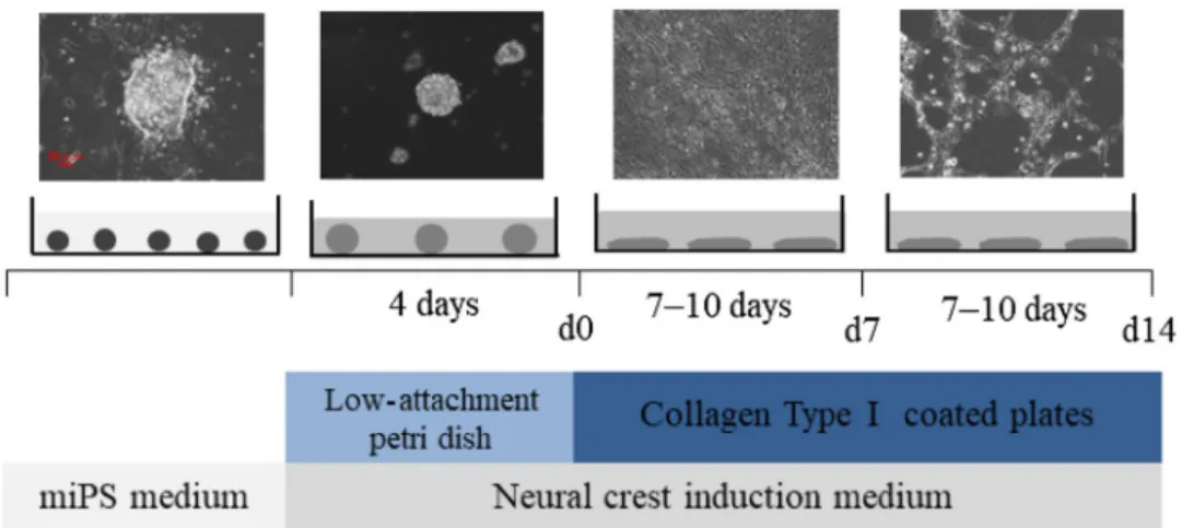
Fig. 1 The experimentalproto-col used to induce the formation of cranial neural crest cells (cNCCs) from mouse-induced pluripotent stem (miPS) cells. The photographs show miPS cells at four different stages: initial miPS cells, embryoid body (EB) on day 0 (d0), and cNCCs on d7 and d14. Small circles represent miPS cells; large circles represent EBs; ellipses represent d7 and d14 cells. Scale bar 50 μm
cell culture plates coated with 1 μg/mL collagen type I at
a density of 1 × 10
5cells/plate to generate d14 cells. This
process was repeated three times. The cells from each of
these passages were collected for RNA extraction.
O9‑1 cell culture
O9-1 cells, a mouse cNCC line, were purchased from
Mil-lipore (Billerica, MA, USA) and cultured as a control, as
previously described [
24
].
RNA extraction and quantitative reverse
transcription polymerase chain reaction analysis
(qRT‑PCR)
The expression of representative NC markers, namely nerve
growth factor receptor (Ngfr), snail family transcriptional
repressor (Snai) 1 and 2, and Sox9 and 10, was analyzed
using qRT-PCR analysis. Total RNA was extracted using
QIAzol
®reagent (Qiagen, Valencia, CA, USA) according to
the manufacturer’s protocol, and RNA purity was assessed
using NanoDrop
®ND-1000 spectrophotometer (Thermo
Fisher Scientific, Waltham, MA, USA). Each RNA
sam-ple exhibited an A260/A280 ratio of > 1.9. Comsam-plemen-
Complemen-tary DNA (cDNA) was synthesized using a high-capacity
cDNA reverse transcription kit (Applied Biosystems, Foster
City, CA, USA), and qRT-PCR analysis was performed with
Premix Ex Taq™ reagent (Takara Bio Inc., Otsu, Japan)
according to the manufacturer’s protocol using Applied
Biosystems
®7500 Fast Real-Time PCR System; the primer
sequences are presented in Table
1
. All samples were
nor-malized to 18S ribosomal RNA levels. Relative expressions
of genes of interest were analyzed using the ΔΔCt method
and were compared among the groups using analysis of
vari-ance, followed by the Bonferroni test when significant
dif-ferences were detected among the groups. A significance
level of p < 0.05 was used for all analyses, and all data were
expressed as mean values and standard deviations.
Immunohistochemistry
The cells were fixed with 4% paraformaldehyde (Wako Pure
Chemical Industries Ltd.) for 15 min followed by methanol
(Wako Pure Chemical Industries Ltd) for 5 min. After
washing, the nonspecific binding of antibodies was blocked
by adding 5% bovine serum albumin (BSA; Wako Pure
Chemical Industries Ltd.) in a phosphate-buffered saline
with 0.5% Triton X-100 (PBST) for 1 h. The cells were then
incubated with the primary antibodies Snai1 1:50 for
Rab-bit polyclonal anti-Snai1 (26183-1-AP; Proteintech Group,
Inc. Chicago, IL, USA.) and Sox10 1:500 for Mouse
mono-clonal anti-Sox10 (AMAb91297; Atlas Antibodies,
Bro-mma, Sweden.) in PBST for 2 nights at 4 °C. We conducted
that the positive control of Snai1 was O9-1 cells (cranial
neural crest cells) and the positive control of Sox10 was
DP cells (dental pulp cells). The negative control of Snai1
and Sox10 was SNL cells (fetus fibroblast cells) (Fig. S1).
They were then incubated in the secondary antibodies
fluo-rescein isothiocyanate Alexa Flour 488-conjugated affinity
purified Goat anti-Rabbit IgG (H&L) (ab150077; Abcam,
Cambridge, MA, USA) at a dilution of 1:500 for Snai1 and
Alexa Flour 568-conjugated affinity purified Goat
anti-Mouse IgG (H&L) (A-11004; Invitrogen) at a dilution of
1:500 for Sox10 in PBST for 1 h. Eventually, the cells were
stained with 4,6-diamidino-2-phenylindole (DAPI; Sigma,
Livonia, MI, USA) to visualize the nuclear DNA.
RNA‑seq
Total RNA from each sample was used to construct
librar-ies with the Illumina TruSeq Stranded mRNA LT Sample
Prep Kit (Illumina, San Diego, CA, USA), according to
the manufacturer’s instructions. Polyadenylated mRNAs
are commonly extracted using oligo-dT beads, following
which the RNA is often fragmented to generate reads that
cover the entire length of the transcripts. The standard
Illu-mina approach relies on randomly primed double-stranded
cDNA synthesis, followed by end-repair, dsDNA adapter
ligation, and PCR amplification. The multiplexed libraries
were sequenced as 125-bp paired-end reads using the
Illu-mina Hiseq 2500 system (IlluIllu-mina). Prior to performing any
analysis, quality of the data was confirmed and read
clean-ing, such as adapter removal and simple quality filterclean-ing, was
performed using Trimmomatic (ver. 0.32). Subsequently,
the paired-end reads were mapped to the mouse genome
reference sequence GRCm38 using the Burrows–Wheeler
Table 1 Primers usedfor quantitative reverse transcription polymerase chain reaction (qRT-PCR)
Gene Forward primer sequence Reverse primer sequence
18S rRNA CGG ACA GGA TTG ACA GAT TG CGC TCC ACC AAC TAA GAA CG
Ngfr (p75NTR) ACT GAG CGC CAG TTA CGC CGT AGA CCT TGT GAT CCA TCG
Snail (Snail) CTT GTG TCT GCA CGA CCT GT AGG AGA ATG GCT TCT CAC CA
Snai2 (Slug) CAT TGC CTT GTC TGC AAG CAG TGA GGG CAA GAG AAA GG
Sox9 GTA CCC GCA TCT GCA CAA C CTC CTC CAC GAA GGG TCT CT
Aligner (ver. 0.7.10). The number of sequence reads mapped
to each gene domain using SAM tools (ver. 0.1.19) was
counted, and the reads per kilobase of transcript per
1 mil-lion mapped reads (RPKM) for known transcripts were
calculated to normalize the expression level data to gene
length and library size, thereby facilitating the comparison
of different samples.
Results
Gene expression profiles
and immunohistochemistry of cNCCs derived
from miPS cells
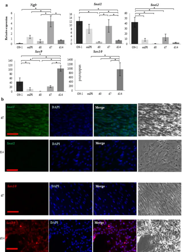
Expressions of the NC markers Ngfr, Snai1, Snai2, Sox9,
and Sox10 were examined by qRT-PCR in cNCCs derived
from miPS cells as well as in O9-1 cells as a control.
Expres-sion of all genes except Ngfr and Sox10 was detected in
O9-1 cells [
24
]. In contrast, expressions of all genes were
detected in cNCCs, with the premigratory NC markers Ngfr,
Snai1, and Snai2 exhibiting the highest expression levels in
d7 cells and the migratory and cranial NC markers Sox9 and
Sox10 exhibiting the highest expression levels in d14 cells
(Fig.
2
a). The strongest immunofluorescent staining was
detected for Snai1 and Sox10 in d7 and d14 cells,
respec-tively (Fig.
2
b).
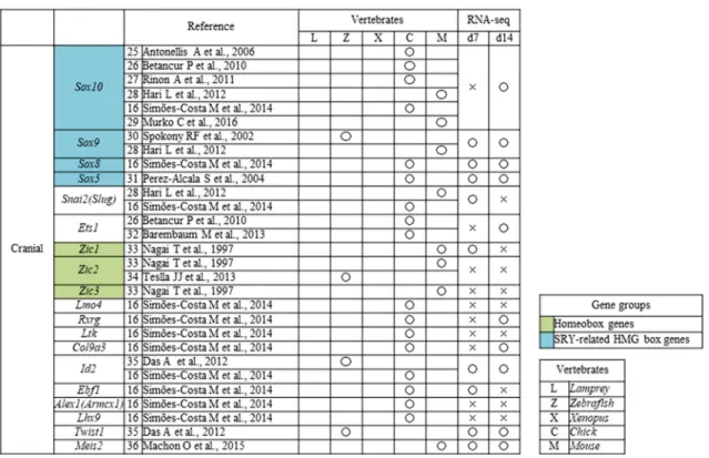
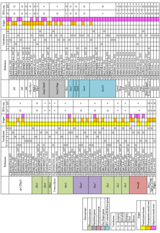
NC specifier transcription factors
We conducted a literature search of NC specifier
transcrip-tion factors identified in vivo [
16
,
25
–
80
] (Tables
2
,
3
) and
compared these reports with our RNA-seq results. The
rela-tive expressions of genes that underwent a significant change
in expression are presented in Fig.
3
a.
The transcription factor AP-2 alpha (Ap2) along with
paired box 3 (Pax3) and zinc finger protein of the cerebellum
1 (Zic1), both of which are regulated by Ap2, were the most
highly expressed genes in d7 cells (Fig.
3
a). Pax6, which
has been reported in human ES and iPS-derived NC cells
(Tables
2
,
3
), was detected in both d7 and d14 cells, whereas
Pax7, which has not previously been reported in the mouse
NC, was detected in the d7 cells (Fig.
3
a). In contrast, the
homeobox genes gastrulation brain homeobox 2 (Gbx2),
msh homeobox 1 (Msx1), distal-less homeobox 3 (Dlx3),
Zic2, and Zic3 were not detected in d7 or d14 cells, and
the homeobox genes Zic1 and Dlx5 were only expressed in
the d7 cells, despite these having been reported in the NC
of a range of species (Table
2
); however, Meis homeobox 2
(Meis2) was expressed in both d7 and d14 cells.
The MYCN proto-oncogenes, bHLH transcription factor
(N-myc) and c-Myc, have been reported in NCCs (Table
3
);
however, c-Myc expression was detected in d7 and d14 cells
(Fig.
3
a), while N-myc was not. Furthermore, there was a
gradual and substantial downregulation of the winged-helix
transcription factor Forkhead box D3 (FoxD3) (Fig.
3
a),
which is an important factor for maintaining the
pluripo-tency of ES cells and a key NC specifier that has been
impli-cated in multiple stages of NC development and NCC
migra-tion in embryos of various species (Tables
2
,
4
).
The premigratory NC markers Ngfr, heart and neural crest
derivatives expressed 2 (Hand2), Snai1, and Snai2 were only
detected in the d7 cells; however, other premigratory NC
markers, such as the platelet derived growth factor receptor,
alpha polypeptide (Pdgfra);
6-phosphofructo-2-kinase/fruc-tose-2,6-biphosphatase 4 (Pfkfb4); inhibitor of DNA binding
2 (Id2), Id3, and Id4; and nestin (Nes) were detected in both
d7 and d14 cells (Fig.
3
a).
Expression of migratory NC markers such as Sox5, -6, -8,
-9, and -10, which encode members of the sex-determining
region Y (SRY)-related high mobility group (HMG)-box
family of transcription factors and are crucial in several
aspects of NCCs, were detected in d7 or d14 cells. Sox10,
a known marker for migratory cNCCs in various species
(Table
2
), was only detected in d14 cells similar to the
other migratory NC markers. Twist family bHLH
tran-scription factor 1 (Twist1), which is activated via various
signal transduction pathways and is crucial for E-cadherin
downregulation, as well as beta-1,3-glucuronyltransferase 1
(B3gat1/Hnk1), which plays a role in the formation of CD57
epitope, was detected in both d7 and d14 cells. In contrast,
the expression of the trunk NC markers lit guidance ligand
1/2 (Slit1/2), which plays an important role in trunk NC cell
migration toward ventral sites, was upregulated only in d7
cells (Fig.
3
a).
Finally, expressions of tenascin C (Tnc), cadherin-6
(Cdh6), and ras homolog family member B (Rhob), all of
which are related to cell adhesion and motility [
81
–
85
], were
significantly increased in both d7 and d14 cells (Fig.
3
b).
Metzincin superfamily zinc proteinase
and protocadherin superfamily
Members of the metzincin superfamily are proteinases that
carry a zinc ion at their active site. This family includes the
matrix metalloproteinases (Mmps), Adam, and Adamts, all
of which have gained attention as factors involved in cancer
cell invasion and migration. Mmp2, -11, -14, -15, -16, -24,
and -28 were significantly upregulated in cNCCs (Fig.
4
a),
all of which except Mmp24 are membrane-bound. Mmp11
and -28 were only expressed in d7 cells, whereas all other
Mmps were detected in both d7 and d14 cells (Fig.
4
a, b).
Only Adam1a, -8, -10, and -12 were upregulated in both
d7 and d14 cells (Fig.
4
c, d); this is contrary to reports that
the members of this family are important in NC migration
and that Adam-10, -12, -15, -19, and -33 are expressed in the
Fig. 2 Comparison between O9-1 cells and cranial neural crest cells (cNCCs) derived from mouse-induced pluripotent stem (miPS) cells using quantitative reverse transcription polymerase chain reaction (qRT-PCR) and immunostaining. a Expression of the premigratory neural crest (NC) markers Ngfr, Snai1, and Snai2 and the migratory NC and cNC markers Sox9 and Sox10. Expressions of the premigra-tory NC markers increased in day 7 (d7) cells, whereas those of the
migratory markers increased in d14 cells. Sox10 was not detected in O9-1 cells. Each experiment was performed in triplicate, with values representing mean ± SD. Groups were compared using ANOVA, fol-lowed by the Bonferroni test: *p < 0.05. b Immunostaining of d7 and d14 cells. Sox10 was more highly expressed in the d14 cells, whereas
mouse NC [
86
]. Moreover, various Adamts family genes,
which are important for connective tissue organization and
cell migration, were upregulated in either d7 or d14 cells
(Fig.
4
c, d). In particular, Adamts1 expression was
mark-edly increased, whereas Adamts2 and -8 expressions, which
are presumably important in cancer cell invasion [
87
–
89
],
increased in the later stages of differentiation.
Most Pcdh genes, which are involved in cell adhesion
[
90
], were upregulated in d7 and d14 cells (Table
5
);
how-ever, Pcdha2, -5, -7, -11, and -12; Pcdhac1; and Pcdhgc5
were only upregulated in d14 cells.
Discussion
In the present study, we derived cells from miPS which are
closely migratory cNCCs genes. Previously, NCCs have
been derived from ES or iPS cells using various approaches
[
91
–
110
], and the protocol used in the present study was
based on the methods outlined by Bajpai et al. [
23
]; however,
few studies have investigated changes in the properties of
cNCCs at different time points after induction.
In the present study, d7 and d14 cells expressed typical
NC markers, such as Ngfr, Snai1, and Snai2. In contrast,
O9-1 cells (controls) did not express Ngfr, suggesting that
cNCCs derived from miPS cells are better than O9-1 cells
for evaluating cNCC characteristics [
24
]. Moreover, unlike
O9-1 cells, d14 cells expressed markedly high levels of
Sox10, which is considered a reliable marker for migratory
cNCCs. Since cNCCs are involved in craniofacial tissue
organization, several reports are available on their gene
expression profiles; however, these reports show varying
results with species and protocols. Moreover, cNCCs
rap-idly differentiate in the embryo [
14
]; thus, it is
consider-ably difficult to synchronize the timing of isolation to a
particular point during their development. Furthermore,
migratory cNCCs intermingle with other cell types in the
embryo, further complicating the isolation and
characteri-zation of a pure cell population. Consequently, there have
been few reports on cNCC markers [
16
,
25
–
36
].
Simões-Costa et al. [
16
] successfully isolated Sox10-positive
cNCCs from chicken embryos and analyzed their gene
profiles. Similarly, we detected Sox10 expression in d14
cNCCs. Reportedly, there are multiple NCC populations
[
11
], and iPS cells can differentiate into numerous
differ-ent NCC populations in the same culture. Therefore, this
diversity in populations may explain the discrepancies in
results; however, under the conditions used in the present
study, c-Myc; Ets1; Sox10; Adamts2; Adamts8; Pcdha2,
-5, -7, -11, and -12; Pcdhac1, and Pcdhgc3 may represent
useful markers for migratory cNCCs. Furthermore, our
results indicated that d7 cells were in the premigratory
Table 2 Neural crest (NC) genes that have previously been examined in vivoOpen circles indicate genes that were upregulated on day 7 (d7) or d14 compared with d0 [log fold change (FC) > 1, p < 0.01, false discovery rate (FDR) < 0.05), whereas crosses indicate genes that were not upregulated
Table 3 N eur al cr es t (N C) tr anscr ip tion f act ors t hat ha ve pr eviousl y been e xamined in viv o Open cir cles indicate g enes t hat w er e upr egulated on da y 7 (d7) or d14 com par ed wit h d0 [log f old c hang e (FC) > 1, p < 0.01, f alse disco ver y r ate (FDR) < 0.05), wher eas cr osses indicate g enes that w er e no t upr egulated
stage despite expressing numerous NC markers.
There-fore, cNCCs derived from miPS cells required > 14 days
to become migratory in vitro, and this duration is
consid-erably longer than that observed in the mouse embryos
in vivo under the same conditions [
111
].
The use of RNA-seq facilitates the normalization of
expression levels of different genes, allowing comparisons
between samples. In our triplicate experiments, none of the
induced cNCCs expressed several homeobox genes
consid-ered to be expressed in the early stages of cNCC
differentia-tion. In particular, we did not observe FoxD3 expression in
either d7 or d14 cells, although it has been recognized as
one of the key transcription factors in cNCCs [
112
]. These
contradictory results suggest that cNCCs derived from miPS
cells express distinct gene regulatory networks. FoxD3, a
pluripotent stem cell marker gene that plays an important
role in maintaining pluripotency, is expressed at different
time points in different cells, but its expression decreases in
a time-dependent manner [
41
], indicating that FoxD3 may
not be a key regulator in iPS-derived cNCCs. However, we
speculate that iPS cells express sufficient levels of FoxD3 to
differentiate into cNCCs.
Protocadherins belong to the cadherin superfamily and
are involved in intercellular interactions [
90
], whereas
metz-incins are key proteinases that facilitate cell migration [
42
].
Unfortunately, the abundances of members of these families
hindered their analysis; however, because RNA-seq enabled
us to comprehensively evaluate the gene expression profiles,
Fig. 3 RNA sequencing results for cranial neural crest cells (cNCCs)differentiated from mouse-induced pluripotent stem (miPS) cells. a Expression of each of the genes listed in Table 2 at day 0 (d0), d7, and d14 after induction. Sex-determining region Y (SRY)-related high mobility group (HMG) box genes showed the highest upregula-tion in d14 cells. The vertical axis reveals reads per kilobase of exon per million mapped reads (RPKM), and the horizontal axis indicates time. Each experiment was performed in triplicate, with values repre-senting mean ± SD. Groups were compared using ANOVA, followed by the Bonferroni test: *p < 0.05. b Expression of genes that have not
been examined during the neural crest stages in vivo. Tnc showed the highest upregulation in d14 cells, whereas Cha6 and Rhob were upregulated in day 7 (d7) cells. The vertical axis indicates reads per kilobase of exon per million mapped reads (RPKM), and the hori-zontal axis indicates time. Open circles indicate genes upregulated in d7 or d14 compared with d0 [log fold change (FC) > 1, p < 0.01, false discovery rate (FDR) < 0.05)]. Each experiment was performed in triplicate, with values representing mean ± SD. Groups were com-pared using ANOVA, followed by the Bonferroni test: *p < 0.05
Table 4 N eur al cr es t (N C) tr anscr ip tion f act ors t hat ha ve pr eviousl y been e xamined in vitr o Open cir cles indicate g enes t hat w er e upr egulated on da y 7 (d7) or d14 com par ed wit h d0 [log f old c hang e (FC) > 1, p < 0.01, f alse disco ver y r ate (FDR) < 0.05), wher eas cr osses indicate g enes that w er e no t upr egulated
we were able to focus on expressions of all procadherin and
metazicin family members. As expected, we observed that
several Adam and Adamts genes were upregulated, with
most of the Admats genes showing significantly increased
expression. The Adam genes with increased expression
in cNCCs were membrane-bound, whereas Adamts genes
which secreted proteinases, indicating that the expression
of various Adamts may allow the matrix to be digested
more efficiently and that each proteinase may be capable
of digesting a different type of extracellular matrix protein
[
42
]. Therefore, the secretion of various Adamts and Pcdh
proteins may play a crucial role in cNCC migration.
Conclusion
In summary, cNCCs derived from miPS exhibited RNA
expression profiles that partly overlap with previously
reported profiles. These cells may be useful for the
regenera-tion of tissue formed by NCCs (osteoblast, melanocyte, and
glial cells). We observed that although the resulting cNCCs
exhibited several NC specifiers, they lacked some of the
specifiers, indicating that a distinct molecular network may
regulate gene expression in miPS-derived cNCCs. Moreover,
our results indicated that c-Myc; Ets1; Sox10; Adamts2 and
-8; Pcdha2, -5,-7, -11, and -12; Pcdhac1; and Pcdhgc3 may
represent appropriate markers for migratory miPS-derived
Fig. 4 RNA sequencing results for the matrix metalloproteinase(Mmp), a disintegrin and metalloproteinase (Adam), and a disinte-grin and metalloproteinase with thrombospondin motifs (Adamts) gene families. a Expressions of Mmp family genes in mouse. Round marks alongside day 7 (d7) or d14 cells indicate that the genes were upregulated compared with d0 [log fold change (logFC) > 1, p < 0.01, false discovery rate (FDR) < 0.05], whereas cross marks indicate lack of upregulation. b Graphical representation of the upregulation of Mmp2, -11, -14, -15, -16, -24, and -28 in d7 or d14 cells. Mmp15 and -16 showed the highest upregulation in d14 cells. The vertical axis indicates reads per kilobase of exon per million mapped reads (RPKM), and the horizontal axis indicates time. Each experiment was performed in triplicate, with values representing mean ± SD. Groups
were compared using ANOVA, followed by the Bonferroni test: *p < 0.05. c Expressions of Adam and Adamts genes in mouse. Round marks alongside d7 or d14 cells indicate that the genes were upregu-lated compared with d0 (logFC > 1, p < 0.01, FDR < 0.05), whereas cross marks indicate lack of upregulation. d Graphical representa-tion of the upregularepresenta-tion of Adam1a and 8–12, and Adamts1–10, -12, and 15–20 in the d7 or d14 cells. Adam2, -4, -7, and -8, and Adamts
9 and -12 showed the highest upregulation in d14 cells. The vertical
axis indicates reads per kilobase of exon per million mapped reads (RPKM), and the horizontal axis indicates time. Each experiment was performed in triplicate, with values representing mean ± SD. Groups were compared using ANOVA, followed by the Bonferroni test: *p < 0.05
cNCCs. Finally, cNCCs expressed a wide spectrum of genes
encoding Adamts family enzymes that may be crucial for
their migration.
Acknowledgements We are grateful to Professor T. Azuma, MD, PhD, Department of Biochemistry, and Professor T. Ichinohe, DDS, PhD, Department of Dental Anesthesiology, for their guidance. We also thank S. Onodera and A. Saito, Department of Biochemistry.
Funding The funding was received by Ministry of Science and Tech-nology (Grant nos. KIBANKENNKYU(B)18H03007 and 18K09753 KIBANKENNKYU(C)).
Compliance with ethical standards
Conflict of interest The authors declare that they have no conflicts of interest.
Open Access This article is distributed under the terms of the Crea-tive Commons Attribution 4.0 International License (http://creat iveco mmons .org/licen ses/by/4.0/), which permits unrestricted use, distribu-tion, and reproduction in any medium, provided you give appropriate
credit to the original author(s) and the source, provide a link to the Creative Commons license, and indicate if changes were made.
References
1. Luan X, Dangaria S, Ito Y, Walker GG, Jin T, Schmidt MK, Galang MT, Druzinsky R (2009) Neural crest lineage segre-gation: a blueprint for periodontal regeneration. J Dent Res 88(9):781–791
2. Malhotra N (2016) Induced pluripotent stem (iPS) cells in den-tistry: a review. Int J Stem Cells 9(2):176–185
3. Knight RD, Schilling TF (2006) Cranial neural crest and develop-ment of the head skeleton. Adv Exp Med Biol 589:120–133 4. Theveneau E, Mayor R (2011) Collective cell migration of the
cephalic neural crest: the art of integrating information. Genesis 49(4):164–176
5. Chai Y, Jiang X, Ito Y, Bringas P Jr, Han J, Rowitch DH, Sori-ano P, McMahon AP, Sucov HM (2000) Fate of the mammalian cranial neural crest during tooth and mandibular morphogenesis. Development 127(8):1671–1679
6. McConnell AM, Mito JK, Ablain J, Dang M, Formichella L, Fisher DE, Zon LI (2018) Neural crest state activation in NRAS
Table 5 Expression of the protocadherin superfamily based on RNA sequencing data
Open circles indicate genes that were upregulated on day 7 (d7) or d14 compared with d0 [log fold change (FC) > 1, p < 0.01, false discovery rate (FDR) < 0.05), whereas crosses indicate genes that were not upregulated
driven melanoma, but not in NRAS-driven melanocyte expan-sion. Dev Biol 449(17):30855–30862
7. Meulemans D, Bronner-Fraser M (2004) Gene-regulatory inter-actions in neural crest evolution and development. Dev Cell 7(3):291–299
8. Steventon B, Carmona-Fontaine C, Mayor R (2005) Genetic net-work during neural crest induction: from cell specification to cell survival. Semin Cell Dev Biol 16(6):647–654
9. Simões-Costa M, Bronner ME (2015) Establishing neural crest identity: a gene regulatory recipe. Development 142(2):242–257 10. Martik ML, Bronner ME (2017) Regulatory logic underlying
diversification of the neural crest. Trends Genet 33(10):715–727 11. Minoux M, Rijli FM (2010) Molecular mechanisms of cranial
neural crest cell migration and patterning in craniofacial develop-ment. Development 137(16):2605–2621
12. Mayor R, Theveneau E (2013) The neural crest. Development 457(1):2247–2251
13. Okuno H, Mihara FR, Ohta S, Fukuda K, Kurosawa K, Akamatsu W, Sanosaka TS, Kohyama J, Hayashi K, Nakajima K, Takahashi T, Wysocka J, Kosaki K, Okano H (2017) CHARGE syndrome modeling using patient-iPSCs reveals defective migration of neu-ral crest cells harboring CHD7 mutations. eLife 6:e21114 14. Simoes-Costa M, Bronner ME (2016) Reprogramming
of avian neural crest axial identity and cell fate. Science 352(6293):1570–1573
15. Milet C, Monsoro-Burq AH (2012) Neural crest induction at the neural plate border in vertebrates. Dev Biol 366(1):22–33 16. Simões-Costa M, Tan-Cabugao J, Antoshechkin I,
Sauka-Spengler T, Bronner ME (2014) Transcriptome analysis reveals novel players in the cranial neural crest gene regulatory network. Genome Res 24(2):281–290
17. Doi D, Samata B, Katsukawa M, Kikuchi T, Morizane A, Ono Y, Sekiguchi K, Nakagawa M, Parmar M, Takahashi J (2014) Isola-tion of human induced pluripotent stem cell derived dopamin-ergic progenitors by cell sorting for successful transplantation. Stem Cell Rep 2(3):337–350
18. Nakane T, Masumoto H, Tinney JP, Yuan F, Kowalski WJ, Ye F, LeBlanc AJ, Sakata R, Yamashita JK, Keller BB (2017) Impact of cell composition and geometry on human induced pluripotent stem cells-derived engineered cardiac tissue. Sci Rep 7:45641 19. Gallego RI, Pai AA, Tung J, Gilad Y (2014) RNA-seq: impact of
RNA degradation on transcript quantification. BMC Biol 12:42 20. Wang Z, Gerstein M, Snyder M (2009) RNA-Seq: a revolutionary
tool for transcriptomics. Nat Rev Genet 10(1):57–63
21. Mortazavi A, Williams BA, McCue K, Schaeffer L, Wold B (2008) Mapping and quantifying mammalian transcriptomes by RNA-Seq. Nat Methods 5(7):621–628
22. Okita K, Ichisaka T, Yamanaka S (2007) Generation of ger-mline competent induced pluripotent stem cells. Nature 448(7151):313–317
23. Bajpai R, Chen DA, Rada-Iglesias A, Zhang J, Xiong Y, Helms J, Chang CP, Zhao Y, Swigut T, Wysocka J (2010) CHD7 coop-erates with PBAF to control multipotent neural crest formation. Nature 463(7283):958–962
24. Ishii M, Arias AC, Liu L, Chen YB, Bronner ME, Maxson RE (2012) A stable cranial neural crest cell line from mouse. Stem Cells Dev 21(17):3069–3080
25. Antonellis A, Bennett WR, Menheniott TR, Prasad AB, Lee-Lin SQ, NISC Comparative Sequencing Program, Green ED, Paisley D, Kelsh RN, Pavan WJ, Ward A (2006) Deletion of long-range sequences at Sox10 compromises developmental expression in a mouse model of Waardenburg-Shah (WS4) syndrome. Hum Mol Genet 15(2):259–271
26. Betancur P, Bronner-Fraser M, Sauka-Spengler T (2010) Genomic code for Sox10 activation reveals a key regulatory
enhancer for cranial neural crest. Proc Natl Acad Sci USA 107(8):3570–3575
27. Rinon A, Molchadsky A, Nathan E, Yovel G, Rotter V, Sarig R, Tzahor E (2011) p53 coordinates cranial neural crest cell growth and epithelial–mesenchymal transition/delamination processes. Development 138(9):1827–1838
28. Hari L, Miescher I, Shakhova O, Suter U, Chin L, Taketo M, Richardson WD, Kessaris N, Sommer L (2012) Temporal control of neural crest lineage generation by Wnt/β-catenin signaling. Development 139(12):2107–2117
29. Murko C, Bronner ME (2016) Tissue specific regulation of the chick Sox10E1 enhancer by different Sox family members. Dev Biol 422(1):47–57
30. Spokony RF, Aoki Y, Germain N, Magner-Fink E, Saint-Jeannet JP (2002) The transcription factor Sox9 is required for cranial neural crest development in Xenopus. Development 129(2):421–432
31. Perez-Alcala S, Nieto MA, Barbas JA (2004) LSox5 regulates RhoB expression in the neural tube and promotes generation of the neural crest. Development 131(18):4455–4465
32. Barembaum M, Bronner ME (2013) Identification and dissection of a key enhancer mediating cranial neural crest specific expres-sion of transcription factor, Ets-1. Dev Biol 382(2):567–575 33. Nagai T, Aruga J, Takada S, Günther T, Spörle R, Schughart K,
Mikoshiba K (1997) The expression of the mouse Zic1, Zic2, and Zic3 gene suggests an essential role for Zic genes in body pattern formation. Dev Biol 182(2):299–313
34. Teslaa JJ, Keller AN, Nyholm MK, Grinblat Y (2013) Zebrafish Zic2a and Zic2b regulate neural crest and craniofacial develop-ment. Dev Biol 380(1):73–86
35. Das A, Crump JG (2012) Bmps and id2a act upstream of Twist1 to restrict ectomesenchyme potential of the cranial neural crest. PLoS Genet 8:e1002710
36. Machon O, Masek J, Machonova O, Krauss S, Kozmik Z (2015) Meis2 is essential for cranial and cardiac neural crest develop-ment. BMC Dev Biol 15:40
37. Mitchell PJ, Timmons PM, Hébert JM, Rigby PW, Tjian R (1991) Transcription factor AP-2 is expressed in neural crest cell line-ages during mouse embryogenesis. Genes Dev 5(1):105–119 38. Shen H, Wilke T, Ashique AM, Narvey M, Zerucha T, Savino
E, Williams T, Richman JM (1997) Chicken transcription fac-tor AP-2: cloning, expression and its role in outgrowth of facial prominences and limb buds. Dev Biol 188(2):248–266 39. Luo T, Lee YH, Saint-Jeannet JP, Sargent TD (2003) Induction
of neural crest in Xenopus by transcription factor AP2alpha. Proc Natl Acad Sci USA 100(2):532–537
40. Sauka-Spengler T, Meulemans D, Jones M, Bronner-Fraser M (2007) Ancient evolutionary origin of the neural crest gene regu-latory network. Dev Cell 13(3):405–420
41. Nikitina N, Sauka-Spengler T, Bronner-Fraser M (2008) Dis-secting early regulatory relationships in the lamprey neural crest gene network. Proc Natl Acad Sci USA 105(51):20083–20088 42. Khudyakov J, Bronner-Fraser M (2009)
Comprehensive spati-otemporal analysis of early chick neural crest network genes. Dev Dyn 238(3):716–723
43. de Crozé N, Maczkowiak F, Monsoro-Burq AH (2011) Reit-erative AP2a activity controls sequential steps in the neu-ral crest gene regulatory network. Proc Natl Acad Sci USA 108(1):155–160
44. Wang WD, Melville DB, Montero-Balaguer M, Hatzopoulos AK, Knapik EW (2011) Tfap2a and Foxd3 regulate early steps in the development of the neural crest progenitor population. Dev Biol 360(1):173–185
45. Powell DR, Hernandez-Lagunas L, LaMonica K, Art-inger KB (2013) Prdm1a directly activates foxd3 and tfap2a
during zebrafish neural crest specification. Development 140(16):3445–3455
46. Yang L, Zhang H, Hu G, Wang H, Abate-Shen C, Shen MM (1998) An early phase of embryonic Dlx5 expres-sion defines the rostral boundary of the neural plate. J Neurosci 18(20):8322–8330
47. Luo T, Matsuo-Takasaki M, Lim JH, Sargent TD (2001) Differen-tial regulation of Dlx gene expression by a BMP morphogenetic gradient. Int J Dev Biol 45(4):681–684
48. Li B, Kuriyama S, Moreno M, Mayor R (2009) The posterioriz-ing gene Gbx2 is a direct target of Wnt signallposterioriz-ing and the earliest factor in neural crest induction. Development 136(19):3267–3278 49. Hill RE, Jones PF, Rees AR, Sime CM, Justice MJ, Copeland NJ,
Jenkins NA, Graham E, Davidson DR (1998) A new family of mouse homeo box-containing genes: molecular structure, chro-mosomal location, and developmental expression of Hox-7.1. Genes Dev 3(1):26–37
50. Suzuki A, Ueno N, Hemmati-Brivanlou A (1997) Xenopus msx1 mediates epidermal induction and neural inhibition by BMP4. Development 124(16):3037–3044
51. Simões-Costa M, McKeown SJ, Tan-Cabugao J, Sauka-Spengler T, Bronner ME (2012) Dynamic and differential regulation of stem cell factor FoxD3 in the neural crest is encrypted in the genome. PLoS Genet 8:e1003142
52. Goulding MD, Chalepakis G, Deutsch U, Erselius JR, Gruss P (1991) Pax-3, a novel murine DNA binding protein expressed during early neurogenesis. EMBO J 10(5):1135–1147
53. Bang AG, Papalopulu N, Goulding MD, Kintner C (1999) Expression of Pax-3 in the lateral neural plate is dependent on a Wnt-mediated signal from posterior nonaxial mesoderm. Dev Biol 212(2):366–380
54. Alkobtawi M, Ray H, Barriga EH, Moreno M, Kerney R, Monsoro-Burq AH, Saint-Jeannet JP, Mayor R (2018) Char-acterization of Pax3 and Sox10 transgenic Xenopus laevis embryos as tools to study neural crest development. Dev Biol 444(17):30693–30700
55. Maczkowiak F, Matéos S, Wang E, Roche D, Harland R, Mon-soro-Burq AH (2010) The Pax3 and Pax7 paralogs cooperate in neural and neural crest patterning using distinct molecular mechanisms, Xenopus laevis embryos. Dev Biol 340(2):381–396 56. Nakata K, Nagai T, Aruga J, Mikoshiba K (1998) Xenopus
Zic family and its role in neural crest development. Mech Dev 75(1–2):43–51
57. Dottori M, Gross MK, Labosky P, Goulding M (2001) The winged helix transcription factor Foxd3 suppresses interneu-ron differentiation and promotes neural crest cell fate. Develop-ment 128(21):4127–4138
58. Kos R, Reedy MV, Johnson RL, Erickson CA (2001) The winged-helix transcription factor FoxD3 is important for estab-lishing the neural crest lineage and repressing melanogenesis in avian embryos. Development 128(8):1467–1479
59. Wilson YM, Richards KL, Ford-Perriss ML, Panthier JJ, Mur-phy M (2004) Neural crest cell lineage segregation in the mouse neural tube. Development 131(24):6153–6162
60. Liu L, Chong SW, Balasubramaniyan NV, Korzh V, Ge R (2002) Platelet-derived growth factor receptor alpha (pdgfr-α) gene in zebrafish embryonic development. Mech Dev 116:227–230 61. Liu KJ, Harland RM (2003) Cloning and characterization
of Xenopus Id4 reveals differing roles for Id genes. Dev Biol 264(2):339–351
62. Figueiredo AL, Maczkowiak F, Borday C, Pla P, Sittewelle M, Pegoraro C, Monsoro Burq AH (2017) PFKFB4 control of AKT signaling is essential for premigratory and migratory neural crest formation. Development 144(22):4183–4194
63. Yang X, Li J, Zeng W, Li C, Mao B (2016) Elongator protein 3 (Elp3) stabilizes Snail1 and regulates neural crest migration in
Xenopus. Sci Rep 6:26238
64. Sefton M, Sánchez S, Nieto MA (1998) Conserved and divergent roles for members of the Snail family of transcription factors in the chick and mouse embryo. Development 125(16):3111–3121 65. del Barrio MG, Nieto MA (2002) Overexpression of Snail family
members highlights their ability to promote chick neural crest formation. Development 129(7):1583–1593
66. Aybar MJ, Nieto MA, Mayor R (2003) Snail precedes Slug in the genetic cascade required for the specification and migration of the Xenopus neural crest. Development 130(3):483–494 67. Nieto MA, Sargent MG, Wilkinson DG, Cooke J (1994) Control
of cell behavior during vertebrate development by Slug, a zinc finger gene. Science 264(5160):835–859
68. Jiang R, Lan Y, Norton CR, Sundberg JP, Gridley T (1998) The Slug gene is not essential for mesoderm or neural crest develop-ment in mice. Dev Biol 198(2):277–285
69. Tien CL, Jones A, Wang H, Gerigk M, Nozell S, Chang C (2015) Snail2/Slug cooperates with Polycomb repressive complex 2 (PRC2) to regulate neural crest development. Development 142(4):722–731
70. Martin BL, Harland RM (2001) Hypaxial muscle migra-tion during primary myogenesis in Xenopus laevis. Dev Biol 239(2):270–280
71. Cheung M, Briscoe J (2003) Neural crest development is regulated by the transcription factor Sox9. Development 130(23):5681–5693
72. Cheung M, Chaboissier MC, Mynett A, Hirst E, Schedl A, Briscoe J (2005) The transcriptional control of trunk neu-ral crest induction, survival, and delamination. Dev Cell 8(2):179–192
73. Honoré SM, Aybar MJ, Mayor R (2003) Sox10 is required for the early development of the prospective neural crest in Xenopus embryos. Dev Biol 260(1):79–96
74. McKeown SJ, Lee VM, Bronner-Fraser M, Newgreen DF, Farlie PG (2005) Sox10 overexpression induces neural crest-like cells from all dorsoventral levels of the neural tube but inhibits dif-ferentiation. Dev Dyn 233(2):430–444
75. Prasad MK, Reed X, Gorkin DU, Cronin JC, McAdow AR, Chain K, Hodonsky CJ, Jones EA, Svaren J, Antonellis A, Johnson SL, Loftus SK, Pavan WJ, McCallion AS (2011) SOX10 directly modulates ERBB3 transcription via an intronic neural crest enhancer. BMC Dev Biol 11:40
76. Baggiolini A, Varum S, Mateos JM, Bettosini D, John N, Bonalli M, Ziegler U, Dimou L, Clevers H, Furrer R, Sommer L (2015) Premigratory and migratory neural crest cells are multipotent in vivo. Cell Stem Cell 16(3):314–322
77. McKinney MC, McLennan R, Kulesa PM (2016) Angiopoietin 2 signaling plays a critical role in neural crest cell migration. BMC Biol 14(1):111
78. Lee HO, Levorse JM, Shin MK (2003) The endothelin receptor-B is required for the migration of neural crest-derived melanocyte and enteric neuron precursors. Dev Biol 259(1):162–175 79. Giovannone D, Ortega B, Reyes M, El-Ghali N, Rabadi M, Sao
S, de Bellard ME (2015) Chicken trunk neural crest migration visualized with HNK1. Acta Histochem 117(3):255–266 80. Zuhdi N, Ortega B, Giovannone D, Ra H, Reyes M, Asención
V, McNicoll I, Ma L, de Bellard ME (2012) Slits affect the timely migration of neural crest cells via robo receptor. Dev Dyn 241(8):1274–1288
81. Chiovaro F, Chiquet-Ehrismann R, Chiquet M (2015) Tran-scriptional regulation of tenascin genes. Cell Adhes Migr. 9(1–2):34–47
82. Taneyhill AL, Coles EG, Bronner-Fraser M (2007) Snail2 directly represses cadherin6B during epithelial-to-mesenchymal transitions of the neural crest. Development 134(8):1480–1490 83. Groysman M, Shoval I, Kalcheim C (2008) A negative
modula-tory role for rho and rho-associated kinase signaling in delamina-tion of neural crest cells. Neural Dev 3:27
84. Vega FM, Thomas M, Reymond N, Ridley AJ (2015) The Rho GTPase RhoB regulates cadherin expression and epithelial cell– cell interaction. Cell Commun Signal 13:6
85. Liu Q, Dalman MR, Sarmah S, Chen S, Chen Y, Hurlbut AK, Spencer MA, Pancoe L, Marrs JA (2011) Cell adhesion molecule cadherin-6 function in zebrafish cranial and lateral line ganglia development. Dev Dyn 240(7):1716–1726
86. Tomczuk M, Takahashi Y, Huang J, Murase S, Mistretta M, Klaffky E, Sutherland A, Bolling L, Coonrod S, Marcinkiewicz C, Sheppard D, Stepp MA, White JM (2003) Role of multiple beta1 integrins in cell adhesion to the disintegrin domains of ADAMs 2 and 3. Exp Cell Res 290(1):68–81
87. Desanlis I, Felstead HL, Edwards DR, Wheeler GN (2018) ADAMTS9, a member of the ADAMTS family, in Xenopus development. Gene Expr Patterns 29:72–81
88. Porter S, Clark IM, Kevorkian L, Edwards DR (2005) The ADAMTS metalloproteinases. Biochem J 386(1):15–27 89. Hubmacher D, Apte SS (2015) ADAMTS proteins as
modu-lators of microfibril formation and function. Matrix Biol 47(2015):34–43
90. Chen WV, Maniatis T (2013) Clustered protocadherins. Develop-ment 140(16):3297–3302
91. Okawa T, Kamiya H, Himeno T, Kato J, Seino Y, Fujiya A, Kondo M, Tsunekawa S, Naruse K, Hamada Y, Ozaki N, Cheng Z, Kito T, Suzuki H, Ito S, Oiso Y, Nakamura J, Isobe K (2013) Transplantation of neural crest-like cells derived from induced pluripotent stem cells improves diabetic polyneuropathy in mice. Cell Transplant 22(10):1767–1783
92. Seki D, Takeshita N, Oyanagi T, Sasaki S, Takano I, Hasegawa M, Takano-Yamamoto T (2015) Differentiation of odontoblast-like cells from mouse induced pluripotent stem cells by Pax9 and Bmp4 transfection stem cells. Transl Med 4(9):993–997 93. Mizuseki K, Sakamoto T, Watanabe K, Muguruma K, Ikeya M,
Nishiyama A, Arakawa A, Suemori H, Nakatsuji N, Kawasaki H, Murakami F, Sasai Y (2003) Generation of neural crest-derived peripheral neurons and floor plate cells from mouse and primate embryonic stem cells. Proc Natl Acad Sci USA 100(10):5828–5833
94. Motohashi T, Aoki H, Chiba K, Yoshimura N, Kunisada T (2007) Multipotent cell fate of neural crest-like cells derived from embryonic stem cells. Stem Cells 25(2):402–412
95. Kawaguchi J, Nichols J, Gierl MS, Faial T, Smith A (2010) Iso-lation and propagation of enteric neural crest progenitor cells from mouse embryonic stem cells and embryos. Development 137(5):693–704
96. Aihara Y, Hayashi Y, Hirata M, Ariki N, Shibata S, Nagoshi N, Nakanishi M, Ohnuma K, Warashina M, Michiue T, Uchiyama H, Okano H, Asashima M, Furue MK (2010) Induction of neural crest cells from mouse embryonic stem cells in a serum-free monolayer culture. Int J Dev Biol 54(8–9):1287–1294
97. Minamino Y, Ohnishi Y, Kakudo K, Nozaki M (2015) Isola-tion and propagaIsola-tion of neural crest stem cells from mouse embryonic stem cells via cranial neurospheres. Stem Cells Dev 24(2):172–181
98. Lee G, Chambers SM, Tomishima MJ, Studer L (2010) Deriva-tion of neural crest cells from human pluripotent stem cells. Nat Protoc 5(4):688–701
99. Wang A, Tang Z, Li X, Jiang Y, Tsou DA, Li S (2012) Deri-vation of smooth muscle cells with neural crest origin from human induced pluripotent stem cells. Cells Tissues Organs 195(1–2):5–14
100. Kreitzer FR, Salomonis N, Sheehan A, Huang M, Park JS, Spin-dler MJ, Lizarraga P, Weiss WA, So PL, Conklin BR (2013) A robust method to derive functional neural crest cells from human pluripotent stem cells. Am J Stem Cells 2(2):119–131
101. Tomokiyo A, Hynes K, Ng J, Menicanin D, Camp E, Arthur A, Gronthos S, Mark Bartold P (2017) Generation of neural crest-like cells from human periodontal ligament cell-derived induced pluripotent stem cells. J Cell Physiol 232(2):402–416
102. Michael D, Wagoner MD, Bohrer LR, Aldrich BT, Greiner MA, Mullins RF, Worthington KS, Tucker BA, Wiley LA (2018) Feeder-free differentiation of cells exhibiting characteristics of corneal endothelium from human induced pluripotent stem cells. Biol Open 7(5):bio032102
103. Pomp O, Brokhman I, Ben-Dor I, Reubinoff B, Goldstein RS (2005) Generation of peripheral sensory and sympathetic neu-rons and neural crest cells from human embryonic stem cells. Stem cells 23(7):923–930
104. Lee G, Kim H, Elkabetz Y, Al Shamy G, Panagiotakos G, Barberi T, Tabar V, Studer L (2007) Isolation and directed differentiation of neural crest stem cells derived from human embryonic stem cells. Nat Biotechnol 25(12):1468–1475
105. Liu Q, Spusta SC, Mi R, Lassiter RN, Stark MR, Höke A, Rao MS, Zeng X (2012) Human neural crest stem cells derived from human ESCs and induced pluripotent stem cells: induction, maintenance, and differentiation into functional schwann cells. Stem Cells Transl Med 1(4):266–278
106. Noisa P, Lund C, Kanduri K, Lund R, Lähdesmäki H, Lahes-maa R, Lundin K, Chokechuwattanalert H, Otonkoski T, Tuuri T, Raivio T (2014) Notch signaling regulates the differentiation of neural crest from human pluripotent stem cells. J Cell Sci 127:2083–2094
107. Karbalaie K, Tanhaei S, Rabiei F, Kiani-Esfahani A, Masoudi NS, Nasr-Esfahani MH, Baharvand H (2015) Stem cells from human exfoliated deciduous tooth exhibit stromal-derived induc-ing activity and lead to generation of neural crest cells from human embryonic stem cells. Cell J 17(1):37–48
108. Avery J, Dalton S (2016) Methods for derivation of multipotent neural crest cells derived from human pluripotent stem cells. Methods Mol Biol 1341:197–208
109. Zhang JT, Weng ZH, Tsang KS, Tsang LL, Chan HC, Jiang XH (2016) MycN is critical for the maintenance of human embryonic stem cell-derived neural crest stem cells. PLoS One 11:e0148062 110. Lovatt M, Yam GH, Peh GS, Colman A, Dunn NR, Mehta JS
(2018) Directed differentiation of periocular mesenchyme from human embryonic stem cells. Differentiation 99:62–69 111. Dennis AR, McLennan R, Jessica MT, Craig LS, Jeffrey SH,
Kulesa PM (2014) The neural crest cell cycle is related to phases of migration in the head. Development 141(5):1095–1103 112. Krishnakumar R, Chen AF, Pantovich MG, Danial M, Parchem
RJ, Labosky PA, Blelloch R (2016) FOXD3 regulates pluripotent stem cell potential by simultaneously initiating and repressing enhancer activity. Cell Stem Cell 18(1):104–117
Publisher’s Note Springer Nature remains neutral with regard to jurisdictional claims in published maps and institutional affiliations.