IRUCAA@TDC : Changes in the homeostatic mechanism of dental pulp with age: expression of the core-binding factor alpha-1, dentin sialoprotein, vascular endothelial growth factor, and heat shock protein 27 messenger RNAs
21
0
0
全文
(2) Abstract Aim: Dental pulp has various characteristics in the pulp chamber, but only a few biological evaluations about the effect of age on dental pulp tissue have been reported. The purpose of this study was to compare dental pulp from young and from adult rats to characterize the homeostatic mechanism. Methodology: Dental pulp cells (DPCs) were obtained from the first molar of rats, weighing 150 g each for the young group and 350 g each for the adult group. Expression of Cbfa-1, VEGF or HSP27 mRNAs by cultured pulp cells was determined using a quantitative real time PCR system after 3, 7 or 14 days. Results: The expression of Cbfa-1 mRNA in the young group was higher than in the adult group. Expression of VEGF and HSP27 mRNAs in the adult group was higher than in the young group. Conclusion The self defense system in young DPCs is undertaken by calcification, but in adult DPCs it is carried out by the expression of self defense proteins and the regeneration of vessels.. Key words: aging; dental pulp; homeostasis; cbfa-1; VEGF; dentin sialoprotein; HSP27; mPCR.
(3) Introduction Dental pulp has various characteristics in the pulp chamber, including fibrosis, atrophy, cellularity loss, dyscalcification and the degeneration of odontoblasts (1, 2). Further, reduction of the chamber size is a typical phenomenon in teeth from older individuals (3). These phenomena affect the dental pulp and root properties, and suggest that aged pulp retains the ability to create dentine but at a diminished rate (4). Further, vital pulpotomy. is. the. method. that. determines. the. characteristics. of. dyscalcification. However, the success ratio of vital pulpotomy is lower in adult pulp than in young pulp. This phenomenon depends on the function of the homeostatic mechanism. Many kinds of cells can be observed in pulp tissue, in which we can confirm odontoblast, fibroblast, endothelial cells, leukocyte, nerve fiber, etc. It is known that mesenchymal cells in pulp have a potential for mineralization from the formation of denticle and secondary or thirdly dentin induced by a variety of stimuli. This phenomenon is known the protective response of pulp against the stimuli. Vascularization plays an important role concerning mineralization and hard tissue formation excluding dystrophic calcification. So, not only expression of self defense protein but also mineralization and vasculariztion are inextricable for the homeostatic mechanism in the pulp. There. are many mechanisms regulating the. homeostasis of dental pulp. Many kinds of bone related protein concern for the mineralization in dental pulp. Core binding factor alfa-1 (Cbfa-1) is known as one of the required transcription factors that are produced when pre-osteoblasts differentiate into osteoblasts (5). Odontoblasts also express Cbfa-1 while differentiating from preodontoblasts (6, 7). In addition to this,.
(4) dentin sialo-protein (DSP) is specific protein of dental pulp for dentin formation. Vascular endothelial growth factor (VEGF) is known as an angiogenic growth factor that elicits cellular responses to hypoxia (8, 9). Induction of VEGF has also been investigated in rat astrocytes and in the heart under hypoxic conditions (10, 11). The development of endothelial cell-based microvascularization and microcirculation is undoubtedly crucial for the preservation of structure and for regeneration (12). Furthermore, heat shock proteins (HSP) are expressed for self-defense against injury. HSP-27 is involved in the cell death of dental pulp cells during stress due to modification and activation (13). To clarify the differences of characteristics between young dental pulp cells (DPCs) and adult DPCs is beneficial for the prognostic of vital pulpotomy. So, the purpose of this study was to evaluate the young and adult DPCs to characterize their cell proliferation and their expression of Cbfa-1, DSP, VEGF or HSP27 mRNAs .. Materials and Methods Cell culture All animal studies were conducted in compliance with the guidelines for the treatment of experimental animals at the Tokyo Dental College. Pulp cells were obtained from 150 g (5-week-old) Wistar male rats as young DPCs, and from. 350 g (15-week-old) Wistar rats as adult DPCs. Animals were. sacrificed with an overdose of thiopental (Ravonal®; Tanabe, Japan). Both.
(5) the upper and lower first molars were extracted from each animal, and were washed for 5 min in ? -minimal essential medium (? -MEM; GIBCO, Carlsbad, CA, USA) containing 500 µg/ml gentamycin and 0.3 µg/ml fungizone. The periodontal ligament was then removed mechanically (using a knife observed through a loupe) and the outer surface of the root was then immersed in. a. solution of sodium. hypochlorite. for avoiding. the. contamination. Following washing with PBS, each tooth was broken using a dental chisel and a mallet. Each pulp was then carefully collected with a dental explorer and a dental scalar using a microscope into a 35 mm culture dish. Cells were cultured in ?-MEM containing 10% fetal bovine serum (FBS, inactivated at 56°C for 35 minutes, GIBCO) and antibiotic (40 µg/ml gentamycin, SIGMA, St Louis, MO, USA) in a humidified atmosphere of 95% air and 5% CO2 at a temperature of 37°C. The culture medium was changed every 2 days. Fourteen days after primary culture, the cells were detached using trypsin / EDTA (0.1% trypsin, 0.02% EDTA, pH 7.2) and were subcultured. Cells from the 4th subculture were then used in the following experiments.. Quantitative RT-PCR Approximately 5×10⁴ DPCs were seeded in 6-well dishes and were cultured. The culture medium was changed every 2 days. Total RNA was extracted from each sample using the acid guanidium thiocyanate / phenol-chloroform method as follows. The culture medium of each dish was removed and cells were then rinsed twice using PBS. The cells were homogenized in 1 ml.
(6) TRIsol® Reagent (Invitrogen, Carlsbad, CA, USA) after 3, 7 or 14 days of incubation. Each solution was transferred to a 1.5 ml tube containing chloroform and was mixed. The tubes were centrifuged at 14,000 rpm at 4°C for 20 minutes, after which the supernatants were placed in 1.5 ml tubes containing 250 µl 100% isopropanol (a half amount of the TRIsol® Reagent) at -80 °C for one hour. After centrifugation at 13200 rpm for 20 minutes at 4 °C, the supernatants were discarded, and the remaining total RNA pellets were washed with 70% cold ethanol. The total RNA pellets were dissolved in 50 µl RNAase-free (DEPC-treated) water. Total RNAs were reverse transcribed and amplified in 20 µl volumes using a Reverse Transcription Kit (Quanti Tect®, Qiagen, MD, USA) containing RNA PCR Buffer, 2 U/µl RNAase inhibitor, 0.25 U/µM reverse transcriptase, 0.125 µM oligo dt-adaptor primer, 5 mM MgCl2 and RNAase-free water. RT-PCR products were analyzed by quantitative real-time RT-PCR in TaqMan® Gene Expression Assays (Applied Biosystems; Foster City, CA, USA) for the target genes: Cbfa-1 (Rn 01512296-m1, 116 bp), DSP (Rn02132391_s1, 87bp), VEGF (Rn 00582935-m1, 75 bp) and HSP 27 (Rn00583001_g1, 136 bp). The TaqMan® Endogenous Control (Applied Biosystems) for the target gene ß-actin (4352340E) was used as an endogenous control. All PCR reactions were performed using a real time PCR 7500 fast system. Gene expression quantitation using TaqMan® Gene Expression Assays was performed as the second step in a two -step RT-PCR. Assays were done in 20 µL singleplex reactions containing TaqMan® Fast Universal PCR Master Mix, TaqMan® Gene Expression Assays, distilled water and cDNA, according to the.
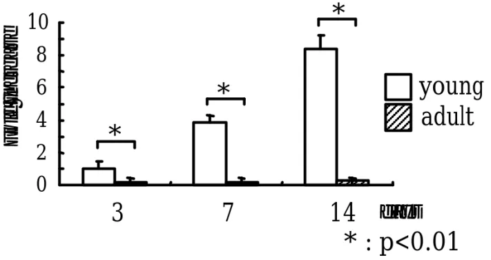
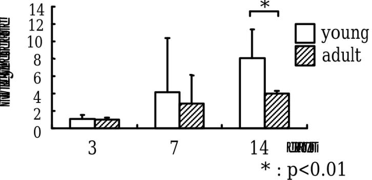
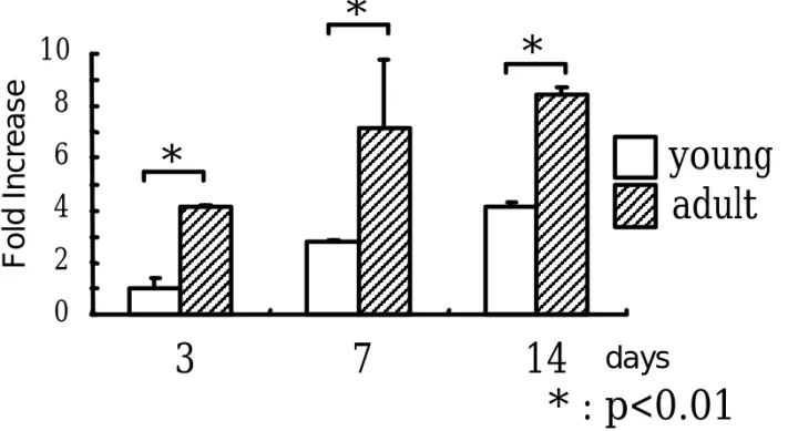
(7) manufacturer's instructions (Applied Biosystems). Reaction conditions consisted of a primary denaturation at 95°C for 20 sec, then cycling for 40 cycles of 95°C for 3 sec and 62°C for 30 sec. PCR data were supplemented by the value of house keeping gene with and were ß-actin reported compared to the corresponding three days of incubation in the young DPCs.. Statistical analysis Three experimental runs were conducted, and data were analyzed using one-way analysis of variance (ANOVA) and a multiple comparison test (Scheffe’s test) (p<0.05).. Results Quantitative RT-PCR Cbfa-1 mRNA expression in young DPCs increased from 3 to 14 days of culture, but in adult DPCs the expression of Cbfa-1 mRNA is very low. Further, the expression of Cbfa-1 mRNA in young DPCs was higher than in adult DPCs. There were significant differences between young and the adult DPCs at all time periods (Fig. 1). DSP mRNA expression in young DPCs also increased from 3 to 14 days of culture, but in adult DPCs the expression of DSP mRNA is slightly increase. DSP mRNA expression in young group at 14 days of incubation was significantly higher than that in adult group (Fig. 2). VEGF mRNA expression in both the young and the adult DPCs tended to increase day by day. The rate of increased VEGF mRNA expression in adult DPCs was higher than in young DPCs. There were significant differences.
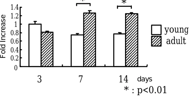
(8) between the young and the adult DPCs at all time periods (Fig. 3). HSP27 mRNA expression in the young group did not vary with culture time, but the expression of HSP27 mRNA in adult DPCs was slightly increased from 3 to 7 days of culture. The HSP27 mRNA expression in adult DPCs at 7 and 14 days of culuter was significantly higher than in young DPCs (Fig. 4).. Discussion It is well known that the volume of the pulp chamber decreases with age, and that this phenomenon occurs as a result of homeostatic mechanisms (14). Only a few biological evaluations about aged dental pulp tissues have been reported (15-17). Further, a reduction of osteocalcin mRNA expression may also be associated with the loss of viability in dental pulp (18). Although Muramatsu et al. evaluated the aged dental pulp tissue from old people, We used pulp cells from the 350 g rats (15-week-old) as not senescence DPCs but adult DPCs in this study. In this study, we analyzed the mRNA expression of several important markers to further evaluate the characteristics of adult dental pulp. Cbfa-1 is a transcription factor that belongs to the runt domain gene family (20-22). Cbfa-1 is a factor that not only regulates osteoblast maturation, but also chondroblast maturation (20, 22). Furthermore, Cbfa-1.
(9) regulates not only the expression of bone and cartilage related genes, but also is involved in dystrophic calcification. Further, DSP is the specific protein for dentin related protein secreted from DPCs and odontoblast. Dentin phosphoprotein (DPP) and dentin sialoprotein (DSP) are two types of dentin non-collagenous matrix proteins. These proteins were originally thought to be dentin-specific until Qin et al. (23) To examine the DSP expression is one of indicators for DPCs’ differentiation. So, a dentin bridge is formed under the necrotic tissue after vital pulpectomy with Ca2OH. Further, the expression of Cbfa-1 in dental pulp tissue is well known (24). In this study, a little expression of Cbfa-1 and DSP mRNAs was seen in adult DPCs, but young DPCs expressed high levels of Cbfa-1 and DSP mRNAs. This phenomenon demonstrates that young DPCs may readily produce dentin bridges or ectopic calcification in the pulp chamber. Angiogenesis plays an important role in maintaining homeostasis. Booth et al. demonstrated that VEGF is involved in angiogenic processes in healthy as well as in diseased periodontal tissues (25). Further, there are some reports about the relationship of VEGF in dental pulp (26, 27). Many studies have investigated angiogenesis in the DPCs, and have gradually clarified its function. However, age-related differences in angiogenesis are less well understood. VEGF is a crucial regulator of vascular development during embryogenesis (vasculogenesis), as well as in blood-vessel formation (angiogenesis) in adult tissues (28). Yoshino et al. reported that mechanical stress stimulates the production of VEGF (29). In this study, the rate of increase in VEGF mRNA expression in the adult group was higher than in.
(10) the young group. Further, it is known that the dental pulp canal becomes stenosed with age which could explain why VEGF mRNA expression of adult DPCs is higher than that of young cells. Heat shock protein (HSP) 27 is a member of the small HSP family that plays a part in regulating cell growth, cell differentiation, wound healing, apoptosis and cell protection against stress (30). Further, HSP serve as molecular chaperones in maintaining homeostasis and are expressed physiologically in response to various types of stress. Furthermore, HSP has remarkable potencies to induce self-defense mechanisms necessary to maintain the homeostatic mechanism in aged tissues. HSP 27 expression can be associated with cellular vulnerability or tolerance to stress and can even be used as an index of cellular stress (31, 32). In this study, the rate of increase in HSP 27 mRNA expression in adult DPCs was higher than in young DPCs, which means that the self defense ability in adult DPCs is high. In conclusion, the self defense system in young DPCs is achieved by calcification, but in adult dental pulp cells it is carried out by the expression of self defense proteins and the regeneration of vessels.. Acknowledgements The authors would like to thank Ms. Saori Takano, Dr. Daisuke Sato and Dr Hideki Soumiya for their technical assistance. This research was in part supported by an Oral Health Science Center Grant HRC7 from the Tokyo Dental College and by a “High-Tech Research Center” Project for Private Universities: matching fund subsidy.
(11) from MEXT (Ministry of Education, Culture, Sports, Science and Technology) of Japan, 2006-2010, and 2007-2010 (No. 19592414)..
(12) References 1. Bernic S, Nedelman C. Effect of aging on the human pulp. J Endod 1975; 1: 88-94.. 2. Ketterl W. Age-induced changes in the teeth and their attachment apparatus. Int Dental J 1983; 33: 262-71.. 3. Oi T, Saka H, Ide Y. Three dimensional observation of pulp cavities in the maxillary first premolar tooth using micro-CT. Int Endodontic J 2004; 37: 46-51.. 4. Woods MA, Robinson QC, Harris EF. Age –progressive changes in pulp widths and root lengths during adulthood: a study of American blacks and whites. Gerodontology 1990; 9: 41-50.. 5. Komori T, Yagi H, Nomura S, et al. Targeted disruption of cbfa-1 results in a complete lack of bone formation owing to maturational arrest of osteoblasts. Cell 1997; 89: 755-64.. 6. Priam F, Ronco V, Locker M, et al. New cellular models for tracking the odontoblast phenotype. Arch Oral Biol 2005; 50: 271-7.. 7. Huang GT, Sonoyama W, Chen J, Park SH. In vitro characterization of human dental pulp cells: various isolation methods and culturing environments. Cell Tissue Res 2006; 324: 225-36..
(13) 8. Ferrara N. The role of vascular endothelial growth factor in pathological angiogenesis. Breast Cancer Res Treat 1995; 36: 127–37.. 9. Carmeliet P, Collen D. Vascular development and disorders: molecular analysis and pathogenic insights. Kidney Int 1998; 53: 1519–49.. 10. Ijichi A, Sakuma S, Tofilon PJ. Hypoxia-induced vascular endothelial growth factor expression in normal rat astrocyte cultures. Glia 1995; 14: 87–93.. 11. Ladoux A, Frelin C. Hypoxia is a strong inducer of vascular endothelial growth factor mRNA expression in the heart. Biochem Biophys Res Commun 1993; 195: 1005–10.. 12. Schmid J, Wallkamm B, Hammerle CH, Gogolewski S, Lang NP. The significance of angiogenesis in guided bone regeneration. A case report of a rabbit experiment. Clin Oral Implants Res 1997; 8: 244-8.. 13. Kitamura C, Nishihara T, Ueno Y, et al. Effects of sequential exposure to lipopolysaccharide and heat stress on dental pulp cells. J Cell Biochem 2006; 99: 797-806.. 14. Gómez PA, Cabrini RL. Anatomic variations of the root canal of the rat according to age. Acta Odontol Latinoam 2004; 17: 39-42.. 15. Muramatsu T, Hamano H, Ogami K, Ohta K, Inoue T, Shimono M..
(14) Reduction of connexin 43 expression in aged human dental pulp. Int Endod J 2004; 37: 814-8.. 16. Uitto MA, Ranta R. Protocollagen proline hydroxylase activity in human dental pulp at various stages of development. Proc Finnish Dental Soc 1973; 69: 254-7.. 17. Van Amerongen JP, Lemmens IG, TOnino GJ. The concentration, extractability and characterization of collagen in human dental pulp. Arch Oral Biol 1983; 28: 339-45.. 18. Muramatsu T, Hamano H, Ogami K, Ohta K, Inoue T, Shimono M. Reduction of osteocalcin expression in aged human dental pulp. Int Endod J 2005; 38: 817-21.. 19. Matsuzaka K, Tsuruoka M, Kokubu E, et al. Age-related differences in expression of vascular endothelial growth factor by periodontal ligament cells in vitro. Bull Tokyo Dental College 2007; 48: 143-6.. 20. de Crombrugghe B, Lefebvre V, Nakashima K. Regulatory mechanisms in the pathways of cartilage and bone formation. Curr Opin Cell Biol 2001; 13: 721-8.. 21. Drissi H, Luc Q, Shakoori R, et al. Transcriptional autoregulation of the bone related CBFA1/RUNX2 gene. J Cell Physiol 2000; 184: 341-50..
(15) 22. Enomoto H, Enomoto-Iwamoto M, Iwamoto M, et al. Cbfa1 is a positive regulatory factor in chondrocyte maturation. J Biol Chem 2000; 275: 8695-702. 23. Qin C, Brunn JC, Cadena E, Ridall A, Tsujigiwa H, Nagatsuka H, Nagai N, Butler WT. The expression of dentin sialophosphoprotein gene in bone. J Dent Res. 2002; 81: 392-4.. 24. Yildirim S, Yapar M, Sermet U, Sener K, Kubar A . The role of dental pulp cells in resorption of deciduous teeth. Oral Surg Oral Med Oral Pathol Oral Radiol Endod 2007; (in press).. 25. Booth V, Young S, Cruchley A, Taichman NS, Paleolog E. Vascular endothelial growth factor in human periodontal disease. J Periodontal Res 1998; 33: 491-9.. 26. Grando Mattuella L, Westphalen Bento L, de Figueiredo JA, Nor JE, de Araujo FB, Fossati AC. Vascular endothelial growth factor and its relationship with the dental pulp. J Endod. 2007; 33: 524-30.. 27. Botero TM, Shelburne CE, Holland GR, Hanks CT, Nor JE. TLR4 mediates LPS-induced VEGF expression in odontoblasts. J Endod. 2006; 32: 951-5..
(16) 28. Olsson AK, Dimberg A, Kreuger J, Claesson-Welsh L. VEGF receptor signaling - in control of vascular function. Nat Rev Mol Cell Biol 2006; 7: 359-71.. 29. Yoshino H, Morita I, Murota SI, Ishikawa I. Mechanical stress induces production of angiogenic regulators in cultured human gingival and periodontal ligament fibroblasts. J Periodontal Res 2003; 38: 405-10.. 30. Leonardi R, Villari L, Caltabiano M, Travali S. Heat shock protein 27 expression in the epithelium of periapical lesions. J Endod 2001; 27: 89-92.. 31. Schlesinger MJ. Heat shock proteins. J Biol Chem 1990; 265: 12111–4.. 32. Amemiya K, Kaneko Y, Muramatsu T, Shimono M, Inoue T. Pulp cell responses during hypoxia and reoxygenation in vitro. Eur J Oral Science 2003; 111: 332-8..
(17) Legends of figures Fig. 1Cbfa-1 mRNA expression Cbfa-1 mRNA expression in the young group increased from 3 to 14 days of culture. Cbfa-1 mRNA expression in adult group did not change at all. The data is shown as the fold increase of corresponding three days of incubation in the young DPCs.. Fig. 2 DSP mRNA expression DSP mRNA expression in both groups increased from 3 to 14 days of culture. However, DSP mRNA expression in adult group increased only slightly. So there is a significant difference between young and adult groups at 14 days. The data is shown as the fold increse of corresponding three days of incubation in young DPCs.. Fig. 3 VEGF mRNA expression VEGF mRNA expression in both the young and the adult DPCs increased day by day. The rate of increase of VEGF mRNA expression in adult DPCs was higher than in the young group. The data is shown as the fold inrease of corresponding three days of incubation in the young DPCs.. Fig. 4 HSP27 mRNA expression HSP27 mRNA expression in young DPCs was not different at various culture times, but HSP27 mRNA expression in adult DPCs was slightly increased from 3 to 7 days of culture. The data is shown as the fold increase of corresponding three days of incubation in the young DPCs..
(18) Fold Increase. 10 8 6 4 2 0. * * * 3. 7. young adult. 14 days * : p<0.01. Fig. 1 Cbfa-1 mRNA expression. 1.
(19) Fold Increase. 14 12 10 8 6 4 2 0. * young adult. 3. 7. 14 days * : p<0.01. Fig. 2 DSP mRNA expression. 2.
(20) *. Fold Increase. 10. *. 8 6. young adult. *. 4 2 0. 3. 7. 14 days * : p<0.01. Fig. 3 VEGF mRNA expression. 3.
(21) Fold Increase. *. 1.4 1.2 1 0.8 0.6 0.4 0.2 0. * young adult. 3. 7. 14 days * : p<0.01. Fig. 4 HSP27 mRNA expression. 4.
(22)
図
関連したドキュメント
Quantitative analysis by real-time Reverse transcription-polymerase chain reaction (RT-PCR) of chronological change in the expression of hepatocyte growth factor (HGF),
Treatment with ONO-1301 increased hepatic HGF mRNA expression, but decreased the expressions of TGF-β1, connective tissue growth factor, α-smooth muscle actin, and type-I and
The method employed to prove indecomposability of the elements of the Martin boundary of the Young lattice can not be applied to Young-Fibonacci lattice, since the K 0 -functor ring
Keywords: continuous time random walk, Brownian motion, collision time, skew Young tableaux, tandem queue.. AMS 2000 Subject Classification: Primary:
We present sufficient conditions for the existence of solutions to Neu- mann and periodic boundary-value problems for some class of quasilinear ordinary differential equations.. We
Answering a question of de la Harpe and Bridson in the Kourovka Notebook, we build the explicit embeddings of the additive group of rational numbers Q in a finitely generated group
Then it follows immediately from a suitable version of “Hensel’s Lemma” [cf., e.g., the argument of [4], Lemma 2.1] that S may be obtained, as the notation suggests, as the m A
Definition An embeddable tiled surface is a tiled surface which is actually achieved as the graph of singular leaves of some embedded orientable surface with closed braid
