INTRODUCTION
Dramatic changes in the endocrine system occur
in women at menopause. The decline in estrogen production results in various degenerative condi-tions, such as osteoporosis and atherosclerosis. Hor-mone replacement therapy (HRT) is widely used to prevent or delay the occurrence of these disor-ders in postmenopausal women. Estrogen modu-lates the production of cytokines since immuno-competent cells possess estrogen receptors (1, 2).
Inflammatory cytokines such as interleukin-1
ORIGINAL
Effects of raloxifene on the production of cytokines in
stimulated whole blood in
ex vivo
and
in vitro
studies
Yuka Kasai
a, Masahiko Maegawa
a, Satoshi Yamamoto
b, Masaharu Kamada
c,
Toshiyuki Yasui
b, Hirokazu Uemura
b, Ayako Kobayashi
b, Masayo Kaneyama
b,
Anna Tani
b, Sumika Matsui
b, Akira Kuwahara
b, Toshiya Matsuzaki
b,
Hiroyuki Furumoto
b, and Minoru Irahara
b aDepartment of Obstetrics and Gynecology, Tokushima Prefectural Central Hospital, Tokushima, Japan ; b
Department of Obstetrics and Gynecology, Institute of Health Biosciences, the University of Tokushima Graduate School, Tokushima, Japan ; and c
Department of Obstetrics and Gynecology, Health Insurance Naruto Hospital, Tokushima, Japan
Abstract : Purpose : The aims of this study were to determine the effects of raloxifene therapy on production of cytokines and in vitro effects of raloxifene on production of cytokines by whole blood cultures. Methods : We obtained samples of peripheral blood from 6 postmenopausal women with osteopenia at baseline and after 3 and 6 months of raloxifene therapy and 10 postmenopausal women who did not receive raloxifene ther-apy. Whole blood from raloxifene-treated women was stimulated with lipopolysaccha-ride (LPS) or phytohemeagglutinin (PHA). Whole blood from postmenopausal women who were not treated with raloxifene was preincubated with raloxifene at concentrations of 10- 10-10- 7M and then stimulated with LPS or PHA. Concentrations of IL-1β, IL-4, IL-6,
IL-12p40, IL-12p70, TNF-α and IFN-γ in the supernatant were measured by respective ELISAs. Results : In ex vivo cultures, raloxifene therapy inhibited LPS-stimulated pro-duction of IL-1β, IL-6, IL-12p40, IL-12p70 and TNF-α, but not PHA-stimulated produc-tion of IL-4 and IFN-γ. In in vitro cultures, raloxifene at a concentration (10-9
M) inhib-ited LPS-stimulated production of IL-1β, IL-6 and IL-12p40 and PHA-stimulated pro-duction of IFN-γ. Conclusions : Raloxifene therapy decreases the production of IL-1β, IL-6, IL-12 and TNF-α but not that of IL-4 and IFN-γ, suggesting that modulation of cytokines could play a role in the mechanisms of the osteoprotective effect of raloxifene. J. Med. Invest. 58 : 110-117, February, 2011
Keywords : raloxifene, cytokine, whole blood assay
Received for publication November 26, 2010 ; accepted January 11, 2011.
Address correspondence and reprint requests to Masahiko Maegawa, M.D., Department of Obstetrics and Gynecology, Tokushima Prefectural Central Hospital, 1 - 10 - 3 Kuramoto - cho, Tokushima 770 - 8539, Japan and Fax : + 81 - 88 - 631 - 2630.
(IL-1), IL-6 and tumor necrosis factor-α (TNF-α) are known to be bone-resorbing cytokines based on many experimental models (3-5). There are many reports demonstrating that estrogen replace-ment decreases the production of IL-1, IL-6, IL-12 and TNF-α by peripheral blood mononuclear cells (PBMC) (6, 7) and whole blood cultures (8). We have also demonstrated that HRT inhibited the pro-duction of IL-1β, IL-4, IL-10, interferon-γ (IFN-γ) and TNF-α in whole blood cultures (9-11) since the whole blood assay is a potentially valuable tool for assessing cellular immune function (12, 13).
Raloxifene, a non-steroidal benzothiophene de-rivative classified as a selective estrogen receptor modulator (SERM), has been used for the treat-ment of postmenopausal osteoporosis. Although raloxifene binds with high affinity to the estrogen receptor, the conformation of the ligand/receptor complex for raloxifene and estrogen differs, result-ing in the differences seen in the pharmacological properties of raloxifene and estrogen. Recently, raloxifene has been reported to have effects on cir-culating cytokines such as IL-6 and TNF-α, but the results of studies on changes in these cytokines caused by raloxifene were inconsistent (14-17). The effects of raloxifene on production of bone-resorbing cytokines were also discrepant because raloxifene treatment inhibited the production of IL-6 and TNF-α in whole blood cultures but not the production of IL-1β in PBMC in postmeno-pausal women (15, 18). Furthermore, the effects of raloxifene therapy on production of cytokines such as IL-4, IFN-γ and IL-12, which acts as a critical bridge between the innate and acquired immune systems, remain to be established.
The aims of this study were to determine the effects of raloxifene therapy for 3 months and 6 months on cytokines in whole blood cultures and to compare with in vitro effects of raloxifene on stimu-lated secretion of cytokines in whole blood cultures.
MATERIALS AND METHODS
Subjects
Sixteen women were enrolled in this study after they gave informed consent. This project had been approved by the Institutional Review Broad of the University of Tokushima, Institute of Health Bi-osciences. The subjects included 10 untreated post-menopausal women (ages, 50 to 65 years ; mean age, 56.6 years) and 6 untreated postmenopausal
women with osteopenia (ages, 52 to 65 years ; mean age, 56.2 years) from the Outpatient Clinic of the De-partment of Obstetrics and Gynecology, Tokushima University Hospital. The subjects had no overt dis-ease and their peripheral blood cell counts, differ-ential leukocyte count, and results of liver and re-nal function tests were within normal limits.
The 6 postmenopausal women with osteopenia received oral administration of 60 mg raloxifene HCl (Evista, Chugai, Japan) every day. Blood sam-ples for cytokine evaluation were drawn into tubes before and 3 to 6 months after the start of raloxifene therapy.
Reagents
Raloxifene (LY139481) was kindly provided by Lilly Research Laboratories (Indianapolis, IN, USA). We used the following reagents : RPMI 1640 (Gibco, Grand Island, NY, USA), lipopolysaccharide (LPS) from Esherichis coli 055 (Difco, Elkhart, IN, USA), phytohemeagglutinin (PHA) (Sigma, St Louis, Mo, USA), and Ficoll-Paque Plus (Amersham Pharma-cia Biotech AB, Uppsala, Sweden).
Ex vivo study
Whole blood cultures were prepared using previ-ously described methods (9, 10). In brief, 1 ml of peripheral blood was withdrawn into a heparinized (10 U/ml) tube, and within 1 h, 0.1 ml of heparinized blood was cultured in a 24-well multicluster plate in 1 ml RPMI 1640 containing 10% fetal calf serum with 1 μg/ml LPS or 10 μg/ml PHA at 37!!for 24 h. The culture supernatants were collected and stored at -40!!until assayed.
In vitro study
The production of cytokines by peripheral blood cells from postmenopausal women was examined by whole blood culture. One ml of peripheral blood was withdrawn into a heparinized (10 U/ml) tube, and within 1 h, 0.1 ml of heparinized blood was cul-tured in a 24-well multicluster plate in 1 ml RPMI 1640 alone (control) or with raloxifene at doses of 10-10, 10-9, 10-8and 10-7 M at 37!!for 12 h. Then
whole blood cultures were stimulated by the addi-tion of 100 ng/ml LPS or 1μg/ml PHA at 37!!for 24 h. The culture supernatants were collected and stored at -40!!until assayed.
Measurement of concentrations of cytokines Concentrations of IL-1β, IL-4, IL-6, TNF-α and IFN-γ in the supernatants were measured using
0 1000 2000 3000 4000 5000 6000
before 3 months 6 months (pg/ml) 0 500 1000 1500 2000 2500
before 3 months 6 months (pg/ml)
A
B
IL-1 䃑 IL-6 0 200 400 600 800 1000 1200 1400 1600 1800before 3 months 6 months (pg/ml)
C
TNF-䃐 䠆 䠆 䠆 䠆 䠆 䠆the respective human ELISA kits (eBioscience, San Diego, CA, USA) according to the manufacturer’s instructions. The sensitivity level of the kits was 4 pg/ml. The intra- and interassay coefficients of variation were less than 10%. The concentration of IL-12p40 was measured using a human ELISA kit (R&D Systems, Minneapolis, MN, USA) according to the manufacturer’s instructions. The sensitivity level of the kit was 15 pg/ml. The intra- and inter-assay coefficients of variation were 4.3% and 9.3%, respectively. The concentration of IL-12p70 was measured using a human ELISA kit (BioSource Internationnal, Camarillo, CA, USA) according to the manufacturer’s instructions. The sensitivity level of the kit was 0.5 pg/ml. The intra- and interassay coefficients of variation were 9.5% and 8.7%, respec-tively.
Assessment of cell viability
Viability of cells treated with raloxifene was de-termined by the trypan blue dye exclusion test. In brief, peripheral blood mononuclear cells (PBMC) were isolated by fractionation on Ficoll-Paque Plus. The interface was removed and washed twice in RPMI 1640. Isolated PBMC were incubated as a density of 5
!
105/ml in RPMI 1640 alone (control)or with different concentrations of raloxifene (10-10,
10-9, 10-8 and 10-7M) at 37"!for 36 h. After the
in-cubation, cells were stained with 0.2% trypan blue for 15 min. The number of stained cells among 200 cells was counted.
Statistical analysis
Statistical analyses comparing treatment groups were performed by the Wilcoxon signed-rank test. Values of p<0.05 were considered to be significant. The StatView 4.1 program (Abacus Concepts, Inc., Berkeley, CA) was used for statistical analysis.
RESULTS
Ex vivo study
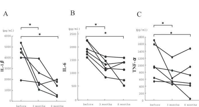
Raloxifene therapy for 3 and 6 months induced a significant decrease in LPS-stimulated production of IL-1β (p<0.05), IL-6 (p<0.05) and TNF-α (p< 0.05) in postmenopausal women (Fig. 1). Raloxifene therapy for 3 and 6 months also induced a significant decrease in LPS-stimulated production of IL-12p40 (p<0.05) and IL-12p70 (p<0.05) (Fig. 2). However, there was no significant change in either IL-4 or IFN-γ after 3 and 6 months of raloxifene therapy (Fig. 3).
Figure 1. Effects of raloxifene therapy on production of IL- 1β (A), IL-6 (B) and TNF-α (C) by 1 μg/ml LPS-stimulated whole blood cells from postmenopausal women with osteopenia. * indicates significance of difference from levels before therapy (p!0.05).
A
0 20 40 60 80 100 120 140before 3 months 6 months
(pg/ml)
B
IL-12p70 0 200 400 600 800 1000 1200 1400 1600before 3 months 6 months
(pg/ml) IL-12p40 䠆 䠆 䠆 䠆 0 2 4 6 8 10 12 14 16 18 20
before 3 months 6 months
(pg/ml) IL-4
A
B
0 200 400 600 800 1000 1200before 3 months 6 months
(pg/ml) IFN-䃒 䠄䠄M䠅 0 20 40 60 80 100 120 140 Basal IL-1 䃑 % of basal 10-7 10-9 10-8
A
10-10 䠆 䠆 䠆 (%) 䠄M䠅 Basal 10-10 10-9 10-8 10-7 IL-6% of basal 0 20 40 60 80 100 120B
䠆 䠆 䠆 (%) 䠄M䠅 Basal 10-10 10-9 10-8 10-7 TNF-䃐 % of basal 0 20 40 60 80 100 120 140 160 䠆 䠆C
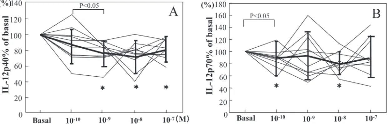
(%) P<0.05 P<0.05 P<0.05 In vitro studyThere was a significant decrease in IL-1β and IL-6 compared to ba-sal levels at raloxifene doses of 10-9, 10-8 and 10-7 M (p<0.05) (Fig.
4A, 4B). There was a significant decrease in TNF-α compared to basal levels at raloxifene doses of 10-8and 10-7M (p<0.05) (Fig. 4C).
There was a significant decrease in IL-12p40 compared to basal lev-els at raloxifene doses of 10-9, 10-8
and 10-7M (p<0.05) (Fig. 5A), and
there was a significant decrease in IL-12p70 compared to basal levels at a raloxifene dose of 10-10and 10-8
M (p<0.05) (Fig. 5B).
There was a significant decrease in IFN-γ compared to basal levels at raloxifene doses of 10-10, 10-9, 10-8
and 10-7M (p<0.05) (Fig. 6A), and
there was a significant decrease in IL-4 compared to basal levels at raloxifene doses of 10-8 and 10-7 M
(p<0.05) (Fig. 6B). Cell viability
The viability of cells treated with raloxifene at doses of 10-10, 10-9, 10-8
Figure 2. Effects of raloxifene therapy on production of IL- 12p40 (A) and IL- 12p 70 (B) by 1μg/ml LPS-stimulated whole blood cells from postmenopausal women with osteopenia. * indicates significance of difference from levels before therapy (p!0.05).
Figure 4. Effects of raloxifene at concentrations of 10-10M to 10-7M on
100 ng/ml LPS - stimulated production of IL- 1β (A), IL-6 (B) and TNF-α (C) in whole blood cultures. Values are percentage of basal (n = 10). Solid lines shows means +/- SD. * indicates sig-nificance of difference from basal (p! 0.05).
Figure 3. Effects of raloxifene therapy on production of IL- 4(A) and IFN-γ (B) by 10μg/ml PHA-stimulated whole blood cells from postmenopausal women with osteopenia. * indicates significance of difference from levels before therapy (p!0.05).
䠄䠄M䠅 Basal 10-10 10-9 10-8 10-7 IL-12p40% of basal 0 20 40 60 80 100 120 140 (%) 䠆 䠆 䠆 Basal 10-10 10-9 10-8 10-7 0 20 40 60 80 100 120 140 160 180 IL-12p70% of basal (%) 䠆
A
B
䠆 P<0.05 P<0.05 䠄䠄M䠅 Basal 10-10 10-9 10-8 10-7 0 40 80 100 120 140 160 180 200 IL-4% of basal 20 60 (%) 䠆 䠆B
Basal 10-10 10-9 10-8 10-7 0 20 40 60 80 100 120 IFN-䃒 % of basal (%) 䠆 䠆 䠆 䠄M䠅A
䠆 P<0.05 P<0.05and 10-7M was not change compared to that of
con-trol cells (data not shown).
DISCUSSION
In the present study, we demonstrated that raloxifene therapy for 3 and 6 months decreases LPS-stimulated production of IL-1β, IL-6, IL-12p40, IL-12p70 and TNF-α but not PHA-stimulated pro-duction of IL-4 and IFN-γ in whole blood cultures, suggesting that modulation of bone-resorbing cy-tokines such as IL-1β, IL-6 and TNF-α could play a role in the mechanisms of the osteoprotective effect of raloxifene. We used the whole blood cul-ture method to determine LPS- or PHA-stimulated cytokine production in ex vivo and in vitro studies. Since isolation of PBMC by hyperosmolar solutions such as Ficoll-Hypaque and their culture ex vivo could modify the function of PBMC compared with that in vivo (19), the whole blood culture method seems to be the best method for mimicking in vivo
conditions of immune reactions (12).
Raloxifene therapy has been reported to have effects on the production or circulating levels of bone-resorbing cytokines such as IL-1β, IL-6 and TNF-α, but results of studies on changes in these cytokines were inconsistent. Gianni et al . reported that the mRNA expression of IL-6 and TNF-α was reduced by raloxifene therapy for 6 months (15). On the other hand, raloxifene treatment did not reduce circulating levels of IL-6 and TNF-α (16, 17), although Walsh et al . reported that raloxifene therapy significantly decreased serum TNF-α level but not IL-6 level (14). Furthermore, Rogers et al . demonstrated that raloxifene therapy did not modu-late 500 ng/ml LPS-stimumodu-lated production of IL-1β by using a whole bloody assay (18). However, we have shown that raloxifene therapy had an inhibi-tory effect on LPS-stimulated production of IL-1β, IL-6 and TNF-α, which is consistent with our pre-vious results using HRT (11). In this study, whole blood cells were stimulated by 1μg/ml LPS, which is a higher concentration than that used by Rogers
Figure 5. Effects of raloxifene at concentrations of 10-10M to 10-7M on 100 ng/ml LPS - stimulated production of IL- 12p40 (A) and
IL- 12p70 (B) in whole blood cultures. Values are percentage of basal (n= 10). Solid lines shows means +/- SD. * indicates signifi-cance of difference from basal (p!0.05).
Figure 6. Effects of raloxifene at concentrations of 10-10M to 10-7M on 1μg/ml PHA-stimulated production of IL-4(A) and IFN-γ
(B) in whole blood cultures. Values are percentage of basal (n= 10). Solid lines shows means +/!SD. * indicates significance of dif-ference from basal (p<0.05).
et al . Since LPS has strong stimulatory effects that could outweigh any potential inhibitory effect of raloxifene, the reason for the difference in the pro-duction of IL-1β is unclear.
In the in vitro study, raloxifene at concentrations of 10-9-10-7M significantly inhibited LPS-stimulated
production of IL-1β and IL-6 and raloxifene at con-centrations of more than 10-8M significantly
inhib-ited LPS-stimulated production of TNF-α. Although we did not observe a dose-dependent response of raloxifene on IL-1β, IL- 6 and TNF-α production, a raloxifene dose of 10-9M was required to show a
sig-nificant response. It is estimated that a raloxifene dose of 10-9M is equivalent to the circulating
con-centration in raloxifene-treated women because the maximum plasma concentration after multiple dosing of raloxifene at 60 mg/d is 1.36 ng/ml, which is equivalent to 2.6
!
10-9M (20). Taranta et al .dem-onstrated that raloxifene inhibited the mRNA ex-pression of IL-1β and IL-6 at a low concentration of 10-10M in in vitro murine osteoblasts (21).
How-ever, in human osteoblastic cells, treatment with raloxifene at doses (10-8, 10-7, 10-6 M), but not at
doses (10-12-10-9 M), resulted in a reduction in the
spontaneous production of IL-1β and IL-6 (22). Fur-thermore, Rogers et al . demonstrated that raloxifene does not modulate spontaneous production of IL-1β in vitro by using a whole bloody assay (18). In our study, whole blood cells were stimulated by the addition of 100 ng/ml LPS after preincubation with raloxifene for 12 h because simultaneous ad-ministration of raloxifene and LPS might negate the inhibitory effect of raloxifene due to the strong stimulatory effect of LPS. The discrepancy in re-sults of in vitro studies may be explained by differ-ences in the cells cultured and the method with or without stimulation by LPS.
To the best of our knowledge, this is the first study showing that raloxifene inhibited the pro-duction of IL-12 ex vivo and in vitro. IL-12 is known to be an inflammatory cytokine produced primarily by phagocytic cells and antigen-presenting cells. IL-12 plays a role in the early inflammatory re-sponse to infection and in the generation of T helper type 1 (Th1) cells while inhibiting the gen-eration of Th2 cells (23). IL-12 is composed of a heavy chain (p40) and a light chain (p35), forming a disulfite-linked heterodimer (p70) (24). IL-12p 40 is produced primarily by activated monocytes/ macrophages, while IL-12p35 is produced con-stantly. In this study, raloxifene therapy for 6 months significantly reduced LPS-stimulated production
of IL-12p40 and IL-12p70, although a concentration (10-9 M) of raloxifene inhibited LPS-stimulated
production of IL-12p40 but not that of IL-12p70. On the other hand, it has been shown that 17β-estradiol at a pregnancy concentration significantly inhibited the production of IL-12p70 in whole blood stimu-lated with a mixture of 1 μg/ml LPS and 5 μg/ml PHA (8). Elenkov et al . also demonstrated that whole blood IL-12 production in pregnant women was decreased 2-fold compared with that in women during the postpartum period (25), suggesting that raloxifene as well as 17β-estradiol has an inhibitory effect on the production of IL-12.
Th1 cells have been found to produce IFN-γ and IL-2, whereas Th2 cells produce different cytoki-nes such as IL-4 and IL-5. It has been reported that raloxifene therapy caused a decrease in serum IL-4 levels (26). In our in vitro study, raloxifene inhib-ited 1 μg/ml PHA-stimulated production of IL-4 and IFN-γ in a dose-dependent manner. However, in our ex vivo study, raloxifene therapy did not modulate 10μg/ml PHA-stimulated production of IL-4 and IFN-γ by whole blood cells. In previously published articles, we have shown that HRT had inhibitory effects on the production of IL-4 and IFN-γ in 10 μg/ml PHA-stimulated cultures (9, 10). Since 10μg/ml PHA might negate the inhibitory fect of raloxifene due to the strong stimulatory ef-fect of PHA, further study is needed to clarify the effect of raloxifene therapy on PHA-stimulated pro-duction of IL-4 and IFN-γ.
In conclusion, we have shown that raloxifene ther-apy and a raloxifene compound have inhibitory ef-fects on the production of bone-resorbing or proin-flammatory cytokines including IL-1β, IL-6, IL-12 and TNF-α, suggesting that raloxifene may play a role in bone resorption and formation due to sup-pression of the production of these cytokines.
ACKNOWLEDGMENTS
We would like to thank Lilly Research Laborato-ries for providing raloxifene (LY139481). This study was supported in part by Grants-in-Aid for Scien-tific Research from the Ministry of Education, Sci-ence and Culture, Japan.
REFERENCE
Abdou NI : Peripheral blood T cells and mono-cytes and B-cell lines derived from patients with lupus express estrogen receptor transcripts similar to those of normal cells. J Rheumatol 25 : 1305-1312, 1998
2. Straub RH : The complex role of estrogens in inflammation. Endocrine Rev 28 : 521-574, 2007 3. Jilka RL, Hangoc G, Girasole G, Passeri G,
Williams DC, Abrams JS, Boyce B, Broxmeyer H, Manolagas SC : Increased osteoclast de-velopment after estrogen loss : mediation by interleukin-6. Science 257 : 88-91, 1992 4. Kitazawa R, Kimble RB, Vannice JL, Kung VT,
Pacifici R : Interleukin 1 receptor antagonist and tumor necrosis factor binding protein de-crease osteoclast formation and bone resorp-tion in ovariectomized mice. J Clin Invest 94 : 2397-2406, 1994
5. Lorenzo JA, Naprta A, Rao Y, Alander C, Glaccum M, Widmer M, Gonowicz G, Kalinowski J, Pilibeam CC : Mice lacking the type 1 interleukin-1 receptor do not lose bone mass after ovariectomy. Endocrinology 139 : 3022-3025, 1998
6. Ralston SH, Russell RG, Gowen M : Estrogen inhibits release of tumor necrosis factor from peripheral blood mononuclear cells in post-menopausal women. J Bone Miner Res 5 : 983-988, 1990
7. Rogers A, Eastell R : The effect of 17β-estradiol on production of cytokines in cultures of pe-ripheral blood. Bone 29 : 30-34, 2001
8. Matalka KZ : The effect of estradiol, but not progesterone, on the production of cytokines in stimulated whole blood, is concentration-depedent. Neuroendocrinol Lett 24 : 185-191, 2003
9. Kamada M, Irahara M, Maegawa M, Ohmoto Y, Murata K, Yasui T, Yamano S, Aono T : Tran-sient increase in the level of T-helper 1 cy-tokines in postmenopausal women and the ef-fects of hormone replacement therapy. Gyne-col Obstet Invest 52 : 82-88, 2001
10. Deguchi K, Kamada M, Irahara M, Maegawa M, Yamamoto S, Ohmoto Y, Murata K, Yasui T, Yamano S, Aono T : Postmenopausal changes in production of type 1 and type 2 cytokines and the effects of hormone replacement therapy. Menopause 8 : 266-273, 2001
11. Uemura H, Kamada M, Maegawa M, Ohmoto Y, Murata K, Kuwahara A, Matsuzaki T, Yasui T, Takeji T, Irahara M : Effect of hormone
replacement therapy on the production of bone-resorbing cytokines by peripheral blood cells in postmenopausal women. Horm Metab Res 37 : 226-230, 2005
12. Petrovsky N, Harrison LC : Cytokine-based human whole blood assay for the detection of antigen-reactive T cells. J Immunol Meth 186 : 37-46, 1995
13. Petrovsky N, Harrison LC : Diurnal rhythmicity of human cytokine production : a dynamic dise-quilibrium in T helper cell type 1/T helper type 2 balance? J Immunol 158 : 163-168, 1997 14. Walsh BW, Cox DA, Sashegyi A, Dean RA,
Tracy RP, Anderson PW : Role of tumor ne-crosis factor- and interleukin-6 in the effects of hormone replacement therapy and raloxifene on C-reactive protein in postmenopausal women. Am J Cardiol 88 : 825-828, 2001 15. Gianni W, Ricci A, Gazzaniga P, Brama M,
Pietropaolo M, Votano S, Patane F, Agliano AM, Spera G, Marigliano V, Ammendola S, Agnusdei D, Migliaccio S, Scandurra R : Raloxifene modulates interleukin-6 and tumor necrosis factor-alpha synthesis in vivo : results from a pilot clinical study. J Clin Endocrinol Metab 89 : 6079-6099, 2004
16. Ozmen B, Kirmaz C, Aydin K, Kafescller SO, Guclu F, Hekimsoy Z : Influence of the selec-tive oestrogen receptor modulator (raloxifene hydrochloride) on IL-6, TNF-α, TGF-β1 and bone turnover markers in the treatment of postmenopausal osteoporosis. Eur Cytokine Netw 18 : 148-153, 2007
17. Yasui T, Uemura H, Hyodo S, Yamada M, Yamamoto S, Maegawa M, Tsuchiya N, Noguchi M, Yuzurihara M, Kase Y, Irahara M : Raloxifene reduces circulating levels of interleukin-7 and monocyte chemoattractant protein-1 in post-menopausal women. Atherosclerosis 204 : 471-475, 2009
18. Rogers A, Clowes JA, Pereda CA, Eastell R : Different effects of raloxifene and estrogen on interleukin-1β and interleukin-1 receptor an-tagonist production using in vitro and ex vivo studies. Bone 40 : 105-110, 2007
19. De Groote D, Zangerle PF, Genaert Y, Fassotte MF, Beguin Y, Noizat-Pirenne F, Pirenne J, Gathy R, Lopez M, Dehart I : Direct stimula-tion of cytokines (IL-1β, TNF-α, IL-6, IL-2, IFN-γ and GM-CSF) in whole blood. I. Com-parison with isolated PBMC stimulation. Cy-tokine 4 : 239-248, 1992
20. Hochner-Celnikier D. Pharmacokinetics of raloxifene and its clinical application : Eur J Obstet Gnyecol Reprod Biol 85 : 23-29, 1999. 21. Taranta A, Brama M, Teti A, De Luca V,
Scandurra R, Spera G, Agnusdei D, Termine JD, Migliaccio S : The selective estrogen re-ceptor modulator raloxifene regulates osteo-clast and osteoblast activity in vitro. Bone 30 : 368-376, 2002
22. Cheung J, Mak YT, Papaioannou S, Evans BAJ, Fogelman I, Hampson G : Interleukin-6 (IL-6), IL-1, receptor activator of nuclear factorκB ligand (RANKL) and osteoprotegerin produc-tion by human osteoblastic cells : comparison of the effects of 17-β oestradiol and raloxifene. J Endocrinol 177 : 423-433, 2003
23. Hsieh CS, Macatonia SE, Tripp CS, Wolf SF, O’Garra A, Murphy KM : Development of TH1 CD4+T cells through IL-12 produced by
Listeria-induced macrophages. Science 260 : 547-549, 1993
24. Wolf SF, Temple PA, Kobayashi M, Young D, Dicig M, Lowe L, Dzialo R, Fitz L, Ferenz C, Hewick RM : Cloning of cDNA for natural killer cell stimulatory factor, a heterodimeric cytokine with multiple biologic effects on T and natural killer cells. J Immunol 146 : 3074-3081, 1991
25. Elenkov IJ, Wilder RL, Bakalov VK, Link AA, Dimitrov MA, Fisher S, Crane M, Kanik KS, Chrousos GP : IL-12, TNF-α and hormonal changes during late pregnancy and early post-partum : Implication for autoimmune disease activity during these times. J Clin Endocrinol Metab 86 : 4933-4938, 2001
26. Kumru S, Yildiz FM, Godekmerdan A, Kutlu S, Yilmaz B, Gurates B : Effects of raloxifene and hormone replacement therapy of serum Th2 and Th3 type cytokine concentrations in healthy postmenopausal women : a randomized controlled trial. Arch Gynecol Obstet 277 : 489-493, 2008