Effects of L2 Immersion Experiences on Translation Task
Performance through a Brain-imaging Technique of
Functional Near-Infrared Spectroscopy
Hideyuki Taura, Aoi Nasu, Daisuke Abe, Sayaka Hirai, Maiya Inoue, Asami Nakagawa, Takashi Nakamura, Mayumi Oga, Ryoko Takamura∗
Abstract
The present study aims to explore, using fNIRS, (1) whether L2 immersion experiences impact brain activation in lexical translation processing when compared to L2 learners without immersion, and (2) what regions of the frontal lobe is most affected by such experiences. 24 Japanese learners of L2 English participated in the timed L1/L2 translation tasks. The results show (1) that overall activation is lowered in all of the three regions in the E-J condition, (2) that as one's L2 improves, the level of activation in Broca's area stays the same but the middle region becomes less activated and more activation takes place in the right region in the J-E condition, and (3) the order of oxy-Hb flow level follows these patterns; [Broca < Right < Middle] in the E-J task, but this order changes from [Middle < Broca = Right] to [Middle < Broca < Right] as one's L2 progresses.
∗
Hideyuki Taura : Professor, Graduate School of Language Education and Information Science Aoi Nasu, Daisuke Abe, Sayaka Hirai, Maiya Inoue, Asami Nakagawa, Takashi Nakamura, Mayumi Oga, Ryoko Takamura : Graduate students, Graduate School of Language Education and Information Science
Effects of L2 Immersion Experiences on Translation Task Performance
Chapter 1 Introduction
1.1 Study abroad programmeStudy abroad programmes abound in the world today reinforcing the view that learning a foreign/second language is easier or quicker when immersed in the target language environment. The effectiveness of such experiences has been extensively explored to date (see Dufon and Churchill, 2006 for an overview). The literature, however, lacks in longitudinal studies, with the majority of them examining programmes that span only for a few weeks to months. The first aim of this study lies in investigating how effective such L2 language experiences are when the programme lasts for over a year.
The focus of studies on study abroad programmes, have been predominantly conducted from linguistic, sociocultural, and motivational perspectives (e.g., Dufon and Churchill, 2006; Isabelli-Garcia, 2006; Yang, 2011; Yashima, 2002). No study using neuroimaging has yet been carried out to look into the effectiveness of study abroad programmes. This newly developed technique of neuroimaging is now used in the SLA discipline, for instance, to identify which cerebral region is activated by L2 tasks. This data can be compared to the region used when one engages in an L1 task, to see if it is differerent. This is where our second aim of this study comes into play.
1.2 Neuroimaging studies on L2
One of the earliest works that looked into which part of the brain becomes the most activated with the link to L2 proficiency in Japanese students, was undertaken by Ohishi (2006). She used functional near-infrared spectroscopy (fNIRS, henceforth) to observe cerebral blood flow in Warnicke's area, the angular gyrus, and the supramarginal gyrus when Japanese L2 learners of English engaged in English listening and reading comprehension tasks. The results showed a U-shaped developmental change in brain activation—little activation in novice learners, hyper-activation in intermediate learners, and selective (reduced) activation in advanced learners. Ishikawa (2009), while criticizing this study for allowing so many variables, focused on lexical and phonological aspects alone using a different type of brain neuroimaging known as functional magnetic resonance imaging (fMRI, hereafter). Uchibori et al. (2009) also found limitations in Ohishi's study for not covering the most linguistically important brain area—Broca's area, and conducted a 24-channelled fNIRS study looking at Broca's area in intermediate Japanese learners of English for syntactic activation. Taking into consideration the shortcomings of these studies, we placed our focus on the lexical aspect alone in terms of brain activation and chose the frontal lobe covering Broca's area and its homologous stie in the right hemisphere, and used a 42-channelled fNIRS for examination.
Differential cerebral activation between L1 and L2 has been discussed in
neuroimaging studies, with reference to onset age (OA) when the person was first exposed to a language and the qualitatively dissimilar make-up of the language faculty in the brain (i.e., Kovelman et al., 2008; Paradis, 2003; Wartenburger et al's study in syntax, 2003; Weber-Fox and Neville, 1996a). Additionally it has also been researched in terms of one's L2 proficiency (i.e., Newman-Norlund et al., 2006; Perani et al., 1998; Wartenburger et al's study in sematics, 2003). Thus, research findings on differential cerebral activation are indeterminate as to whether it is induced by onset age or proficiency. In order to explore if it is due to one's L2 proficiency,
Effects of L2 Immersion Experiences on Translation Task Performance
we compare two Japanese groups of English learners who are divided depending upon their L2 proficiency.
1.3 Brain imaging techniques
Among the variety of brain imaging techniques, the non-invasive functional neuro-imaging method known as fNIRS is said to be easier to use and sensitive to detecting small substance concentrations, with a high temporal resolution (Toga and Mzaaiotta, 2002). In principle, fNIRS measures brain activation using changes in the intensity of light detected by source and detector probes that are attached to the head with a harmless light penetrating the brain. Other brain imaging techniques including fMRI, PET (positron emission tomography), and MEG (magnetoencephalography) require large and bulky instruments which make it difficult for small children to be examined. In comparison, fNIRS uses light via fiber optics which can even allow babies to be examined (Miyai et al., 2001). In addition, fNIRS is sensitive to a very low substance concentration using fluorescence methods and similar to PET scans without the disadvantage of radioactive tracers. Temporal and spatial resolution differ vastly from one brain imaging method to another: high temporal resolution is obtained using the MEG eletrophysiological method (starting from 1 m.s.) while hemodynamics-based techniques such as fMRI (starting from 1 mm) provide a greater spatial resolution. fNIRS offers a smaller, easier to use option to monitor vascular, metabolic-cellular, and neuronal responses with a high temporal resolution of about 1,000 m.s. despite its low spatial resolution of about 3 cm. It is also known that fNIRS data are consistent with fMIR data in predicting hemispheric dominance in linguistic tasks (e.g., Kennan et al., 2002). fNIRS has been widely used and proved to be useful in various disciplines (see Kovelman et al., 2008 for summary).
1.4 Research questions
Research question 1: Does the experience of immersion in the L2 environment for
over a year have an impact on brain activation in lexical processing when compared to L2 learners without such an immersion?
Research question 2: If the answer to the first question is affirmative, which part
of the frontal lobe is affected the most in each proficiency group - Broca's area, its homologous site in the right hemisphere or the middle of the prefrontal region?
Research question 3: Does fNIRS prove to be useful in the linguistic query to find
the answers to research questions 1 and 2?
Chapter 2 Methodology
2.1 ParticipantsA total of 24 university students divided into two groups, participated in this study. They had all learned their L2 (English) in a formal school setting but some of them had the additional experience of residing in and being immersed in L2 communities for one to eight years (Group 1), Group 2 being the non-immersion students. The participants' demographic data are summarized in Table 1. The two groups were matched in number (N=12) and age (mean age of 22, t(22)=1.84, p>.05). They were also matched in left or right handedness, which is one of the most important attributes that has to be considered when conducting a brain-imaging study,
Effects of L2 Immersion Experiences on Translation Task Performance
as seen in the Edinburgh 'handedness inventory' (Oldfield, 1971. Appendix 1). In this study, 11 in each group were right-handed while one in both groups was left-handed.
Table 1. Participants' demographic data
We asked each participant to self-evaluate the level of their own four skills (speaking, listening, reading and writing) in their L1 (Japanese) and L2 (English) out of three with '3' being superior, '2' equal, and '1' inferior to monolinguals (Appendix 2). Table 2 summarizes the results. There are no differences between the two groups in Japanese writing and listening skills or English reading. Meanwhile, Group 1's opinion on their own L2 (English) writing, listening, and speaking skills was statistically better than Group 2. Additionally, Group 2 also rated their L1 (Japanese) speaking and reading skills as better than Group 1. Perhaps these two groups are not matched when evaluating on their own skills in the L2, due to first group's L2 immersion experience.
Table 2. Self-evaluation on four skills in L1 and L2
Prior to the experiment, a signed consent form was obtained from all 24 participants (Appendix 3), which was approved by the institutional ethics committee of Ritsumeikan University, Japan.
2.2 Stimuli
The stimuli was presented to the participants in five sets of 10 words, 50 in total, that were taken from the easiest 2,000 words (Levels 1 and 2) of JACET 8,000 which is a list based on 100 million words in the British National Corpus. It has been compiled to provide the
Effects of L2 Immersion Experiences on Translation Task Performance
so-called most important 8,000 words for Japanese learners of English to use for effective communication in the current globalized world. In order to offset the translation sequence effect, that is, whether translation is chosen from Japanese to English or from English to Japanese, two sets of tasks were devised. Those participants who began their task in Japanese received (1) 10 Japanese words to read aloud, (2) 10 Japanese words to translate into English, (3) 10 English words to read aloud, (4) 10 English words to translate into Japanese, and (5) 10 English words to read aloud. On the other hand, those who started their task in English received (1) 10 English words to read aloud, (2) 10 English words to translate into Japanese, (3) 10 Japanese words to read aloud, (4) 10 Japanese words to translate into English, and (5) 10 Japanese words to read aloud. In order to counter-balance the language sequence choice, six participants in each group were given (without a choice) the Japanese-English sequence tasks while the rest were given the English-Japanese sequence tasks. The words that were used for Japanese presentation (reading aloud or translating into English) were translated from the original pool of 50 words taken from the JACET 8,000 Level 1 and Level 2 lists.
2.3 Procedure
Participants were individually shown into a laboratory and seated in a comfortable chair with their eyes approximately 30 cm away from the monitor (Apple MacBook Pro 17"). While a flexible cap with 27 fiber probes was being placed on their head, information was gathered on their language background, handedness, and self-assessment of their four skills (speaking, listening, writing, and reading) in the two languages. Once the cap was appropriately placed and connected, photos were taken to record the position of the cap from three
angles—from the left, right and front. Then, a video clip was shown to inform them of what the experiment involved including an explanation of what was expected. When participants were made fully aware of the task in front of them, they were told that it was their right to stop it any time they felt uncomfortable, and the experiment started. An instruction slide was followed by five sets of 10 words. The explanation, for example, shown on one slide said 'read aloud the animal', and this came before 10 English words were individually presented for the reading aloud task. Similarly, the slide 'translate house into Japanese', was shown before 10 English words were presented on the computer as a translation task. Each word slide was projected onto the monitor screen for 3 seconds. The task itself, had 50 task slides and 5 instruction slides, and lasted about 3 minutes, during which time both video and audio data were collected for
performance accuracy analysis to be conducted afterwards. When the experiment was completed, each participant was given an appropriate token of gratitude—a book voucher.
A blocked design of two language sequences are shown in Figure 1.
Figure 1. Blocked design for the experiment
Effects of L2 Immersion Experiences on Translation Task Performance
2.4 The fNIRS instrument
A multichannel continuous wave optical imager (Shimadzu FOIRE-3000) was used. This device has 13 emitters and 14 receivers, 3 centimeters apart from each other to detect 42 areas (channels). The fNIRS machine uses three wavelengths of near-infrared light (780±5nm, 805±5nm, 830±5nm). According to the International 10/20 system (Jasper, 1958), emitters and receivers of fNIRS were placed bilaterally over the frontal lobe, including Broca's area in the left inferior frontal cortex, with the lowest probes being positioned along the T3-Fp1-Fz-Fp2-T4 line (Figure 2). Receivers #8 and #7 were aligned with the Cz-Nz (right above the nose over the Frontal, Parietal, and Occipital lobes) line.
Our decision to examine the frontal lobe in both hemispheres is based on findings from Just et al. (1996) and Yamadori (1998) that in a language task, not only the left laterosuperior temporal cortex (Wernicke's area) and the left inferior frontal gyrus (Broca's area) but also their homologous right-hemisphere areas are found to be activated, though the right areas had much smaller volumes of activation.
Effects of L2 Immersion Experiences on Translation Task Performance
2.5 Data analysis
Data from the fNIRS were processed using Shimadzu FOIRE (a functional optical imagery software for research) and PASW SPSS software. The first stage involved identifying in the individual participants, the three distinct areas of the brain for this study, namely, Broca's area, its homologous area in the right hemisphere, and the middle region of the prefrontal lobe (forehead region). Video and still-photo data were carefully examined to identify these three areas, together with the field notes taken during the experiment by the researcher. Diagrams displaying changes in oxyhemoglobin (oxy-Hb), deoxyhemoglobin (deoxy-Hb), and total hemoglobin (total-Hb) using the FOIRE software (Figure 3) were produced, in order to discard any data at particular channels that showed the involvement of artefacts which may have been due to a jerking movement of the participant or probes popping out of the holder sockets. Despite its excellent temporal resolution, fNIRS lacks spatial resolution (Toga and Marital, 2002), so we needed to include 3 to 5 channels of data to cover the designated three regions of the brain. Broca's area included channels 17, 25, and 34 while its right hemispheric counterpart included channels 9, 18, and 26. The five channels of 21, 22, 30, 38, and 39 covered the mid pre-frontal region.
FOIRE-3000 shows three different fNIRS parameters (oxy-Hb as the red line in Figure 3, deoxy-Hb as the blue line, and total-Hb as the green line). Based on the findings that fNIRS oxy-Hb exhibits a high correlation with the blood oxygeneration level dependent (BOLD) signal used in fMRI (e.g., Ryo et al., 2008; Shimoyama et al., 2006; Toronov et al., 2007), and the data produced may therefore be used to represent all three parameters (Fukuda, 2009), this study focuses solely on oxy-Hb. The oxy-Hb change in Figure 3, for instance, can be interpreted in the following way: (1) that the oxy-Hb red line stays stable for the first 33 seconds (one instruction plus ten stimuli slides, each lasting for 3 seconds) of simply reading aloud L2 words, (2) in the next 33 seconds it goes up in the L2-L1 translation task, (3) in the next 33 seconds it gradually goes down when the task is to simply read aloud L1 words, and (4) in the final 33 seconds when the task is to translate L1 words into L2 it goes up again but declines towards the end of the task, which continues into the rest task at the end.
Figure 3. Trend diagram of oxy-Hb, deoxy-Hb, and total-Hb (the x-axis representing
Effects of L2 Immersion Experiences on Translation Task Performance
For the second stage of analysis, the fNIRS data during the translation task were subtracted from the fNIRS data in the preceding (reading-aloud) rest-task, which was originally devised by Peterson et al. (1988) using PET scanning and has since been widely used in neuroimaging studies. In this way the precise difference in fNIRS between the reading-aloud and translation tasks could be pinpointed. This is based on the premise that everything else in speech production is the same, that is, visually recognizing a stimulus item, accessing one's mental lexicon for the meaning, seeking a translation equivalent either in L1 or L2 (mostly observed in the inferior frontal gyrus in the left hemisphere of the brain; 19, 39, 37, 42, 22, and 40 on the Brodmann map of the brain), and verbally articulating it (BA 19, 39 and 44).
For an example, the fNIRS data from a 30-second task of translating 10 Japanese words into English were subtracted from the fNIRS data taken from a 30-second task to simply read aloud 10 Japanese words and were identified as Data 1. In other words, the difference between the two tasks in fNIRS data was calculated. Data 2 were obtained in exactly the same way but using a reverse translation sequence: the fNIRS data from a 30-second task of
translating 10 English words into Japanese were subtracted from the data from a 30-second task of reading out 10 English words. Data 1 therefore represented what it cost the brain to translate L1 Japanese into L2 English whereas Data 2 signified what the brain required to translate L2 English into L1 Japanese. A comparison of these two sets of data in three different regions of the brain was carried out (repeated measures in general linear models using PASW SPSS software) to reveal if there was any obvious impact on the brain in attempting L1-L2 tasks, from
participants' immersion experiences in L2 environments.
Chapter 3 Results
3.1 Behavioural dataTable 3 shows the number of words that were correctly responded to during the experiment, and these were compared between the two groups for each task, resulting in no statistical differences at all.
Table 3. Behavioural data
Effects of L2 Immersion Experiences on Translation Task Performance
Participants in both groups read out or translated the printed words with over 90% of accuracy, irrespective of the task sequence. This performance is rather impressive, considering the time constraint imposed on participants with each stimulus word shown on the computer screen for only 3 seconds at a time. This equally high performance, suggests the same level of task difficulty for both groups, eliminating the possibility that any difference in brain activation is caused by the task itself.
A repeated measures of ANOVA was also carried out to examine any accuracy differences on the four tasks between the two groups. The results revealed neither a main effect (F(3, 20)=2.516, p>.05, partial eta squared=.274) nor an interaction effect (F(3, 20)=.519, p>.05, partial eta squared=.072). This denotes that there were no task differences between the groups in terms of accuracy.
3.2 fNIRS data 3.2.1 An overall trend
The descriptive data (see an example of fNIRS data from a participant in Appendix 4) are summarized in Table 4.
A GLM (mixed between-within subjects ANOVA) revealed statistically significant main effects among task types (F(5, 500)=1470.22, p<.01, partial eta squared=.936). This signifies that when both groups' fNIRS data were examined, the following result became apparent. First, the J-E (L1-L2) translation task in the right region of the brain required the most hemoglobin flow. This was followed by the J-E (L1-L2) task in the left region, the E-J (L2-L1) task in the middle region, the J-E (L1-L2) task in the middle region, the E-J (L2-L1) task in the right region, and the E-J (L2L1) task in the left region as displayed in Figure 4. In other words, the English into Japanese (L2-L1) translation task required a much lower amount of cerebral blood flow than the Japanese into English (L1-L2) task in the left and right regions of the brain whereas the opposite tendency was the case with the middle brain region.
Table 4. fNIRS descriptive data
Effects of L2 Immersion Experiences on Translation Task Performance
Interaction effects (tasks*groups) were also observed (F(5, 500)=72.76, p<.01, partial
eta squared=.421). A post-hoc Tukey revealed no statistical differences in fNIRS data between the middle and right regions of the brain (Figure 5). This signifies that Broca's area is the least activated when compared with the middle and right regions where no statistical differences to one other were observed.
These results are based on the data taken from both Group 1 and Group 2 in their translation tasks and in order to examine the fNIRS data in more depth, within-group and between-group analyses were carried out as described below.
Figure 4. fNIRS data (mM-mm) among tasks
Figure 5. fNIRS data in three brain regions
3.2.2 Within-group analysis 3.2.2.1 Group 1
Group 1 includes 12 participants with the experience of being immersed in the L2 milieu for over one year. Repeated measures of ANOVA showed significant differences in the
Effects of L2 Immersion Experiences on Translation Task Performance
three brain-regions when subjects undertook the two translation tasks: F(5, 248)=2153.89, p<.01, partial eta squared=.978. A post-hoc Bonferroni found statistical differences among all six fNIRS data—the greatest being the J-E task (the right region of the brain > the left region), followed by the middle region (E-J task > the J-E task), then the E-J task (the right region > the left) as shown in Figure 6.
Figure 6. G1 within-group analysis
A few distinct features can be seen in the Bonferroni analyis. First, the right
hemisphere displays more activation than Broca's area, irrespective of the type of translation. The task of translating Japanese words into English (J-E or L1-L2) requires more hemoglobin flow than the reverse translation task (E-J or L2-L1) in both regions of the brain. A completely opposite tendency was observed, however, in the middle of the frontal lobe where the L2-L1 (E-J) translation task requires more cerebral blood flow than the L1-L2 (J-E) task.
3.2.2.2 Group 2
Group 2 includes 12 participants with the experience of immersion in the L2
environment for less than a few months or none at all. Repeated measures of ANOVA showed significant differences among the three brain regions and two translation tasks: F(5,
248)=490.69, p<.01, partial eta squared=.908. A post-hoc Bonferroni revealed no statistical differences among the following three tasks of the J-E task in the left region, the E-J task in the middle region, and the J-E task in the right region in fNIRS. The above three tasks, however, required more brain activation than the E-J task in the left and right regions along with the J-E task in the middle brain region. In addition, the J-E task in the middle region needed more hemoglobin flow than the E-J task in the right region which in turn needed more hemoglobin flow than the E-J task in the left region (Figure 7).
Three distinct features were observed in the results: (1) higher fNIRS data were recorded in the J-E translation task than in the E-J translation task in both hemispheres, which signifies that L2 production costs more energy than L1 production, (2) the middle region in the pre-frontal lobe is affected differently by the translation tasks, than Broca's area and its right hemispheric counterpart. In other words, in the middle brain region when observed on the E-J translation task, requires more hemoglobin flow than the J-E translation task, and (3) the E-J
Effects of L2 Immersion Experiences on Translation Task Performance
translation task in the middle brain region produced the same level of oxy-Hb as J-E tasks in Broca and its right hemispheric counterpart in G2, whereas in G1 the fNIRS data in the J-E task in the middle region was significantly lower than the results from the J-E task from both sides of the brain.
Figure 7. G2 within-group analysis
3.2.3 Between-group analysis
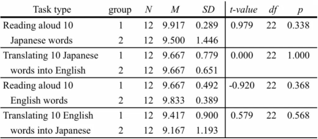
The fNIRS data of G1 and G2 are compared in the three brain regions on two types of translation tasks by carrying out six independent-samples t-tests. The results are presented in Table 5 and Figures 8 to 10.
Table 5. Between-group comparison in each task and brain region
An overall trend shows that more brain activation was seen in G2 than in G1 with two exceptions. The first exception involved no difference between the groups in the translation task of Japanese into English in Broca's area (Left J-E G1 = G2 in Figure 8). The second exception
Effects of L2 Immersion Experiences on Translation Task Performance
concerned a higher amount of cerebral blood flow in G1 than G2 on the task of translating Japanese words into English in the right region (Right J-E G1 > G2 in Figure 8).
Figure 8. A comparison of two groups on all tasks and brain regions
Figure 9. A comparison of oxy-Hb fNIRS data between G1 (left) and G2 (right) with the top
colour patch being the middle region, the left being Broca's area, and the right its homologous site in the right hemisphere on the English (L2) into Japanese (L1) task (red indicating the most activated area and dark blue the least activated)
Effects of L2 Immersion Experiences on Translation Task Performance
Figure 10. A comparison of oxy-Hb fNIRS data between G1 (left) and G2 (right)
in the Japanese (L1) into English (L2) task
Chapter 4 Discussion
While behavioural results showed a comparable performance in terms of accuracy rates between the two groups, the fNIRS data showed both similarities and differences between the two groups examined (Figure 9). For both Group 1 with the L2 immersion experience for over a year and Group 2 with less than a few months immersion experience, the fNIRS results in Broca's area and its right hemispheric counterpart were identical in both groups where increased cerebral blood oxygenation patterns were observed in the J-E (L1-L2) task when compared to the E-J (L2-L1) task. The two groups were also similar in the greater amount of activation in the middle region of the frontal lobe during the E-J (L2-L1) task than the activation seen during the J-E task. An additional similarity revealed itself in the fNIRS data: the J-E task > Mid J-E > Right E-J > Left E-J. The last similarity shared by the two groups was the degree of fNIRS data in the L2-L1 translation task—the middle frontal lobe showed the highest oxy-Hb flow, followed by the right hemisphere and then Broca's area.
Four differences between the two groups arose as well. The first difference was that increased oxy-Hb was detected more often in Group 2 than Group 1 with two exceptions: (1) there were no group differences in the J-E task in Broca's area (Left J-E G1 and Left J-E G2 in Figure 11) and (2) there was more brain activation in Group 1 (Right J-E G1) than Group 2 in the J-E task in the right hemisphere (Right J-E G2). The second difference appeared in the fNIRS data in the J-E task where Broca's area (Left J-E G1) needed less oxy-Hb than its right hemispheric counterpart in Group 1 (Right J-E G1) while there was no difference between the hemispheres in Group 2 (Left J-E G2 and Right J-E G2). The third difference was that for Group 1 during the E-J task, the middle pre-frontal lobe (Mid E-J G1) was less activated than during the J-E task in Broca's area (Left J-E G1) whereas there was no such difference in Group 2 (Mid E-J G2 and Left J-E G2). Fourthly, compared to Group 2, the group with a longer L2 immersion period needed less activation energy in all the three regions of the brain during E-J (L2-L1) conditions.
Effects of L2 Immersion Experiences on Translation Task Performance
Figure 11. Rearrangement of fNIRS data in Figure 8
If we take the stance that Group 1 with its longer L2 immersion experience, is the more advanced L2 learners' group it is possible to interpret the above results from a developmental perspective in the following manner. Initially, L2 learners (Group 2) find the task of translating an L2 (English) word into L1 (Japanese) easier than the reverse. During the relatively easier E-J task, the activation level is the lowest in Broca's area, followed by the right hemispheric region and then the middle region (highest activation). Meanwhile, in undertaking the more demanding task of L1-L2 (J-E) translation, the activation of Broca's area is equivalent to that of its right hemispheric counterpart, both results being statistically higher than that of the middle region (Broca = right > middle). After being immersed in an L2 environment for more than a year, L2 learners from Group 1 still find the E-J task easier than the J-E task and the comparative activation level order remains the same as before: middle > right > Broca's area. Despite the activation order remaining the same, the actual fNIRS data showed a significant decrease in all of the three regions of the brain on the E-J task. This signals that the three brain regions needed less oxy-Hb in an easy E-J translation task as the L2 improves. Meanwhile, in the more demanding task of translating Japanese into English, the activation level order changes from [Middle < Broca = Right] to [Middle < Broca < Right] as one's L2 skill progresses. A careful look at the fNIRS data discloses a decrease of activation in the middle regions, no change in Broca's area, and an increase in the right region.
This summary highlights a sharp contrast in brain activation, depending on which translation type we look at. When the focus is placed on the task of translating L2 words into L1, a better L2 leads to a lower overall level of activation in all three regions of the brain, leaving the activation level order unchanged, that is [Broca < Right < Middle]. Meanwhile, as L2 skills progress, under the J-E translation task the activation level order undergoes a slight change from [Middle < Broca = Right] to [Middle < Broca < Right]. This is due to the differential
involvement in the J-E task by the three brain regions, that is, there is more right hemispheric involvement, less in the middle area and no change in Broca's area. Simply put, as one's L2
Effects of L2 Immersion Experiences on Translation Task Performance
improves, Broca's area's level of activation stays the same but the middle region becomes less activated and more activation takes place in the right region in the J-E condition whereas overall activation is lowered in all of the three regions in the E-J condition. The order of oxy-Hb flow level stays at [Broca < Right < Middle] in the E-J task, but in the J-E task the order changes from [Middle < Broca = Right] to [Middle < Broca < Right] as one's L2 progresses.
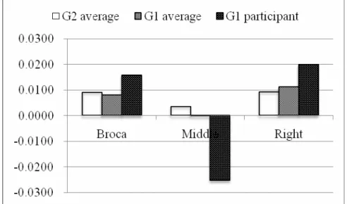
So far this research has examined group averages, but in the second language acquisition and attrition field huge individual differences exist among L2 learners (i.e., De Bot, 2005, and Skehan, 1989). In order to address this issue, one participant in the supposedly more advanced group (Group 1) has been chosen in an attempt to compare his results with the Group 2 average - Group 2 being composed of L2 learners without extensive exposure to L2 in an immersion situation. We chose an extreme case (participant) whose onset age (OA) for L2 was the earliest and the length spent in the L2 environment (LOR) the longest, in an attempt to explore whether more advanced developmental traits were observed in this participant in relation to the Group 1 and Group 2 averages. Figure 12 summarizes the comparison in the J-E task.
Figure 12. Comparison of a G1 participant with G1/G2 averages
Figure 12 shows that in the middle region of the brain, there is less developmental activity in the fNIRS result in Group 2 (less advanced L2 learner group) than in Group 1 (more advanced group) similarly reflected in the more advanced L2 learner's fNIRS data from Group 1. A similar developmental change is also seen in the right region of the brain, though the trend is in an increasing direction. However, Broca's area, which is linguistically recognized to be the most crucial area, did not show the same activation level in both groups, nor a similar
developmental change as exhibited by the other two regions. In short, this individual case represented a similar trend observed to Group 1 and Group 2 averages and was this further supported by the data from the middle and right regions of the brain, but was not the same in Broca's area. This calls for studies involving more advanced L2 learners to confirm our results. It also draws our attention towards individual differences as opposed to group averages.
Effects of L2 Immersion Experiences on Translation Task Performance
Another issue to be further investigated is the role played by the middle region of the brain. Our data in this study (Figure 11) are inconclusive in deciding whether this region of the brain serves some linguistic function or not. This is partly because the middle region displayed a developmental trend similar to Broca's area and its right hemispheric counterpart. This trend was shown in a decreased oxy-Hb flow in the more advanced L2 learners on a relatively easier E-J (L2-L1) task. Additionally the middle region showed brain activation levels in both tasks completely different to those in the other two brain regions: the J-E (L1-L2) task required more oxy-Hb flow than during the E-J (L2-L1) task on both sides of the brain while the fNIRS data in the middle region was in the order of the E-J task (more flow) and J-E, irrespective of the L2 proficiency. In order to clarify the inconsistency revealed in this study, we need to perform both linguistic and non-linguistic tasks on the same participants to collect data in the middle region of the frontal lobe. At the same time, the results of a task that involve only one language may provide some clues on this matter, since Dehaene et al. (1997) report in their fMIR study that the anterior cingulate region (a slightly deeper area of the brain than the middle region we
examined) is active only in foreign language processing, suggesting the conscious use of metalinguistic knowledge by L2 learners.
Chapter 5 Conclusion
This study demonstrates that the impact L2 immersion exerts on translation task performance varies, depending on what aspect of the performance we examine. When performance accuracy comes into focus, the L2 immersion experience makes virtually no impact at all. However, when the focus is shifted to the amount of brain activation, such experience contributes to significantly less energy being expended in all brain regions observed in the translation task of L2 English to L1 Japanese. This suggests that immersion in the L2 region for over a year leads to the automatization of translating L2 words into L1. This positive impact, however, gains only partial support from the results of the L1-L2 (J-E) translation task where the brain is less activated in the middle region alone. Broca's area and its homologous site in the right hemisphere, however, show the same amount of activation or sometimes a
heightened amount. Thus, our answers to the first two research questions are not straightforward:
Research Question 1: Does the experience of immersion in the L2 environment for over
a year have an impact on brain activation in lexical processing when compared to L2 learners without such an immersion? Performance accuracy is not affected by immersion.
Research question 2: If the answer to the first question is affirmative, which part of the
frontal lobe is affected the most in each proficiency group - Broca's area, its homologous site in the right hemisphere or the middle of the prefrontal region? All three regions in the brain in the L2-L1 task as well as the middle region alone in the L1-L2 task are affected positively and were activated more easily. Broca's area and the right hemispheric area displayed little effect from L2 immersion experiences in the L1-L2 task.
Effects of L2 Immersion Experiences on Translation Task Performance
In the query into L2 immersion effects on brain activation, a few incidental findings were made. The first involves the relative easiness of the L2-L1 over the L1-L2 translation task. The second finding concerns the relation of the task type and activation level in different brain regions: in the L2-L1 task, brain activation is the highest in the middle region followed by the right and left hemispheric regions for all L2 learners in this experiment, while in the J-E task the order changes from [Middle < Broca = Right] to [Middle < Broca < Right] as one's L2
progresses. This leads to the third finding that in the linguistic task of translation, not only Broca's area but also the right hemisphere and the middle brain area play some role. This does not imply that each region's involvement is similar to each other in quality because the activation of the middle region is the highest on the relatively easier task of translating L2 into L1 but the lowest in the more demanding L1-L2 task, while the right hemispheric activation is either equal to or more than that of Broca's area which is central to human speech control.
Despite these findings, the current study is not without shortcomings. Considering the fact that the middle region in the brain is relatively less explored in the literature (i.e., De Groot, 2011; Hagoot, 2005) and that our fNIRS data in the middle region did not show the same consistency as the data from the left and right hemispheres, it is possible that the middle region of the brain plays a qualitatively different role to the other areas of the brain that we looked at. This implies that data from the middle region should perhaps not be used in a comparison to Broca's area data, for instance. Secondly, some neuroimaging (both fNIRS and fMRI) studies such as Buchweitz et al. (2009), Dehaene et al. (1997), Kovelman et al. (2008), Ryo et al. (2008), Tan et al. (2001) report right hemispheric involvement in linguistic processing of various degrees in bilinguals as well as monolinguals using logographic languages such as Chinese and Japanese. These findings should be scrutinized further to help explain the query generated in this study—why more advanced L2 learners display an enhanced activation in the right hemisphere in the L1-L2 translation task. Thirdly, in order to reach a firmer conclusion, we need to test an additional group of more advanced participants whose onset age is earlier and length of time spent in the L2 environment longer. If such participants were to show a decrease rather than an increase in oxy-Hb in the right brain region as we observed in Group 1,
(remembering that the oxy-Hb shows a U shape from the novice to intermediate and advanced L2 learners), it could be interpreted in the following way. Before automatization, the brain activation in intermediate L2 learners may become even more heightened than in L2 novice or advanced learners as reported by Ohishi (2006) and Ishikawa (2009). The fourth shortcoming to consider is that the indicators of participants' L2 proficiency such as TOEFL scores should be obtained in addition to the participants' self-evaluations used in this study, so that target groups could be more easily identified as novice, intermediate, or advanced L2 learners for a higher validity. Lastly concerned is the research design: the present study used a cross-sectional design by comparing groups, but perhaps an extended longitudinal investigation addressing the course of L2 acquisition is also necessary to reveal real-life mechanisms through different the fields of psycholinguistics and neuro-psycholinguistics.
Thus, in answer the third research question of whether or not fNIRS proves to be useful in the linguistic query to find the answers to research questions 1 and 2–the present study illustrates the validity of how fNIRS, a brain-imaging technique, can be utilized in exploring a linguistic query from a different perspective. fNIRS is therefore a particularly promising tool for linguists in that it can be more easily used with young children whose language and cognitive development are still underway, as a safe, non-invasive alternative which still allows participants
Effects of L2 Immersion Experiences on Translation Task Performance
to be able to move (unlike other brain scannning techniques) as seen in fNIRS experiments on infants (e.g., Meek et al., 1998; Pena et al, 2003; Taga et al., 2003; Watanabe et al., 2010).
Acknowledgements
We are grateful to the participants in this study and also to the research promotion grant awarded by Ritsumeikan University in the academic year of 2010.
Appendix 1 An example of the Edinburgh Handedness Inventory
Effects of L2 Immersion Experiences on Translation Task Performance
Effects of L2 Immersion Experiences on Translation Task Performance
Appendix 3 Letter to parents and assent form
Seeking volunteers to participate in brain-language research project
Hideyuki Taura, Ph.D Ritsumeikan University
Sep 1, 2010 To the parents of XXXXXXXXXXXXXXX
LEIS (Language Education and Information Science post-graduate course at Ritsumeikan University, Kyoto) has been awarded an intra-university research grant to undertake a new project to explore the bilingual language acquisition mechanism using newly developed brain technology - near-infrared spectroscopy (we have been granted permission for this research project by Review Committee for Research Ethics Involving Human Subjects,
Ritsumeikan University).
This project is unique in that it is the first of its kind in the world to explore bilingual language acquisition using NIRS. NIRS has already been utilized in Japan on over 1,000 monolingual Japanese children attending local Tokyo elementary schools. The research findings at XXX and XXX would no doubt contribute to the field of bilingual language acquisition along with providing feedback to your institution in terms of effective language learning.
Both headmasters - Messrs. XXXXXXXXX - have agreed to conduct this research at XXXXX as long as parents' consent is granted. It would be greatly appreciated if your children who have been exposed to two languages from early on, i.e. before the age of 10 (G1 to G12 at XXX, or either returnee or international marriage bilingual students at XXX) could participate in this project.
If your children are willing to volunteer, please fill in the separate assent form and drop it into a box at the information centre by 4pm Sep 10 (Fri), 2010. We will contact you to notify the exact time and date of your appointment. We are looking forward to seeing as many children as possible from XXXXX.
[research]
<place> XXXXX (Room 101, used to be called SSC) <experiment> Approximately 1 hour in length
1. Questionnaire on child's language background and right or left handedness 2. Language dominance test (5 minutes)
3. NIRS experiment (a 10-minute long word association game with probes on)
*NIRS is the safest brain scanning device which observes blood flow by projecting totally harmless near-infrared light onto one's head, and research has been even done with newly-born babies with no side effects whatsoever.
4. We'll immediately stop the experiment if your child feels uncomfortable. In case of anything unforeseen arising on the way to school or during the experiment, we will cover insurance costs for each child.
<time> 2010.9.13 - 2010.10.3 (2 weeks) & 2010.10.23 - 2010.10.31(except for Oct 30) <token of gratitude> 5,000 yen worth of book vouchers (including transportation)
<privacy> Collected data are publicized as group averages, never in a form of a particular individual being identified.
[contact] Any questions or queries may be directly addressed to Prof. Hideyuki Taura. Language Education and Information Science, Ritsumeikan University.
Effects of L2 Immersion Experiences on Translation Task Performance