九州大学学術情報リポジトリ
Kyushu University Institutional Repository
ヒト型ハイブリドーマのモノクローナル抗体生産性 増強に関する研究
菅原, 卓也
九州大学農学研究科食糧化学工学専攻
https://doi.org/10.11501/3065533
出版情報:Kyushu University, 1992, 博士(農学), 課程博士
- --- -----------------
Studies on Enhancentent of Productivity of Monoclonal Antibodies by
Huntan-Huntan Hybridontas
TAKUYA SUGAHARA
1993
CONTENTS
PAGE
CHAPTER I GENERAL INTRODUCTION 1
CHAPTER II IMMUNOGLOBULIN PRODUCTION STIMULAT- ING FACTOR-IT IN NAMALWA CELLS 6
1. Introduction 6
2. Materials and methods 7
2-1. Cells and cell cui ture 7
2-2. Preparation of cultured supernatant and lysate of Namalwa
cells 7
2-3. Determination of IPSF activity 8
2-4. Fractionation of Namalwa cell lysate using salting out 8
3. Results 9
3-1. Effect of Namalwa cell culture supernatant and lysate on
IgM production of HB4C5 cells 9
3-2. Fractionation of Namalwa cell lysate by salting out using ammonium sulfate
4. Discussion 5. Conclusion
CHAPTER III PURIFICATION OF IMMUNOGLOBULIN PRODUCTION STIMULATING FACTOR -II a
9 12 13
(IPSF-IIa} DERIVED FROM NAMALWA CELLS 14
1. Introduction 15
2. Materials and methods 15
2-1. Cells and cell culture 15
2-2. Assay of IPSF activity on hybridomas 15 2-3. Purification of IPSF-Ila from Namalwa cells 15 2-4. Polyacrylamide gel electrophoresis of IPSF-II a 15
3. Results 16
3-1. Hydrophobic interaction chromatography of Namalwa cell
lysate 16
3-2. Gel filtration of Fraction A obtained by hydrophobic interaction column chromatography
3-3. Analysis of composition of IPSF-Ila
17 17
1
4. Discussion 5. Conclusion
CHAPTER IV CHARACTERIZATION OF IPSF-IIa
1. Introduction
2. Materials and methods
2-1. Cells and cell culture 2-2. Assay of IPSF activity 3. Results
3-1. Temperature and pH stabilities of IPSF-II a
3-2. Enhancement of IgM productivity of HB4C5 cells by IPSF- IIa
3-3. Dose response of IPSF-II a
3-4. Effect of serum on IPSF activity of IPSF-II a
3-5. Specific stimulation of IgM productivity of hybridomas by IPSF-IIa in serum-free medium
4. Discussion 5. Conclusion
CHAPTER V IDENTIFICATION OF ACTIVE SUBUNIT
22 23
24 24 24 24 25 25 25
26 26 26
27 34 36
PEPTIDE OF IPSF-IIa 37
1. Introduction 37
2. Materials and methods 37
2-1. Identification of active subunit of IPSF-II a 37 2-2. Analysis of amino acid sequence of N-terminus of IPSF-IIa 37
3. Res ults 38
3-1. Identification of active subunit of IPSF-IIa 38 3-2. N-terminal amino acid sequence of 36 KD subunit of IPSF-
IIa 38
4. Discussion 42
5. Conclusion 43
CHAPTER VI IPSF ACTIVITY OF GLYCERALDEHYDE-3-
PHOSPHATE DEHYDROGENASE 44
1. Introduction 44
2. Materials and method 44
2-1. Immunoglobulin production stimulating factor-IIa 44 2-2. Glyceraldehyde-3-phosphate dehydrogenases (GPDs) 45
11
2-3. Assay of IPSF activity 2-4. Assay of GPD activity 3. Result
3-1. GPD activity of the 36 KD subunit in IPSF-Ila 3-2. IPSF activity of GPDs
4. Discussion 5. Conclusion
CHAPTER VII MODES OF ACTION OF IPSF-lla 1. Introduction
2. Materials and methods 2-1. Assay of IPSF activity
2-2. Assay of enzymic activity of GPD 2-3. 125I-labeling of GPD
2-4. Cell-free translation of immunoglobulin 2-5. Quantitation of IgM-mRNA in HB4C5 cells 3. Results
3-1. Relations between enzymic activity and IPSF activity 3-2. Time course of GPD on antibody production
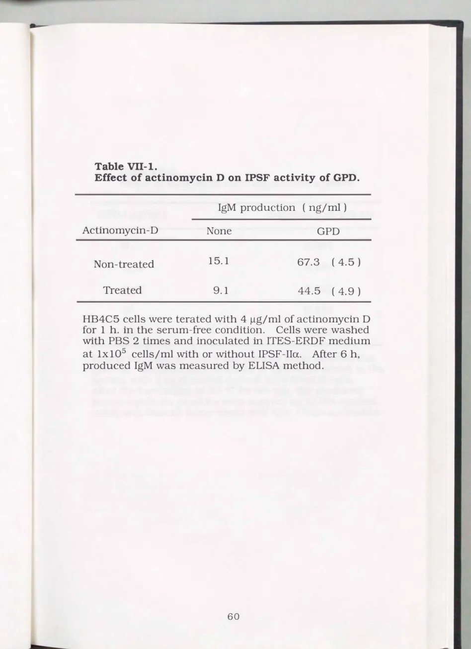
3-3. Effect of actinomycin D treatment of hybridomas on IPSF activity
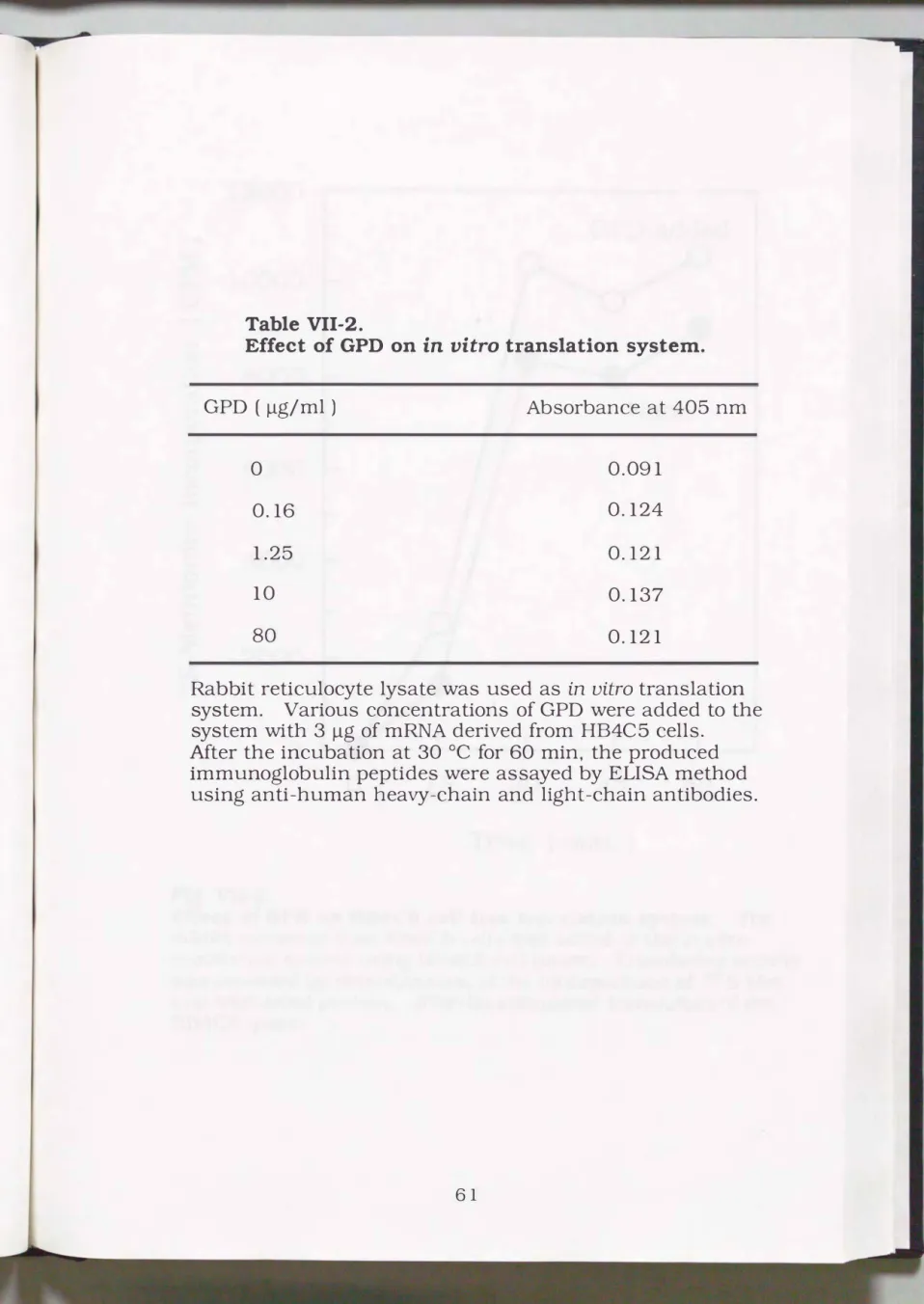
3-4. Effect of GPD on cell-free translation system 3-5. Association of GPD on HB4C5 cells
3-6. Effect of IPSF-II a to the IgM specific mRNA level in HB4C5 cells
4. Discussion 5. Conclusion
CHAPTER VIII PURIFICATION OF IMMUNOGLOBULIN
45 45 46 46 46 49 51
52 52 52 52 53 53 53 54 54 54 55
55 56 56
56 65 69
PROSUCTION STIMULATING FACTOR-lip 70
1. Introduction 70
2. Materials an d me thods 70
2-1. Cells and cell culture 70
2-2. Assay of IPSF activity 71
2-3. Purification of IPSF-IIP from Namalwa cell lysate 71
3. Results 71
3-1. Hydrophobic interaction chromatography of Namalwa cell
lysate 71
111
3-2.
Anion-exchange chromatography of IPSF-IIP fraction
obtained from hydrophobic interaction column
723-3.
Gel filtration of IPSF-IIP fraction and molecular weight
analysis by SDS-PAGE
724. Discussion 77
5. Conclusion 78
CHAPTER IX IDENTIFICATION OF IPSF-II
P
1. Introduction
2. Materials and methods
2-1.
Analysis of amino acid sequence of IPSF-np
2-2.
Assay of enolase activity
3. Results
3-1.
Partial amino acid sequence of IPSF-np
3-2.
IPSF activities of IPSF-IIP and enolase
3-3.
Enolase activity of IPSF-liP
4. Discussion 5. Conclusion
CHAPTER X CHARACTERIZATION OF IPSF-II
p
1. Introduction
2. Materials and methods 2-1.
Cells and cell culture
3. Results
3-1.
Heat and pH stability of IPSF activity of IPSF-np
3-2.
Time-course of IPSF-IIP activity
3-3.
Effect of IPSF- np on MAb productions with various
79 79
79 79 80 80 80 80 84 85
86 86 86 86 87 87 87
hybridomas
874. Discussion 93
5. Conclusion 95
CHAPTER XI IPSF ACTIVITIES OF NUCLEIC ACID BINDING PROTEINS AND POLY-BASIC AMINO ACIDS 96
1. Introduction 96
2. Materials and methods 97
2-1.
Materials
972-2.
Assay of IPSF activity on hybridomas
97IV
3. Results 97 3-1. Effects of histones on IgM production of HB4C5 cells 97 3-2. Effects of protamines on IgM production of HB4C5 cells 98 3-3. Effect of lactate dehydrogenase on IgM production of
HB4C5 cells' 98
3-4. Effects of poly-basic amino acids on IgM production of
HB4C5 cells 98
4. Discussion 103
5. Conclusion 1 04
CHAPTER XU VIRUS CONTAINMENT CONT INUOUS
CULTURE OF ANIMAL CELLS 1 05
1. Introduction 105
2. Materials and methods 1 05
2-1. The BMM hollow fiber 1 05
2-2. Cell culture system 106
2-3. Cell and cell culture 106
3. Results 1 08
3-1. Semi-continuous Filtration of spent medium from high-
density continuous culture of HB4C5 cells 108 3-2. Protein recovery rate of the BMM hollow fiber
4. Discussion 5. Conclusion
CHAPTERXlli
1. Introduction
LARGE-SCALE, IDGH-DENSITY FREEZIN G OF ANIMAL CELLS AN D ITS APPLICATION TO HIGH-DENSITY CULTURE
2. Materials and methods 2-1. Materials
2-2. Cell and cell culture
2-3. Freezing and thawing of cells 2-4. High-density culture
3. Results
3-1. High-density culture of Namalwa cells
3-2. Inoculation of Namalwa cells from frozen bag 4. Discussion
5. Conclusion
108 111 112
113 113 113 113 114 114 115 115 115 115 119 121
v
CHAPTER XIV CONCLUSION
ACKNOWLEDGMENT
REFERENCE
122
127
128
Vl
CHAPTER I GENERAL INTRODUCTION
Cells in vivo produce a variety of bioactive substances to maintain body homeostasis, many of which are highly promising for use of as drugs for therapeutic or diagnostic purposes. After the establishment of recombinant DNA techniques, it was believed that these proteins could be produced by procaryotes in large quantities. Some proteins are simple enough for procaryotes to express. However, many of these bioactive proteins in mammals are more complex and need post
translational modifications such as glycosylation in the lumen of the endoplasmic reticulum or in the Golgi apparatus. Recently, apoprotein of human interferon a-2a expressed in E. coli by the recombinant DNA technique has been found to be antigenic to the human immune system. Moreover, proteins from which their glycoside chains have
been partially removed are usually very unstable when administered in vivo. Deglycosylated proteins cannot be accepted by the target cells as the receptors on the cells recognize the glycosidic residues of the protein. The receptors usually distinguish the species of those glycosylated proteins may not have sufficient in vivo activity. Of course, carbohydrates are highly polar, hydrophilic molecules. Hence, they increase the water-solubility of glycoproteins and assure that a protein region containing an attached oligosaccharide faces an aqueous medium. Thus carbohydrate units stabilize glycoproteins. For example, the oligosaccharide units of antibodies protect them from digestion by proteases. Furthermore, carbohydrates can serve as markers for the uptake and subsequent destruction of proteins, as in the removal of aging plasma glycoproteins by the liver. Glycoproteins contain oligosaccharide units linked to either asparagine side chains by N-glycosidic bands or to serine and threonine side chains by 0-
glycosidic bonds. N-linked oligosaccharides, though diverse, contain a common pentasaccharide core consisting of three mannose and two N
acetylglucosamine residues. In animal cells, these oligosaccharides are combined with the growing polypeptide chain on the lumenal side of the endoplasmic reticulum membrane. We must obtain these
bioactive glycoproteins which need complex modification with mammals. Animal cell culture is therefore the only practically available means for producing bioactive glycosylated proteins adaptable to therapeutic uses.
The antibodies are one of the glycoproteins. The origosaccharide units of antibody molecules not only protect them from digestion but also alter their reactivities (Tachibana et al.,
1992).
So, an antibody must be produced in an animalcells. Developments in the production
and application of rodent monoclonal antibodies (MAbs) in science and medicine have produced at a rapid pace since Kohler and Milstein first demonstrated that specific antibody-secreting cell lines could be obtained by the hybridization of immune mouse spleen and myeloma cells, followed by cultivation in a selective medium (Kohler et la.,1975).
This has resulted in new and improved methods for assaying, isolating and characterizing a wide exquisite probes for studying complex cellular interactions and novel reagents for the investigation and treatment of a variety of diseases. Many attempts have been made to obtain MAbs reactive to human cancer for diagnostic and therapeutic uses (Colcher et al.,1981,
Cuttitta et al.,1981
and Imai et al.,1981).
Though these mouse MAbs are useful for diagnosis of human cancers and studies on cancer specific antigens, their therapeutic use is limited because of their antigenic activity in human bodies. To overcome this problem, the use of human MAbs is preferable. Many
produce MAbs specific for various types of cancers such as breast, lung, stomach, etc.
The first serum-free medium of cultured cells for producing biopharmaceutical proteins in a large quantity was formulated in 1982 for the successful cultivation of many hybridomas to produce MAbs (Murakami et al., 1982). Using this serum-free medium, sufficient amounts of MAb produced by hybridomas came to be available in extreme purity, free from a large amount of ambiguous serum components. The serum-free medium was also modified to maintain and maximize such cellular functions as growth and productivities of MAbs (Kovar, 1988, Murakami, 1989 and Franek et al., 1991). Serum
free and protein-free media were soon applied to large scale industrial cell cultures for various biopharmaceutical productions, as these media were available in large amounts at low cost. This outcome rapidly stimulated competing constructions of industrial bioreactor systems such as serum-free or protein-free media. Serum-free culture also revolutionized the purification system of cellular products. In serum
supplemented medium, MAb concentrations of cultured medium are only one hundredth of the total proteins due to the enormous amount of serum proteins. However, in serum-free medium the ratio usually rises to only 30-40 °/o. As mentioned above, serum-free medium is useful for the production of biopharmaceutical products. However, in serum-free medium, the productivity of MAb of hybridomas is worse than that in serum-supplemented medium. We must overcome this problem of the effective production of MAb, because more MAbs are recently required. The total worldwide production of MAb in 1980 was 1 g, though in 1988 this was in the region of 50 kg or higher (Griffiths, 1990). Much more MAbs will be necessary, if MAbs are to be applied to therapeutic uses and other purposes in the future. Thus,
further sophistication of production systems for obtaining large quantities of MAbs are needed.
There are three basic technologies to produce MAb in large quantities, large scale culture, high-density culture and increase in cellular productivity. In the first two technologies production of large quantities of the protein can be accomplished by culturing large number of cells. Consistent with this idea, various types of large scale bioreactors using stirred, airlift, spin filter, encapsulation and porous beads systems which hold 100-10,000 L medium are in operation in several countries. High-density culture systems can also culture a
large number of cells and make it possible to use smaller bioreactors to obtain a required amount of MAb, which diminishes the cost of investment for the construction of production facilities. However, a greater advantage of high-density culture is the lowered requirement for growth and regulatory factors in the serum-free medium (Takazawa et al., 1988 and Yoshioka et al., 1990). This significantly reduces the cost of the medium, because growth factors are usually much more expensive than amino acids and vitamins, and also facilitates purification of MAb.
In conjunction with large scale culture, high-density culture will achieve production of large quantities of MAb to some extent, even if MAb productivities of cells are still at the conventional levels. It will obviously obtain a far greater amount of MAbs, if increased productivities of cells are introduced in the high-density culture system. But is there evidence to support the hypothesis that cells can potentially produce a large amount of protein? A plasma cell (matured B lymphocyte) in lymph nodes secretes 2000 antibody molecules/ sec (Warner, 197
4),
i. e. 1. 7x 1 os moleculesI
day, corresponding to43
pgMAb productivity. Lactoferrin and casein in milk enhanced IgM production of human-human hybridoma (Yamada et aZ., 1989b, 1990a and 1990c). Dexamethasone, synthetic glucocorticoid, stimulated IgG production of human myeloma lymphoblasts cell line (Manzer et al., 1988). Recombinant human Interleukin-6, which induces the final maturation of B cells into antibody secreting cells, enhanced IgG-type MAb productivity of mouse-mouse hybridoma line (Terada et al., 1992).
Some cellular materials which enhanced MAb productivity of human
human hybridoma were found in our laboratory. We named these factors as immunoglobulin production stimulating factor (IPSF). IPSF was first found together with some inhibiting factors in the cell extracts of human lung adenocarcinoma PC-8 cells (Shinmoto et aZ., 1988). IPSF-I isolated from the cultured medium of human lymphoblastoid H0-323 was a complex protein of 410 KD (Toyoda et al., 1990). IPSF-II was found in human Burkitt's lymphoma Namalwa cells. In this thesis, IPSF-Ils were purified, characterized and identified from Namalwa cell lysate. The modes of action of IPSF-Ila, one of the IPSF-Ils, was also examined. Some attempts were also made to efficiently perform a large-scale culture.
CHAPTER II
1. Introduction
IMMUNOGLOBULIN PRODUCTION
STIMULATING FACTOR-II IN NAMALWA CELLS
Since mouse-mouse hybridomas secreting monoclonal antibody (MAb) was produced (Kohler et al.,
1975),
many attempts have been made to obtain MAbs reactive to human cancer for diagnostic and therapeutic usages (Colcher et al.,1981,
Cuttitta et al.,1981
and Imaiet al.,
1981).
These mouse MAbs are useful for diagnosis of human cancers and studies on cancer specific antigens. However, their therapeutic use is limited because of their antigenic activity in human bodies. To overcome this problem, the use of human MAb is preferable.To produce human-human hybridomas efficiently, an excellent fusion partner is important. Thus, many human fusion partners were established by Murakami and his coworkers (Murakami et al.,
1982,
Ohashi et al.,1986,
Kawahara et al.,1990
and Kawahara et al.,1992).
Most hybridomas derived from the fusion partners proliferate and secrete immunoglobulin in serum-free medium, as well as in serum
supplemented medium (Aihara ei al.,
1988,
Hashizume et al.,1986,
Murakami et al.,1985
and Murakami et al.,1987).
These hybridomas can be grown in serum-free, high density suspension cultures, and large amounts of MAb are recovered easily from the serum-free medium. However, immunoglobulin productivities of these humanhuman hybridomas are still lower than those of mouse-mouse hybridomas, and larger amounts of MAb are necessary for clinical
which were screened for immunoglobulin productivity of human
human hybridomas in serum-free cultures, were found in various cells.
IPSF was first found together with some inhibiting factors in the cell extracts of human lung adenocarcinoma PC-8 cells (Shinmoto et al., 1988). The other IPSF, named IPSF-I, was isolated from a human lymphoblastoid H0-323 cells. In this chapter, we mentioned about the third IPSF, IPSF-II, derived from human Burkitt's lymphoma Namalwa cells.
2. Materials and methods
2-1. Cells and cell culture
Namalwa cells are human lymphoblastoid cells derived from Burkitt's lymphoma. Namalwa cells were maintained in ERDF medium supplemented with 5 °/o feral bovine serum (FBS). Human-human hybridoma HB4C5 cells were fusion products of human lymphocytes and a human fusion partner NAT-30 cells, derived from Namalwa cells.
HB4C5 cells produce human lung cancer specific MAb of IgM class.
HB4C5 cells were maintained serum-free ERDF medium supplemented with 10 �g/ml insulin, 35 �g/ml transferrin, 10 �M ethanolamine and 2.5 nM sodium selenite (ITES) (Murakami et al., 1982).
2-2. Preparation of cultured supernatant and lysate of Namalwa cells
Namalwa cells were inoculated into ITES-ERDF medium at 1x10⁶ cells/ml, and then incubated for 3 days. The cultured supernatant was isolated by centrifugation at 400 x g for 5 min. The cell pellet obtained was suspended in a phosphate buffered saline (PBS) and sonicated at 1.1x1 06 cells/mi. The lysate was centrifuged at 6000 x g for 10 min to remove insoluble materials and filtered through a 0.22
J.lffi
nitrocellulose filter for sterilization. The protein content of Namalwa cell lysate thus obtained was 110 J.lg/ml.
2-3.
Determination of
IPSFactivity
Hybridomas were inoculated into ITES-ERDF medium supplemented with cultured medium or Namalwa cell lysate and cultured for 3 days. Cultured medium of the hybridomas were recovered by centrifugation at 400 x g for 5 min, and immunoglobulin content in the supernatant was determined by enzyme-linked immunosorbent assay (ELISA). To determine human IgM contents in HB4C5 cell supernatant, 96- well immunoplate (NUNC, Denmark) was coated with goat F(ab)2 anti-human IgM suspended in
1 °/obovine serum albumin (BSA), reacted with cultured medium of hybridoma for
1
h at 37 °C, reacted with peroxidase conjugate of goat F(ab)2 anti
human IgM for
1h at 37 oc , and then reacted with substrate solution containing 0.003
°/oH202, 0.1 M citrate buffer (pH 4.0) and 0.3 mg/ml p-
2, 2'azino-di- (3-ethylbenzothiazoline- 6- sulfonic acid) diammoni urn salt.
2-4.
Fractionation of Namalwa cell lysate using salting out
Ammonium sulfate was added to the ice-cold Namalwa cell lysate
up to 50
o/osaturation. Leaving overnight at 4 °C, the solution was
centrifuged at 8000 x g for 20 min to separate precipitation. The
ammonium sulfate concentration of the obtained supernatant was rised
to 80
°/osaturation for further fractionation. After separation of
precipitate and supernatant by centrifugation, the obtained 4 fractions,
precipitate and supernatant of 50
°/osaturation and 80
°/osaturation
respectively, were then dialyzed against 10 mM sodium phosphate
3. Results
3-1. Effect ofNamalwa cell culture supernatant and lysate on IgM
production of HB4C5 cells
Immunoglobulin production stimulating activity of the supernatant of Namalwa cells became stronger when the cells grew to saturation density and their viability had begun to decrease (Data not shown).
This suggested that larger amounts of IPSF were present in Namalwa cells. Thus the IPSF activity of Namalwa cell lysate was examined. As shown in Table Il-l, Namalwa cell supernatant stimulated IgM production of HB4C5 cells. The stimulatory effect of Namalwa cell lysate on IgM production was much higher than that of the supernatant
(about 15-fold increase of
IgMproductivity).
3-2. Fractionation of Namalwa cell lysate by salting out using ammonium sulfate
Namalwa cell lysate was fractionized by salting out using ammonium sulfate at 4 °C. IPSF active substance was concentrated in supernatant of 50 °/o saturation (Table II-2}. When the concentration of ammonium sulfate of the supernatant was raised to 80 °/o saturation, IPSF was mainly collected in the supernatant fraction. About 70 °/o of proteins contained in Namalwa lysate were precipitated by 50 °/o saturation of ammonium sulfate. And then the supernatant of 80 °/o saturation contained only 20 °/o of total protein content. These results suggest that the purity of IPSF-II in supernatant of 80 °/o saturation was 5 times higher than that in Namalwa cell lysate.
Table 11-1.
IPSF activity in cultured supernatant and lysate of Namalwa cells.
ITES-ERDF
Cultured supernatant
PBS
Lysate
I
gM ( ng/ml)
30.9 99.0 31.0 1017.0
Table 11-2.
Relative IPSF activity of Namalwa cell lysate fractionized by ammonium sulfate precipitaion.
Ammonium Sulfate Cone.
(Ofo
saturation)0 0-50 50-80
Relative IPSF activity
Precipitate Supernatant
1
0.2 1.7
1.3 4.4
4. Discussion
Immumoglobulin production stimulating factor was searched to enhance immunoglobulin productivity of human-human hybridomas in serum-free cultures. Cell lysate of human lymphoblastoid Namalwa cells stimulated IgM production of human-human hybridoma HB4C5 cells derived from NAT-30 cells. a human fusion partner screened from Namalwa cells. IPSF-II derived from Namalwa cells were not secreted in cultured medium, but contained in the cells. The IPSF-II solution obtained by homogenation of Namalwa cells was fractionized with salting out using ammonium sulfate. As the result of that, IPSF-II was concentrated in supernatant of 80 °/o saturation and the purity was enhanced 4.5-fold higher than that of Namalwa cell lysate. It was
suggested that this salting out was useful for fractionantion of IPSF-II previous to purification using liquid chromatography.
In consideration of recovery rate of IPSF-II, Namalwa cell lysate should be pre-fractionized by 50 °/o saturation of ammonium sulfate prior to liquid chromatography.
5. Conclusion
IPSF-II was mainly contained in Namalwa cells and not secreted in the culture medium. So, we must purify IPSF-II from Namalwa cell lysate which contain a large quantity of various proteins in comparison with cultured medium.
Namalwa cell lysate must be pre-fractionized before purification using liquid chromatography for the reason mentioned above. Then the salting out with ammonium sulfate was applied to the cell lysate.
IPSF-II was mainly fractionized in supernatant of 80 °/o saturation.
Though the recovery rate of IPSF- II was dropped at that condition, the per-fractionation of the lysate would be performed at 50 °/o saturation.
CHAPTER
ill
PURIFICATION OF IMMUN OGLOBULIN PROD UCTION STIMULATING FACTOR -II a (IPSF-IIa) DERIVED FROMNAMALWA
CELLS
1. Introduction
Immunoglobulin production stimulating factor (IPSF) was found in cell lysate of human Burkitt's lymphoma Namalwa cells. IPSF derived from Namalwa cells named IPSF-II stimulated monoclonal antibody (MAb) production of human-human hybridoma HB4C5 cells. The cell lysate was fractionized by salting out using 50 °/o and 80 °/o saturation of ammonium sulfate as mentioned in
CHAPTER
II. IPSF-II was concentrated in the supernatant of 80 °/o saturation 4.5-fold than the Namalwa cell lysate. However, to purify IPSF, the supernatant of 50 °/o saturation was applied to liquid chromatography because of yield of IPSF-II. As the result of the choice of separation mode of liquid chromatography, hydrophobic interaction chromatography was most suitable. There were a few reasons for choice of the separation mode.One was that the supernatant of 50 °/o saturation could be applied to the column without dialysis. Another reason was that IPSF-II adsorbed the hydrophobic interaction column, even though a half of the proteins contained in the supernatant could not adsorb the column. The hydrophobic interaction column could easily enhance the purity of IPSF-II.
In this chapter, IPSF-II was purified by the serial use of ammonium sulfate fractionation, hydrophobic interaction chromatography and gel filtration, and estimated its molecular mass.
2. Materials and methods
2-1. Cells and cell culture
Namalwa cells. derived from human Burkitt's lymphoma, were cultured to prepare cell lysate at a high density ( 2 x 10⁷ cells/ml ) in ERDF medium (Kyokuto Pharmacy Industrial Co.) supplemented with 20/o fetal bovine serum (FBS) and ITES (insulin, 10 J..lg/ml; transferrin.
20 J..lg/ml; ethanolamine, 20 J..lM; sodium selenite, 25 nM) using a high density culture system (Shimadzu. SHC-1 type). To assay IPSF activity, a human-human hybridoma cell line. HB4C5 cells were used. HB4C5 cells. which produce human lung cancer specific MAb of IgM class, were fusion products of human lymphocytes and a human fusion partner NAT-30 cells (Murakami
et al.. 1985). HB4C5
cells were maintained in ITES-ERDF medium to assay of IPSF activity.2-2. Assay of IPSF activity on hybrldomas
IPSF activity was examined by measuring amounts of antibodies secreted by hybridomas in the medium by ELISA method as described previously (CHAPTER II). HB4C5 cells
(l
x 105 cells/ml) suspended in ITES-ERDF medium were inoculated in 96 well plate in the presence of IPSF-II for 6 h. After 6 hours. antibody concentration of cultured medium was determined.2-3. Purification of IPSF-IIa from Namalwa cells
All the procedures are described in the Results section.
2-4. Polyacrylamide gel electrophoresis of IPSF-IIa
The purified IPSF-II was electrophoresed using a So/o polyacrylamide gel as a running gel in non-denaturation condition as
described previously (Toyoda et al.,
1990,
Davis,1964).
Briefly, samples were dissolved in the buffer (62.5 mM Tris-HCl, pH 6.8) containing10 °/o
glycerol and electrophoresed in the running buffer (25 mM Tris -190
mM glycine, pH 8.3) for about40
min at 25 mA current. Molecular weight of the IPSF was determined by SDS-polyacrylamide gel electrophoresis. Samples were dissolved in the buffer (62.5 mM Tris-HCl, pH
6.8)
containing10 °/o
glycerol, 2°/o
SDS and with or without 5°/o
2-mercaptoethanol. Mter boiling for4
min, the samples were electrophoresed in the running buffer (25 mM Tris-190
mM glycine-2°/o
SDS, pH8.3)
with SDS-polyacrylamide gel containing12 °/o
acrylamide (Laemmli,1970).
Mter electrophoresis, proteins in the gel were fixed by 20°/o
trichloroacetic acid for more than 1 h and visualized by silver-staining (Merril,1981).
3. Results
3-1.
Hydrophobic interaction chromatography of Namalwa cell lysate
Namalwa cells
(109
cells) were ultrasonically homogenized in10
mM sodium phosphate buffer (pH 7.4). Ammonium sulfate was added to the cell lysate to 50 °/o
saturation at 4 °C. Mter standing at4
°C overnight, the precipitate was separated from the supernatant by centrifugation.The supernatant was further purified by hydrophobic interaction column chromatography. A hydrophobic interaction column (BU1YL TOYOPEARL 650M, 22 mm x 25 em, TOSOH) was equilibrated with
10
mM sodium phosphate buffer (pH7.4)
containing 2 M ammonium sulfate. The supernatant fraction obtained by a 50 °/o
ammonium sulfate saturation was applied to the column and eluted with the10
mMadsorbed substances were eluted with linear gradient of ammonium sulfate from 2 to 0 M in 10 mM sodium phosphate buffer (pH 7.4).
IPSF activities were detected in the protein fractions eluted broadly at the concentration of ammonium sulfate less than about 1 M (Fig. III-1). Since the IPSF activities were broadly distributed, active fractions were grouped into A, B and C and fraction A was further purified here.
3-2.
Gel filtration of Fraction
Aobtained by hydrophobic interaction column chromatography
Fraction A obtained by hydrophobic interaction column chromatography was gel-filtrated with TSKgel G3000SW (7.5 mm I.D. x 60 em;
TOSOH).
After equilibration with 200 mM sodium phosphate buffer (pH 6.0), the IPSF fraction was applied to the column. The fractionized solution was dialyzed against 10 mM sodium phosphate buffer (pH 7.4) and IPSF activity was assayed. Although two peaks were detected by measuring absorbance at 280 nm (Fig. III-2A), strong IPSF activity was shown in a first sharp single peak (Fig. III-2B). From the standard curve of molecular weight the molecular size of the IPSF was estimated at about 110 KD. The IPSF was named IPSF-IIa.IPSF-Ila purified by gel filtration was electrophoresed on a polyacrylamide gel. A single band was detected in non-denaturation condition, showing that the protein was highly purified (Fig. III -2C).
3-3.
Analysis of composition of IPSF-IIa
IPSF-II a purified by gel filtration was analyzed for its subunit structure. The SDS-polyacrylamide gel electrophoresis of the IPSF-Ila revealed two bands of 40 KD and 36 KD irrespective to the presence of 2-mercaptoethanol (Fig. III -3A). This indicated that these subunits
were not linked with disulfide bonding and that each subunit were composed of a single peptide. It was shown by densitometry that the ratio of 40 KD to 36 KD bands was 1 to 2, taking the molecular size of IPSF-Ila ca. 110 KD into account (Fig. III-3B). These results suggest that IPSF-Ila is a 112 KD protein which has a subunit structure composed of a 40 KD polypeptide chain and two 36 KD polypeptide chains as shown in Fig. III-3C.
80
70
l
60
8
' 50 'oJ)� 40 C) � 0 30 u
:;E
� -'oJ) 20
c.o
10
0
1.0
0.8
............................................
0
§
00 N 0.6
1-J ('lj
···
···
···
···
I·
A·I·
B·I·
c ..1
···
···
2
C1) C)
... 0
-8 §
0 0.4(fJ
�
0.2
0
10 20 5 10 15 20 25 30 35
Fraction Number
Fig. DI-1.
Purification of IPSF-D from cell lysate using hydrophobic interaction column (BUTYL
TOYOPEARL
650M).
A 50°/o ammonium sulfate saturated supernatant from Namalwa cell lysate was applied to the column. Adsorbed materials were eluted with 300 ml of linear gradient of ammonium sulfate. Each fraction (10 ml) was dialysed against 10 mM sodium phosphate buffer (pH 7 .4) and IPSF activity was assayed. Solid line and dotted line indicate UV absorbance at 280nm and ammonium sulfate concentration, respectively. Open circles40
�
C) � u 0
1-J C1)
� "3
rJ)
s
;:j ....j
�0.04
A c
110
KDs �
0 0 00 C'l
.+-J
cd
0.02
Q) C,.)
-e §
0r.n
�
Davis-PAGE
0
30 B
�
...'o[)
£ 20
C) 0 0 u
:E
'o[)10
-
0
0 15 20 25 30
Elution Volume
(ml) Fig.
111-2.Gel Filtration of IPSF
-IIapurified by a hydrophobic interaction column using TSKgel G3000SW.
One ml of the fraction A purified by a hydrophobic interaction column was applied to the column and eluted with200
rnM sodium phosphate buffer (pH6.0).
A, elution pattern of UV-absorbent materials; B, IPSF activity; C,5°/o
polyacrylamide gel electrophoresis of IPSF-II a in a fraction exhibiting maximum IPSF activity in a Davis' non-denaturation condition.
N
1--'
40 KD---..
36 KD---..
Fig.
111-3.A B c
SDS-polyacrylamide gel electrophoresis of IPSF-IIa. IPSF-Ila purified by gel filtration was electrophoresed on a
SDS-polyacrylamide gel containing
12°/oacrylamide. After
silver-staining, the density of each band was determined by
densitometry. (A) SDS-polyacrylamide gel electrophoregram of
IPSF-Ila. (B) a model ofiPSF-Ila.
4. Discussion
Low immunoglobulin productivity of human-human hybridomas in serum-free medium compared to that in serum-supplemented medium prompted us to screen immunoglobulin production stimulating factors.
Cellular IPSF was first found together with some inhibiting factors in the cell extracts of human lung adenocarcinoma PC-8 cells (Shinmoto
et al.,1988). IPSF-I isolated from a human lymphoblastoid
H0-323cells was a complex protein of 410
KD(Toyoda
et al.,1990). In this chapter, IPSF-Ila from Namalwa cell lysate was completely purified by usage of liquid chromatography. The IPSF-Ila was estimated as a 112 KD protein composed of a 40
KDand two
36 KDsubunits by gel filtration and SDS polyacrylamide gel electrophoresis. The structure of IPSF-II a was very simple compared to that of IPSF-I which had
3isomers and many subunits. The
36 KDsubunit exclusively showed IPSF activity. Since 40 and
36 KDsubunits were dissociated by SDS gel electrophoresis without treatment with such a reducing agent as
2-mercaptoethanol to break disulfide bonds, 40 and
36 KDsubunits may be associated by non-covalent bonds. Both 40 and
36 KDsubunits were composed of single peptides, which had no intramolecular disulfide bondings.
It is necessary to investigate which subunit has IPSF activity.
Characterization and identification of IPSF-II a are also necessary for
discussing IPSF activity of IPSF-II a. Furthermore, there are some
evidences of the existence of another IPSF, IPSF-II�. IPSF-II� must be
purified and identified.
5. Conclusion
In this chapter, IPSF-Ila from Namalwa cells was completely purified from supernatant of 50 °/o saturation of ammonium sulfate.
IPSF-Ila was estimated as a 112 KD protein composed of a 40 KD and two 36 KD subunits.
As the result of liquid chromatography of Namalwa cell lysate, the existence of another IPSF was supposed. Purification and identification of another IPSF, IPSF-II�, were mentioned in CHAPTER VIII and IX.
CHAPTER IV CHARACTERIZATION OF IPSF-IIa
1. Introduction
The substances enhanced immunoglobulin production of hybridomas were found in various cells. The substance was named immunoglobulin production stimulating factor (IPS F). IPSF found in Namalwa cell lysate was named IPSF-11 and the IPSF-11 contained a few IPSFs. IPSF-IIcx, one of IPSF-IIs, was purified from Namalwa cell lysate by ammonium sulfate precipitation, hydrophobic interaction chromatography and gel filtration. IPSF-IIcx was a 112 KD protein composed of a 40 KD and two 36 KD subunits by gel filtration and SDS
PAGE as mentioned CHAPTER III.
In this chapter, various features of IPSF-IIcx were investigated.
Heat and pH stabilities of IPSF-IIcx were examined. The effect of IPSF
IIcx on monoclonal antibody (MAb) productions of various human-human and mouse-mouse hybridomas. Moreover, the time-course effect of IPSF-IIcx on cell growth, antibody production and antibody productivity of hybridoma HB4C5 was examined. These biochemical and physicochemical features of IPSF-IIcx may contribute to investigation of the identification of the peptide and the mechanisms of the stimulation.
2. Materials and methods
2-1. Cells and cell culture
Human-human hybridomas tested here were fusion products of regional lymphocytes from cancer patients and human malignant fusion
HF10B4. H15F1 cells were obtained by fusion with a fusion partner H0-323 cells (Ohashi et al., 1986). HB4C5 and HF10B4 cells were derived from another fusion partner NAT-30 (Murakami et al., 1985).
IgG-producing human-human hybridomas were K-1.5, HB731 and HB732. K-1.5 cells were derived from H0-323 cells. HB731 and HB732 cells were obtained by fusion with a new fusion partner HK- 128, which was recently established for making IgG producing hybridomas (Kawahara et al., 1990). Among mouse-mouse hybridomas, F5 and D1 cells were IgM producers, 4F12 and 4C10B6 were IgG producers. All mouse-mouse hybridomas used here were derived from a fusion partner, P3-X63 -Ag8 (P3U 1) cells. These cells were maintained in ERDF medium supplemented with 5 °/o FBS at 37 oc under humidified 5 °/o C02/95 °/o air.
2-2. Assay of IPSF activity
The methods explained in CHAPTER II and III in detail.
3. Results
3-1. Temperature and pH stabilities of IPSF-IIa
IPSF -Ila purified by hydrophobic interaction column was incubated in 10 mM phosphate buffer (pH 7 .4) at varying temperatures for 30 min. After cooling, the IPSF activities of the samples were assayed immediately. As shown in Fig. IV-1, IPSF-Ila lost its activity at the temperatures higher than 60 °C.
For analysis of pH stability, IPSF-Ila was incubated for 20 h at 4 oc in buffer solutions of various pH. IPSF-Ila was fairly stable in such alkaline conditions as pH 13, as indicated in Fig. IV-2. It was unstable in acidic solution and the activity was completely lost below pH 4.
3-2. Enhancement of IgM productivity of HB4C5 cells by IPSF-IIa The time-course of the IPSF-II a activity was examined on the IgM production of HB4C5 in serum-free medium. The hybridoma cells
immediately started to enhance IgM production by the addition of 25 j.lg/ml IPSF-Ila (Fig. IV-3A). After 72 h, the IgM deposited in the IPSF-Ila containing medium attained to 2.6 llg/ml which was more than 20-fold higher than that produced without the IPSF-Ila. During the cultivation, IPSF-II a showed no growth stimulating activity to the hybridoma cells (Fig. IV -3B). This clearly showed that the IPSF-II a increased the cellular productivity of IgM. The IgM productivity per 105 cells was stimulated more than 20-fold by IPSF-Ila and the effect was maintained during 72 h (Fig. IV-3C), though the IgM productivities of the hybridoma cells with or without IPSF-II a were varied during the culture period.
3-3. Dose response of IPSF-IIa
Dose-response curves of IPSF-II a activity on the IgM productivity of HB4C5 cells were also assessed. As shown in Fig. IV-4, IPSF-Ila stimulated the IgM production of HB4C5 cells dose-dependently.
Productivity was maximized up to 20-fold by addition of 25 llg/ml of this protein. Acceleration of the cellular growth was not detected even by the addition of elevated concentrations of IPSF-Ila.
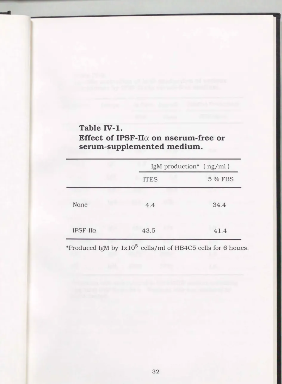
3-4. Effect of serum on IPSF activity of IPSF-IIa
Effect of serum on the IPSF-Ila activity was examined. HB4C5 cells (1 x 10⁵ cells/ml) were inoculated in ITES-ERDF medium or in 5 o/o FBS supplemented medium with or without 10 llg/ml of IPSF-Ila.
HB4C5 cells secreted 34.4 ng/ml of IgM in serum supplemented
secreted was only 4.4 ng/ml in serum-free medium. In the presence of IPSF-IIa, the hybridomas secreted 43.5 ng/ml of IgM in serum-free medium. IPSF-IIa enhanced IgM productivity of the cells in serum
free medium as much as in serum supplemented medium. Whereas, HB4C5 cells secreted 41.4 ng/ml of IgM in serum supplemented medium containing IPSF-II a. The enhancing effect of IPSF-II a on IgM production of hybridomas was largely masked in serum supplemented medium.
3-5. Specific stimulation of IgM productivity of hybrldomas by IPSF
IIa in serum-free medium
The activity of IPSF-IIa (5 �g/ml ) was examined for various hybridomas derived from both human and mouse cells. IPSF-Ila enhanced IgM productivity of hybridomas derived from both human and mouse cells (Table IV-2). Hl5Fl cells, a human-human hybridoma derived from H0-323 cells showed a 15 fold-enhanced IgM productivity by addition of IPSF-II a. IgM productivities of HB4C5 and HF10B4 cells derived from NAT-30 cells were enhanced 4- and 8-fold by the IPSF-Ila, respectively. IPSF-Ila enhanced IgM productivity of mouse-mouse hybridomas ca. 2 fold. However, IPSF-Ila enhanced the IgG productivity of neither human-human nor mouse-mouse hybridomas in the experimental condition.
100
80
-
:::R
0.__
0 60
·s:
:p�
(l).2::
-+-J
� ro
& 40
20
0
0 20 40 60 80 100
Temperature ( °C)
Fig. IV-1.
Heat-stabiliy of IPSF-IIa. The purified IPSF-Ila was incubated in 10 mM phosphate buffer (pH 7.4) at
varying temperature for 30 min. After cooling to 0 °C,
IPSF activity was immediately assayed with HB4C5
cells. Relative activity was calculated by dividing the
activity of IPSF-Ila treated at each temperature by the
control activity of IPSF-Ila treated at 0 °C.
,._
�
0 ..__.0
·�>
:;j
�
a).?:;
-+-J
...o4 rn
&
100
80
60
40
20
0
2 4
Fig. IV-2.
6 8
pH
10 12 14
PH sensitivity of IPSF-IIa. The purified IPSF-IIa
was incubated at indicated pH for 20 h at 4 °C.
Mter dialysis against 10 mM sodium phosphate
buffer (pH 7.4), IPSF activity was assayed. The
results were indicated as relative activities.
s
...1x106
::::::l rJJ
Q,)
5x1d
CJ
b 'Cii
c::
1x1d
Cl Q,) ... 3 v u
2
a
...�
c:: 0 :.::1 u :::1
"'0 0 1-o
0..
§
1
0
Fig.
IV-3..c
<D
?
500v
u 100
1./l 0 ...
... 'o.() 10
c
0 1.0
�
u::l 0.1
"0 0 1-.
0..
::E
'o.()
� �
� �
f- 0 0
f-
I I I I I I I
0 12 24 48 72
Time (hour)
0
61
2 18 24 48Time (hour)
A
72
Time-course of IPSF-IIa activity on the IgM productivity of
HB4C5cells.
HB4C5 cells were incubated with 25 11-g/ml of the purified IPSF-Ila in
ITES-ERDF medium and IgM concentration in the medium was assayed. A shows the accumulation of IgM. B indicates the growth curve of HB4C5 cells in the presence or absence of IPSF-IIa. C shows the IgM productivity per l 0 5 cells for 6 h.
0;
medium containing no IPSF-Ila, �;medium containing IPSF-Ila.50
40
a
...30
'of)
�
C) � u 0 20
� 'of)
�
10
0
Fig. IV-4.
0 0.5 1 5 10 50 100
Protein Cone. ( )lg/ml )
Effect of IPSF-Ila. concentration on the stimulation of IgM production of HB4C5 cells in serum-free medium. HB4C5 cells (1 x 10 5 cells/ml) were cultured for 6 h in ITES-ERDF
medium containing various amounts of the purified IPSF -Ila. and IgM in the medium was assayed.
Table IV-1.
Effect of IPSF-IIa on nserum-free or serum-supplemented medium.
IgM production* ( ng/ml)
ITES 5
°/oFBS
None 4.4 34.4
IPSF-Ila 43.5 41.4
*Produced IgM by
lxl05cells/ml of HB4C5 cells for
6houes.
Table IV-2.
Specific activation of MAb production of various hybridomas by IPSF-IIa in serum-free medium.
Hybridoma lsotype Ig Cone. (ng/rnl) Relative Productivity
IPSF None IPSF/None
Human
HB731 IgG 14.5 17.6 0.8
HB732 IgG 10.7 11.9 0.9
K-1-5 IgG 8.3 7.9 1.1
Hl5Fl IgM 21.4 1.4 15.3
HB4C5 IgM 205 55.3 3.7
HF10B4 IgM 823 103 8.0
Mouse
4F12 IgG 454 474 1.0
4C10B6 IgG 503 515 1.0
F5 IgM 6270 3950 1.6
D2 IgM 6810 3730 1.8
Hybridoma cells were cultured in ITES-ERDF medium containing 5 �g/ml of IPSF-IIa for 24 h. Produced MAb was measured by ELISA method.
4. Discussion
Although IgM productivity of human-human hybridomas was much lower in serum-free medium than in serum-supplemented medium, IPSF-Ila enhanced IgM productivity of hybridomas in serum-free medium as much as in serum supplemented medium. IPSF-Ila did not remarkably enhance IgM productivity of hybridomas in serum
supplemented medium. A maximum enhancement of IgM productivity of HB4C5 cells were observed by the addition of 25 j.lg/ml of this protein. IPSF-Ila immediately started to enhance immunoglobulin productivity after its addition to medium and deposition of MAb was increased up to 72 h cultivation. After 72 h cultivation, MAb production with IPSF-Ila ceased due to the deficiencies of nutrients in the medium. IPSF-II a showed no growth stimulating activity to the hybridoma cells. These results indicate that IPSF-II a increased cellular productivity of the MAb. This is clearly shown in Fig. III-7C, in which cellular MAb productivity per unit time is elevated 20-fold by addition of IPSF-Ila for more than 72 h. Cellular MAb productivities with or without IPSF-Ila gradually increased up to 24 h cultivation, though the reason was still ambiguous. The decrease in IgM
productivities after 24 h may be due to the deficiency of nutrients and accumulation of inhibitory metabolites for the protein production, such as cytotoxins.
Although IPSF-Ila stimulated IgM productivities of various kinds of
hybridomas derived from human and mouse in serum-free medium about 4-15 and 2-fold, respectively, no stimulation of IgG production of hybridomas by IPSF-II a was observed. Similar result had been obtained in the case of IPSF-I. A possibility still remains that IPSF-Ila
may enhance IgG production of hybridomas in other experimental conditions.
Since IPSF-II a showed no growth stimulating activity, it will be
advantageous to add it to a medium for production of MAb in stationary phase in large scale continuous culture. MAb production of some hybridomas is enhanced by the addition of phosphate (Sato et al.,
1989),
polyethylenglycol (Shintani et al.,1988)
and glutamine (Geaugey et al.,1989),
thus, there is a possibility that MAb productivity of hybridomas may be more enhanced using IPSF-II a together with these factors. However, it is obviously difficult to obtain enough amount of IPSF-Ila from Namalwa cells to be used for large scale culture of hybridomas. There should be two practical ways to use IPSF-Ilcx for enhancing MAb productivity. One is to obtain a large amount of IPSFIlcx by bacteria in which the IPSF gene is introduced. Another is to transfer the gene of IPSF- II a directly into hybridoma cells so that exogenous IPSF-II a need not to be supplied.
5. Conclusion
IPSF-Ila enhanced IgM production of human-human hybridoma HB4C5 cells up to 20-fold by addition of 25 ).lg/ml.
IPSF-Ila specifically affected on IgM production of human-human
and mouse-mouse hybridomas. This result supposes that there are differences in regulation of immunoglobulin production between IgM and IgG in hybridomas. There are differences of the effect of IPSF-II a between hybridoma clones.
IPSF- II a was stable in alkaline condition but unstable in acidic condition below pH 4. IPSF-II a was lost its IPSF activity at temperatures higher than 60 °C.
IPSF-II a enhanced the IgM production of HB4C5 cells as much as
in serum supplemented medium. Even in serum supplemented condition, IPSF-Ila ba rely showed enhancing activity of IgM production.
CHAPTERV
1. Introduction
IDENTIFICATION OF ACTIVE SUBUNIT PEPTIDE OF IPSF-IIcx
IPSF-II a was completely purified from Namalwa cell lysate as mentioned
CHAPTER III.IPSF-Ila was 1 12
KDprotein composed of a 40
KDand two 36
KDsubunits. Which peptide has IPSF activity 40
KDpeptide or 36
KDpeptide? To answer this question, each peptide was extracted from polyacrylamide gel after SDS-PAGE and examined its IPSF activity.
In this chapter, identification of IPSF-Ila active subunit and amino acid sequence analysis of the subunit were performed.
2. Materials and methods
2-1. Identification of active subunit of IPSF-IIa
Subunit proteins of IPSF-II a were extracted electrophoretically from the SDS-polyacrylamide gels by the method of Hashizume
et al.( 1984 ). The scheme was illustrated in Fig. V-1. The electrophoretical extraction performed at 300 volt for 3.5 h. After
extraction, SDS was removed from the extract using Dowex 1-X2 gel (Weber
et al.,197 1) to assess the IPSF activity of each subunit protein.
2-2. Analysis of amino acid sequence of N-terminus of IPSF-IIa
IPSF-II a was electrophoresed according to Laemmli ( 1970). After
electrophoresis, the IPSF-II a was electroblotted onto PVDF membrane
from polyacrylamide gel and stained with coomassie blue (Matsudaira,
1987). The 36
KDband was cut out with a razor. The membrane was
centered on the teflon seal and placed in the cartridge block of the sequencer. Sequencer used here was the Applied biosystems model 4 77 A protein sequencer.
3. Results
3-1.
Identification of active subunit of IPSF-IIa
After SDS-polyacrylamide gel electrophoresis, proteins in each band were extracted from the polyacrylamide gel and IPSF activity of each IPSF-Ila subunit was examined (Table V-1). The 36 KD subunit exclusively showed IPSF activity. The 40 KD protein showed neither IPSF activity nor the synergistic effect for IPSF activity of 36 KD subunit. Thus, the 36 KD subunit protein was responsible for IPSF activity of IPSF-II a.
3-2.
N-tenninal amino acid sequence of
36 KDsubunit of IPSF-IIa
The sequence of twenty amino acids from N-terminus of 36 KD subunit of IPSF-Ila was shown in Fig.V-2. The sequence was highly homologous to glyceraldehyde-3-phosphate dehydrogenase (GPD; EC 1.2.1.12) from various origins which is a key enzyme in glycolytic pathway, representing the single oxidative step in that fundamental pathway. With regard to the amino acids sequence, IPSF-Ila was completely homologous to GPD from human liver. In human muscle GPD, only asparagine-8 (8-N) of IPSF-Ila was substituted for aspartic acid (8-D). GPD from B.stearothermophilus revealed low homology in the sequence with IPSF-Ila.
Silicon tube
ylon mesh
: : :Cellulose
:: : : · ·
embrane
::::::::::::: .. :· ... :::::::
ffi
·+
···::• • 0 • 0 • 0 • • • 0 • • • • • • • 0 0 0 • 0 0 • • • 0 •
• • 0 • 0 • 0 • • • 0 • 0 • • 0 • 0 • 0 0 • 0 • 0 • • • •
• 0 • • 0 0 • • • • 0 • • • 0 • • • • 0 • • • • 0 • • • • •
• 0 • • • 0 • • 0 • • • • • • 0 • • • 0 • • • • • 0 • • 0 0 • • •
Fig. V-1.
2.5mMTris 1 OmM Glycine
(pH 8.5)
378mMTris 124mM HCl 10°/o Saccarose
(pH 8.5)
Apparatus of electrphoretical extraction.
Table V-1.
IPSF activity of IPSF-IIa subunits.
Control 33KD 40KD
33 KD+40 KD
IgM Cone.
(ng/ml)
6.8 21.5 9.5 18.4
Relative Productivity
1.0 3.2 1.4 2.7
IPSF activity of each subunit electrophoretically eluted from a SDS-polyacrylamide gel was assayed after removal of SDS by using Dowex1-X2 gel as described in the Materials and
methods section.
� �
IPSF-Ila Human liver
Human muscle Pig Chicken Lobster
Yeast
B. stearothermophilus
2
Fig. V-2.
1 10 20
G-K-V-K-V-G-V-N-G-F-G-R-1-G-R-L-V-T-R-A
Hom.
�-�-Y-�-Y-�-Y-N-�-E-�-R-1-�-B-�-Y-I-B-A lOOo/o
2 10 20
�-�-Y-�-Y-�-y-o-�-E-�-B-1-�-B-�-Y-I-B-A 95.0o/o
10 20
Y-�-Y-�-y-o-�-E-�-B-1-�-B-�-Y-I-B-A 94.4o/o
1 10 18
Y-K-Y-�-Y-N-�-�-�-E-I-�-E-L-Y-�-E-A lOOo/o
10 18
S-K-I-�-I -D-.G-I:-�-E-.I-.G-E-L-Y-L-E-A 76.5 °/o
1 10 18
I-R-I-A-I -N-�-�-�-B-I.-�-E-L-Y-L-B 66.7 °/o
2 10 18
Y-�-Y-�-I-H-G-�-�-B-1-G-B-N-Y-F-B-A 83.3o/o
10 19
Partial amino acid sequence of IPSF-IIa and its homology to various
glyceraldehyde-3-phosphate dehydrogenase. The 36 KD subunit of IPSF -Ila had high homology to GPD derived from various origins. Especially IPSF-Ila was
completely homologous to GPDs derived from human liver and chicken.
4. Discussion
IPSF-Ila, completely purified as mentioned CHAPTER III, composed a
40 KD
and two 36KD
subunits. Since40
and 36KD
subunits were dissociated by SDS-PAGE without treatment with 2- mercaptoethanol to break disulfide bonds,40
and 36KD
subunits may be associated by non-covalent bond such as hydrogen bond. In this chapter, it was investigated that which subunits(40
or 36KD)
had IPSF activity. Each peptide was extracted from polyacrylamide gel after SDS-PAGE and investigated its IPSF activity. As shown in Table V-1, 36KD
peptide retained IPSF activity. Though the role of40 KD
peptide in unknown, there is a possibility that it may contribute to stabilize or transport the 36 KD protein in vivo.Amino acid sequence of the 36
KD
peptide was investigated, and 20 amino acid sequence from N-terminous was revealed. The sequence was highly homologous to glyceraldehyde-3-phosphate dehydrogenase (GPD; EC 1.2.1.12) derived from various origins, especially from human liver. GPD, a key enzyme in glycolytic pathway, cataryzes the conversion of glyceraldehyde-3-phosphate to 1,3- bisphosphoglycerate. These results suggested that the active subunit of IPSF-Ila was GPD or its isozyme. It is a moot point whether enzymic activity of the enzyme contributes to the IPSF activity. This problem will be solved in CHAPTER VII.5. Conclusion
IPSF active subunit of IPSF-Ila was a 36 KD peptide, but the role of 40 KD peptide for IPSF activity was unknown.
The N-terminal amino acid sequen ce of the 36 KD peptide was highly homologous to glyceraldehyde-3-phosphate dehydrogenase