The Journal of Medical Research 2018; 4(5): 207-212
Research Article
JMR 2018; 4(5): 207-212 September- October ISSN: 2395-7565
© 2018, All rights reserved www.medicinearticle.com Received: 17-09-2018 Accepted: 08-10-2018
*Corresponding author:
Prof Masahiko Kurabayashi Department of Cardiovascular Medicine, Gunma University Graduate School of Medicine, 3-39-15 Showa-machi, Maebashi, Gunma, Japan Email:[email protected]
Prevalence of and Risk Factors for Paroxysmal Atrial Fibrillation in Patients with Cardiovascular Disease but without diagnosed Atrial
Fibrillation
Katsura Niijima1,Masaru Obokata2, Masahiko Kurabayashi2,
1Kan-etsu Chuo Hospital, Cardiovascular Division 71 Kitahara-machi, Takasaki, Gunma 370-3513, Japan
2Department of Cardiovascular Medicine, Gunma University Graduate School of Medicine, 3-39-15 Showa-machi, Maebashi, Gunma 371-8511, Japan
Abstract
Background: Atrial fibrillation (AF) is the most prevalent arrhythmia in the clinical practice, and a range of cardiovascular disease (CVD) is associated with the increased risk of AF. While many epidemiological studies identified risk factors for AF in general population, there have been few studies investigating the prevalence of and risk factors for paroxysmal AF (Paf) in patients with CVD. In this study, we examined the prevalence of Paf and attempted to define the risk factors for Paf in patients with CVD but no known history of AF. Study Design and setting: We selected 342 patients who had prior CVD, or were at risk for CVD, but had not been diagnosed with AF. All patients were followed up with specified tests (e.g., blood tests and urinalysis) at our outpatient clinic. To detect Paf in these patients, a 24-hour Holter electrocardiography (ECG) was performed. Results: Paf was detected in 25 patients (7.2 %) in 342 enrolled patients (mean age, 67.0 years; men, 42.2%). Comparison of clinical features showed that patients with Paf were older (p=0.02), had higher serum brain natriuretic peptide (p=0.04) than those without Paf. More patients with Paf had a history of heart failure (HF) (56.0 % vs.15.7%; odds ratio [OR], 6.81; 95% confidential interval [CI] 2.93 to 15.9;
p<0.0001) and proteinuria (32.0% vs. 15.1%; OR 2.64;95%CI 1.08 to 6.45; p=0.03) as well as with advancing age (OR 1.03; 95%CI 1.00 to 1.06; p=0.03). Multivariate logistic regression analysis revealed that HF was significantly associated with Paf independently of other comorbidities (OR 5.40; 95%CI 2.07 to 14.1; p=0.0006). Conclusion: Our study using 24- hour Holter ECG readily identified Paf in the patients with CVD but without diagnosed AF. HF was most strongly associated with Paf independently of other risk factors established by prior population-based studies. A history of HF should be considered as a leading risk factor for Paf in clinical practice.
Keywords: Arrhythmia, Clinical electrophysiology, Epidemiology.
INTRODUCTION
Atrial fibrillation (AF) is the most common arrhythmia that increases the risk of morbidity and mortality [1-
4]. Clinical outcomes of AF is determined by a combination of hemodynamic alteration, progressive atrial structural and autonomic remodeling, impaired atrial-ventricular contractile interaction, ischemic stroke, and systemic thromboembolism[5]. Over the past decades, several prospective cohort studies revealed the individuals with nonvalvular AF, the most common form of AF, is associated with 5-fold increase in the risk for stroke, a 2-fold increase in the risk for allcause mortality, and a 3-fold increase in the risk for heart failure (HF), and therefore AF poses a major public health problem[ 2, 6, 7].
While AF may occur in the absence of known structural or electrophysiological abnormalities, many population studies have identified various risk factors associated with AF. Long-established risk factors for AF include advancing age, male sex, a history of myocardial infarction, heart failure (HF), hypertension, diabetes, and chronic kidney disease (CKD) [5, 8-12]. Therefore, the high prevalence of AF in the patients with any types of cardiovascular disease (CVD) should always be considered in everyday clinical practice.
However, AF is often transient, asymptomatic and consequently undiagnosed even in the patients with established CVD who regularly visit outpatient department and undertake routine ECG at the follow-up of their disease.
Prevailing notion about natural history of AF is that AF begins with paroxysmal episodes that increase in frequency and duration, then progress to more persistent AF subtypes [13 14-16]. Therefore, the understanding the risk factors for Paf may help us to detect AF onset before the progression to persistent AF in clinical practice. In contrast to the increasing number of population-based studies which identified the association of persistent or permanent AF with clinical risk factors, there have been few studies which determine the risk factors for Paf in patients with CVD [17].
Since Paf is likely to be asymptomatic in patients with a variety of comorbidities including advancing age, the prevalence of Paf is often underestimated in the patients with CVD [18].
In this study, we aimed to determine the prevalence of Paf by utilizing 24-hour Holter ECG and to define the risk factors associated with Paf in the patients who had prior cardiovascular disease (CVD), or were at risk for CVD but not being diagnosed with AF on routine standard 12-lead ECG during follow-up. We found that 24-hour Holter ECG can often identify Paf in patients with CVD, and that among many established risk factors for AF, a history of HF is highly associated with Paf..
METHODS
Ethics
All patients gave written informed consent, and the Institutional Review Board of the Kan-etsu Chuo Hospital approved the study protocol.
Data collection
From January 2014 to November 2016, we performed 24-hour Holter ECG in 349 patients with cardiovascular disease but not known AF, who regularly visit outpatients department. Participants are eligible for inclusion if 16 years or older and previously diagnosed as having at least one of the following conditions: HF, myocardial infarction, valvular disease, cardiomyopathy, hypertension, diabetes mellitus, or previous stroke. Major exclusion criteria were acute myocardial infarction, current angina, current decompensated heart failure, accelerated hypertension, a history of AF, current oral anticoagulation treatment, active cancer, and other serious illness.
Definition of paroxysmal atrial fibrillation
All enrolled patients underwent 24-hour Holter ECG, and the presence or absence of Paf episodes was determined. In this study, Paf was defined as AF persisted 30 seconds or more and resolved within 7 days
[6]. When an AF episode lasting ≥6 minutes was detected, oral anticoagulation was initiated.
Risk factors
All participants completed a questionnaire concerning demographic and medical history information. Blood was drawn to measure total cholesterol, low-density lipoprotein cholesterol (LDL-C), high-density lipoprotein cholesterol (HDL-C), triglyceride (TG), fasting glucose, hemoglobin A1c, and serum creatinine. Hypertension was defined as
>140/90 mmHg or use of antihypertensive medication. Participants were considered to have type 2 diabetes if a fasting plasma glucose levels>126mg/dL, 2h post-loaded or casual glucose levels >200mg/dL, hemoglobin A1c >6.5%, or current use of oral hypoglycemic agents or insulin. Dyslipidemia was defined by LDL-C>140mg/dL, and/or HDL- C<40mg/dL, and/or TG>150mg/dL, or current use of anti- hyperlipidemic agents. Estimated glomerular filtration rate (eGFR) was calculated using MDRD study equation modified for Japanese population:194 x serum creatinine-1.09 x Age-0.287 x 0.739 (if female) [19]. Proteinuria was defined as a urine dipstick protein result of >+1.
Clinical characteristics of patients with Paf were compared to those of patients without Paf to identify relevant risk factors. For comparison, all patients underwent physical examination, blood tests, and urinalysis.
indicated. Categorical variables were summarized by count and percentage. Brain natriuretic peptide (BNP) values were log transformed to approach a normal distribution. Characteristics of the patients with Paf or not were compared using Student’s t test and the Mann-Whitney U test for continuous data and the chi-square test for categorical data.
Univariate logistic regression analysis was performed on age, eGFR<60, proteinuria, and HF to calculate odds ratio (OR). Then, multivariate logistic regression analysis was performed on these risk factors in order to evaluate the strength of the association. Differences in the predicted values were determined by the area under the curve (AUC). The level of significance was 5% and all p values and confidence intervals were 2- sided. All analyses were conducted using SPSS version 21.0 (SPSS, Chicago, IL, USA).
RESULTS
Clinical Characteristics
Our study had 349 patients with cardiovascular disease including HF, coronary artery disease, valvular disease (excepting for mitral stenosis), hypertension, and cardiomyopathy, but without AF diagnosis. The patients with CKD at stage 5, and the patients receiving anticoagulation therapy were excluded. An overall mean age was 67.0 years, and 42.4%
of the patients were men.
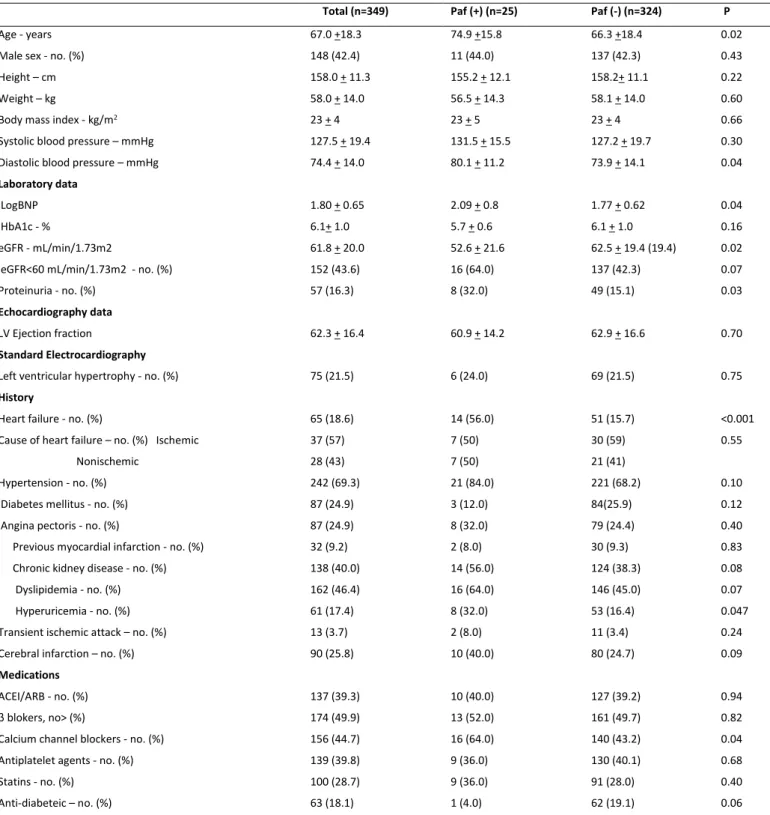
In this cross-sectional study, Paf was detected in 25 (7.2%) of the 349 patients by a single 24-hour Holter ECG examination. Table 1 provides the clinical characteristics of patients with and without Paf. Patients with Paf were more likely to be older (p=0.02), had more frequent history of HF (p<0.001), compared with patients without Paf. There were no difference in the prevalence of history of hypertension, angina pectoris, myocardial infarction, diabetes, CKD, and dyslipidemia between patients with and without Paf.
Mean of log transformed BNP concentrations was higher (p=0.04) and mean eGFR was lower (p=0.02) in patients with Paf compared with those without Paf. Notably, patients with Paf were more likely to have proteinuria (p=0.03). Systolic blood pressure, glycosylated hemoglobin (HbA1c), prevalence of left ventricular hypertrophy as evaluated by ECG, and left ventricular ejection fraction were similar between two groups. There were no significant difference in the use of angiotensin converting enzyme inhibitors (ACEI) or angiotensin receptor blockers (ARB), blockers and statins, but patients with Paf more often used calcium channel blockers (CCB) (p=0.04).
Risk factors for Paf
Among the cardiovascular risk factors listed in Table 1, advancing age, proteinuria, and a history of HF were significantly associated with Paf.
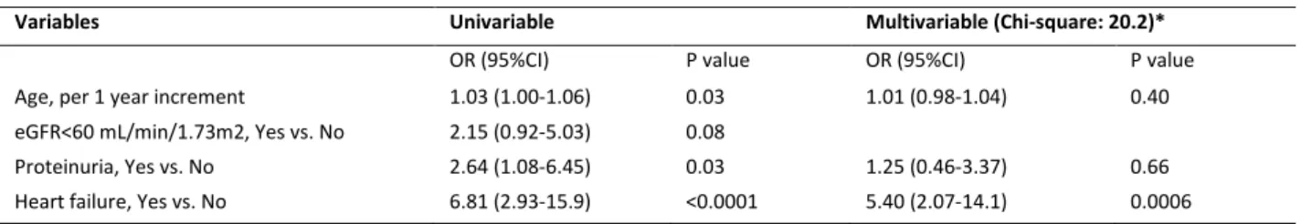
Table 2 shows the univariate logistic regression analysis of odds ratios (ORs) for Paf and the respective p values. Age was highly associated with Paf (OR 1.03 per 1 year increase, 1.03; 95% confidence interval (CI) 1.00-1.06, p=0.03). HF was strongly related to Paf (OR 6.81;95% CI, 2.93-15.9, p<0.0001). Patients with proteinuria were associated with 2.64-fold increased risk for AF (95%CI 1.08-6.45, p=0.03). OR for eGFR
<60mL/min/1.73m2 was 2.15, but did not reach statistical significance (p=0.08). These results are also shown in forest plots in Fig.1. No significant differences on ORs were observed between two groups for the other clinical features examined including male sex, body mass index (BMI), LogBNP, HbA1c, left ventricular hypertrophy evaluated by
Table 1. Comparison of clinical manifestations between Paf (+) and Paf (-) patients*
Total (n=349) Paf (+) (n=25) Paf (-) (n=324) P
Age - years 67.0 +18.3 74.9 +15.8 66.3 +18.4 0.02
Male sex - no. (%) 148 (42.4) 11 (44.0) 137 (42.3) 0.43
Height – cm 158.0 + 11.3 155.2 + 12.1 158.2+ 11.1 0.22
Weight – kg 58.0 + 14.0 56.5 + 14.3 58.1 + 14.0 0.60
Body mass index - kg/m2 23 + 4 23 + 5 23 + 4 0.66
Systolic blood pressure – mmHg 127.5 + 19.4 131.5 + 15.5 127.2 + 19.7 0.30
Diastolic blood pressure – mmHg 74.4 + 14.0 80.1 + 11.2 73.9 + 14.1 0.04
Laboratory data
LogBNP 1.80 + 0.65 2.09 + 0.8 1.77 + 0.62 0.04
HbA1c - % 6.1+ 1.0 5.7 + 0.6 6.1 + 1.0 0.16
eGFR - mL/min/1.73m2 61.8 + 20.0 52.6 + 21.6 62.5 + 19.4 (19.4) 0.02
eGFR<60 mL/min/1.73m2 - no. (%) 152 (43.6) 16 (64.0) 137 (42.3) 0.07
Proteinuria - no. (%) 57 (16.3) 8 (32.0) 49 (15.1) 0.03
Echocardiography data
LV Ejection fraction 62.3 + 16.4 60.9 + 14.2 62.9 + 16.6 0.70
Standard Electrocardiography
Left ventricular hypertrophy - no. (%) 75 (21.5) 6 (24.0) 69 (21.5) 0.75
History
Heart failure - no. (%) 65 (18.6) 14 (56.0) 51 (15.7) <0.001
Cause of heart failure – no. (%) Ischemic 37 (57) 7 (50) 30 (59) 0.55
Nonischemic 28 (43) 7 (50) 21 (41)
Hypertension - no. (%) 242 (69.3) 21 (84.0) 221 (68.2) 0.10
Diabetes mellitus - no. (%) 87 (24.9) 3 (12.0) 84(25.9) 0.12
Angina pectoris - no. (%) 87 (24.9) 8 (32.0) 79 (24.4) 0.40
Previous myocardial infarction - no. (%) 32 (9.2) 2 (8.0) 30 (9.3) 0.83
Chronic kidney disease - no. (%) 138 (40.0) 14 (56.0) 124 (38.3) 0.08
Dyslipidemia - no. (%) 162 (46.4) 16 (64.0) 146 (45.0) 0.07
Hyperuricemia - no. (%) 61 (17.4) 8 (32.0) 53 (16.4) 0.047
Transient ischemic attack – no. (%) 13 (3.7) 2 (8.0) 11 (3.4) 0.24
Cerebral infarction – no. (%) 90 (25.8) 10 (40.0) 80 (24.7) 0.09
Medications
ACEI/ARB - no. (%) 137 (39.3) 10 (40.0) 127 (39.2) 0.94
β blokers, no> (%) 174 (49.9) 13 (52.0) 161 (49.7) 0.82
Calcium channel blockers - no. (%) 156 (44.7) 16 (64.0) 140 (43.2) 0.04
Antiplatelet agents - no. (%) 139 (39.8) 9 (36.0) 130 (40.1) 0.68
Statins - no. (%) 100 (28.7) 9 (36.0) 91 (28.0) 0.40
Anti-diabeteic – no. (%) 63 (18.1) 1 (4.0) 62 (19.1) 0.06
*Plus-minus values are mean + SD. P-value was established by using the Fisher’s exact test, t-test or Mann–Whitney U-test. Abbrevalation:
Multivariable-adjusted ORs for Paf are shown in Table 2. The strong association between HF and Paf persisted after adjusting for age and proteinuria (OR was 5.40, 95% CI 2.07-14.1, p=0.0006), while neither the association between Paf and age nor the association between Paf
and proteinuria were statistically significant after multivariable adjustment (p=0.40, p=0.66, respectively).
Figure 1: Odds ratios (OR) and 95% confidence intervals (CI) of Paf
The horizontal bars represent 95% confidence intervals. Date are adjusted for vrialbes showing the significant association by univariate regression analysis.
Table 2. Univariable and multivariable logistic regression for the association with Paf
Variables Univariable Multivariable (Chi-square: 20.2)*
OR (95%CI) P value OR (95%CI) P value
Age, per 1 year increment 1.03 (1.00-1.06) 0.03 1.01 (0.98-1.04) 0.40
eGFR<60 mL/min/1.73m2, Yes vs. No 2.15 (0.92-5.03) 0.08
Proteinuria, Yes vs. No 2.64 (1.08-6.45) 0.03 1.25 (0.46-3.37) 0.66
Heart failure, Yes vs. No 6.81 (2.93-15.9) <0.0001 5.40 (2.07-14.1) 0.0006
*Data are adjusted for age, proteinuria and HF.
Next, we evaluated the diagnostic accuracy of 4 selected risk factors to discriminate Paf group from Paf (-) group. Age per 1 year increment, proteinuria, and HF could distinguish Paf (+) patients from Paf (-) with AUC of 0.66, 0.58, and 0.70, respectively (Table 3). Notably, the value
of AUC to predict Paf was significantly increased by the presence of HF (p<0.0001), in which sensitivity was 56% and specificity was 84%. These results suggest the importance of HF history to predict Paf.
Table 3. Diagnostic accuracy
Variables AUC P value Sensitivity Specificity
Age, per 1 year increment 0.66 0.01 76%* 53%*
eGFR<60 mL/min/1.73m2, Yes vs. No 0.59 0.07 64% 55%
Proteinuria, Yes vs. No 0.58 0.04 32% 85%
Heart failure, Yes vs. No 0.70 <0.0001 56% 84%
*Cutoff 72 years-old (optimal) DISCUSSION
There have been few studies of the prevalence and the risk factors of Paf in patients with CVD who have many comorbidities contributing to the increased risk of AF. In the present study using 24-hour Holter ECG, we showed that Paf was detected in 25 patients (7.2%) of the 349 patients with established CVD but without known AF, who visited outpatients department. In addition, we found that among many risk factors of AF so far identified in the epidemiological studies, HF was significantly associated with Paf, independent of other conventional risk factors in general and of proteinuria in particular.
Coexistence of AF and HF has long been well acknowledged [1, 8, 20-23], and it is clear that AF confers the increased risk of developing HF, and HF predisposes to AF. However, it remains unclear whether HF is independently associated with Paf in the patients with CVD, who have multiple comorbidities such as advancing age, a history of myocardial infarction, valve disease, left ventricular dysfunction, hypertension,
after adjusting for age and proteinuria, which were significantly associated with Paf in univariate regression analysis. These findings may have important consequence for the management of HF patients.
Several mechanisms may explain the increases risk of Paf in HF patients. Paf usually involves ectopic beats originating the cardiac muscle sleeve around pulmonary veins by rapid focal activity or local re-entry [24-27]. These ectopic firings mechanisms are promoted in HF, because HF prolongs the action potential duration (APD) and enhances Ca2+ uptake into sacroplasmic reticulum, which causes ectopic firing as well as early afterdepolarization (EAD) initiation [27]. A second possible mechanism may involve arterial cell Ca2+ overload and ryanodine receptors (RyR) dysfunction in HF [28]. These changes induce abnormal diastolic release of Ca2+ from sarcoplasmic reticulum, and induce transient inward current that underlie delayed after depolarization (DAD)-related AF[21]. Third, Paf may be caused by excessive activation of neurohumoral system including sympathetic nervous system and renin-angiotensin-aldosterone system (RAAS) in HF. Abnormal
nerve densities are significantly increased in patients with HF[30].Lastly, it has been well known that atrial substrate of AF is generated in HF[27]. The hemodynamic overload induces atrial remodeling characterized by interstitial fibrosis and pro-inflammatory activation with cellular hypertrophy, accelerated apoptosis, and abnormal myocytes Ca2+
handling[31, 32]. These pathophysiological changes can induce Paf- susceptible atrial substrates.
Study Limitations
We acknowledge the limitation of our study. First, this study is subjected to selection bias and confounding because of a small single- center study. Second limitation is that prevalent Paf was evaluated based on a single examination of 24-hour Holter ECG. Obviously, this may have caused an underestimation of the true prevalence of Paf.
Third limitation is the use of dipstick to evaluate protein, which potentially causes measurement bias. In addition, only one measurement of a urine protein may have caused underestimation of the prevalence of proteinuria. Forth, the number of HF was small and causes of HF were heterogeneous, thus we cannot assess cause- specific association between HF and Paf. Lastly, the number of Paf patients were low, which limited the number of predictors (explanatory variables) to be evaluated in the multivariable logistic regression analysis.
CONCLUSION
This study showed that Paf is robustly associated with HF in CVD patients without known AF. This finding suggests that HF patients should be considered a very high risk for Paf and should be recommended for more careful observation for the potential development of Paf in clinical practice.
Funding
This work was supported by grants from Japan Society for the Promotion of Science (M.K.), Ministry of Education, Culture, Sports, Science, and Technology of Japan, and Gunma University Initiative for Advanced Research (to M.K.).
Conflict of Interest Nothing to declare.
Authors’ Contribution
The first author has conceptualized the hypothesis and designed the study. Analysis with interpretation of data and final preparation of the original article are done by all of the authors.
REFERENCES
1. Kannel WB, Wolf PA, Benjamin EJ, Levy D. Prevalence, incidence, prognosis, and predisposing conditions for atrial fibrillation:
population-based estimates. Am J Cardiol. 1998; 82:2N-9N.
2. Nieuwlaat R, Prins MH, Le Heuzey JY, Vardas PE, Aliot E, Santini M et al. Prognosis, disease progression, and treatment of atrial fibrillation patients during 1 year: follow-up of the Euro Heart Survey on atrial fibrillation. Eur Heart J. 2008; 29:1181-9.
3. Schnabel RB, Yin X, Gona P, Larson MG, Beiser AS, McManus DD et al. 50 year trends in atrial fibrillation prevalence, incidence, risk factors, and mortality in the Framingham Heart Study: a cohort study. Lancet. 2015; 386:154-62.
4. Schmidt M, Ulrichsen SP, Pedersen L, Botker HE, Nielsen JC, Sorensen HT. 30-year nationwide trends in incidence of atrial fibrillation in Denmark and associated 5-year risk of heart failure, stroke, and death. Int J Cardiol. 2016; 225:30-36.
5. Staerk L, Sherer JA, Ko D, Benjamin EJ, Helm RH. Atrial Fibrillation:
Epidemiology, Pathophysiology, and Clinical Outcomes. Circ Res.
2017; 120:1501-1517.
6. January CT, Wann LS, Alpert JS, Calkins H, Cigarroa JE, Cleveland JC et al. 2014 AHA/ACC/HRS guideline for the management of patients with atrial fibrillation: executive summary: a report of the American College of Cardiology/American Heart Association Task Force on practice guidelines and the Heart Rhythm Society.
Circulation. 2014; 130:2071-104.
7. Kirchhof P, Benussi S, Kotecha D, Ahlsson A, Atar D, Casadei B et al. 2016 ESC Guidelines for the management of atrial fibrillation developed in collaboration with EACTS. Eur Heart J. 2016;
37:2893-2962.
8. Benjamin EJ, Levy D, Vaziri SM, D'Agostino RB, Belanger AJ, Wolf PA. Independent risk factors for atrial fibrillation in a population- based cohort. The Framingham Heart Study. JAMA. 1994;
271:840-4.
9. Kannel WB, Abbott RD, Savage DD, McNamara PM. Epidemiologic features of chronic atrial fibrillation: the Framingham study. N Engl J Med. 1982; 306:1018-22.
10. Krahn AD, Manfreda J, Tate RB, Mathewson FA, Cuddy TE. The natural history of atrial fibrillation: incidence, risk factors, and prognosis in the Manitoba Follow-Up Study. Am J Med. 1995;
98:476-84.
11. Alonso A, Lopez FL, Matsushita K, Loehr LR, Agarwal SK, Chen LY et al. Chronic kidney disease is associated with the incidence of atrial fibrillation: the Atherosclerosis Risk in Communities (ARIC) study.
Circulation. 2011; 123:2946-53.
12. Baber U, Howard VJ, Halperin JL, Soliman EZ, Zhang X, McClellan W et al. Association of chronic kidney disease with atrial fibrillation among adults in the United States: REasons for Geographic and Racial Differences in Stroke (REGARDS) Study. Circ Arrhythm Electrophysiol. 2011; 4:26-32.
13. Kerr CR, Humphries KH, Talajic M, Klein GJ, Connolly SJ, Green M et al. Progression to chronic atrial fibrillation after the initial diagnosis of paroxysmal atrial fibrillation: results from the Canadian Registry of Atrial Fibrillation. Am Heart J. 2005; 149:489- 96.
14. Saksena S, Hettrick DA, Koehler JL, Grammatico A, Padeletti L.
Progression of paroxysmal atrial fibrillation to persistent atrial fibrillation in patients with bradyarrhythmias. Am Heart J. 2007;
154:884-92.
15. de Vos CB, Pisters R, Nieuwlaat R, Prins MH, Tieleman RG, Coelen RJ et al. Progression from paroxysmal to persistent atrial fibrillation clinical correlates and prognosis. J Am Coll Cardiol.
2010; 55:725-31.
16. Veasey RA, Sugihara C, Sandhu K, Dhillon G, Freemantle N, Furniss SS et al. The natural history of atrial fibrillation in patients with permanent pacemakers: is atrial fibrillation a progressive disease?
J Interv Card Electrophysiol. 2015; 44:23-30.
17. Chiang CE, Naditch-Brule L, Murin J, Goethals M, Inoue H, O'Neill J et al. Distribution and risk profile of paroxysmal, persistent, and permanent atrial fibrillation in routine clinical practice: insight from the real-life global survey evaluating patients with atrial fibrillation international registry. Circ Arrhythm Electrophysiol.
2012; 5:632-9.
18. Lubitz SA, Moser C, Sullivan L, Rienstra M, Fontes JD, Villalon ML et al. Atrial fibrillation patterns and risks of subsequent stroke, heart failure, or death in the community. J Am Heart Assoc. 2013;
2:e000126.
19. Horio M, Imai E, Yasuda Y, Watanabe T, Matsuo S. Modification of the CKD epidemiology collaboration (CKD-EPI) equation for Japanese: accuracy and use for population estimates. Am J Kidney Dis. 2010; 56:32-8.
20. Maisel WH, Stevenson LW. Atrial fibrillation in heart failure:
epidemiology, pathophysiology, and rationale for therapy. Am J Cardiol. 2003; 91:2D-8D.
21. Sanders P, Morton JB, Davidson NC, Spence SJ, Vohra JK, Sparks PB et al. Electrical remodeling of the atria in congestive heart failure: electrophysiological and electroanatomic mapping in humans. Circulation. 2003; 108:1461-8.
22. Nattel S, Burstein B, Dobrev D. Atrial remodeling and atrial fibrillation: mechanisms and implications. Circ Arrhythm Electrophysiol. 2008; 1:62-73.
23. Schnabel RB, Rienstra M, Sullivan LM, Sun JX, Moser CB, Levy D et al. Risk assessment for incident heart failure in individuals with atrial fibrillation. Eur J Heart Fail. 2013; 15:843-9.
24. Haissaguerre M, Jais P, Shah DC, Takahashi A, Hocini M, Quiniou G et al. Spontaneous initiation of atrial fibrillation by ectopic beats originating in the pulmonary veins. N Engl J Med. 1998; 339:659- 66.
25. Hocini M, Ho SY, Kawara T, Linnenbank AC, Potse M, Shah D et al.
Electrical conduction in canine pulmonary veins:
electrophysiological and anatomic correlation. Circulation. 2002;
105:2442-8.
26. Po SS, Li Y, Tang D, Liu H, Geng N, Jackman WM et al. Rapid and stable re-entry within the pulmonary vein as a mechanism initiating paroxysmal atrial fibrillation. J Am Coll Cardiol. 2005;
45:1871-7.
27. Iwasaki YK, Nishida K, Kato T, Nattel S. Atrial fibrillation pathophysiology: implications for management. Circulation. 2011;
124:2264-74.
28. Yeh YH, Wakili R, Qi XY, Chartier D, Boknik P, Kaab S et al. Calcium- handling abnormalities underlying atrial arrhythmogenesis and contractile dysfunction in dogs with congestive heart failure. Circ Arrhythm Electrophysiol. 2008; 1:93-102.
29. Chang CM, Wu TJ, Zhou S, Doshi RN, Lee MH, Ohara T et al. Nerve sprouting and sympathetic hyperinnervation in a canine model of
atrial fibrillation produced by prolonged right atrial pacing.
Circulation. 2001; 103:22-5.
30. Nguyen BL, Fishbein MC, Chen LS, Chen PS, Masroor S.
Histopathological substrate for chronic atrial fibrillation in humans. Heart Rhythm. 2009; 6:454-60.
31. Burstein B, Nattel S. Atrial fibrosis: mechanisms and clinical relevance in atrial fibrillation. J Am Coll Cardiol. 2008; 51:802-9.
32. Yue L, Xie J, Nattel S. Molecular determinants of cardiac fibroblast electrical function and therapeutic implications for atrial fibrillation. Cardiovasc Res. 2011; 89:744-53.