Original
Toll-like receptor 4-mediated signaling activated by lipopolysaccharide suppresses transforming growth factor-beta-induced nerve growth factor expression
in periodontal ligament-derived fibroblasts
Maiko OHTA1, 2#, Akira NEMOTO3#, Naoyuki CHOSA1, Seiko KYAKUMOTO1, Seiji YOKOTA1, Masaharu KAMO1, Toshiyuki SHIBATA4, Shigeharu JOH5, Kenichi SATO2, and Akira ISHISAKI1
1Division of Cellular Biosignal Sciences, Department of Biochemistry, Iwate Medical University
(Chief: Prof. Akira ISHISAKI)
2Division of Dental Anesthesia, Department of Reconstructive Oral and Maxillofacial Surgery, Iwate Medical University
(Chief: Prof. Kenichi SATO)
3Division of Operative Dentistry and Endodontics, Department of Conservative Dentistry, Iwate Medical University
(Chief: Prof. Mamoru NODA)
4Department of Oral and Maxillofacial Surgery, Gifu University Graduate School of Medicine
(Chief: Prof. Toshiyuki SHIBATA)
5Division of Oral and Dysphasia Rehabilitation, Department of Prosthodontics, Iwate Medical University
(Chief: Prof. Shigeharu JOH)
#These authors Contributed equally to this work.
[Received:December 15 2017:Accepted:December 22 2017]
Abstract:Sensory neurons in the periodontal ligament (PDL) transmit the impulses, which are generated by the mechanical stimulation of the tooth, into the trigeminal ganglion, resulting in the excitement of the nucleus ventralis posteromedialis of the thalamus. When sensory neurons are injured, the neurites become atrophied and degenerated. Nerve growth factor (NGF) belonging to neurotrophic factors plays important roles in neurite extension and regeneration of injured sensory neurons. We evaluated how the major component of the outer membrane of gram-negative bacteria, lipopolysaccharide (LPS), affected transforming growth factor-beta1 (TGF- β 1)-induced NGF expression in rat PDL-derived fibroblasts SCDC2 cells. qRT-PCR and ELISA analyses showed that LPS suppressed TGF- β 1-induced NGF synthesis through the activation of Toll-like receptor 4
(TLR4). In addition, inhibitor of κ B (I- κ B) kinase-2 (IKK-2) inhibitor, TPCA-1, abrogated LPS- induced suppression of TGF- β 1-promoted NGF expression. Intriguingly, western blotting showed that LPS inhibited TGF- β 1-induced activation of p38 mitogen-activated protein kinase (MAPK) that mediated TGF- β 1-induced intra-cellular signal transduction for NGF expression. These results suggested that TLR4-mediated signaling activated by LPS suppresses TGF- β 1-induced NGF expression in PDL fibroblasts by inhibiting TGF- β 1-induced p38 MAPK activation in a nuclear
Introduction
Mechanosensitive (MS) neurons in the periodontal ligament (PDL) that respond to the mechanical stimulation of teeth are known to transmit the output signals into the neurons of the trigeminal mesencephalic nucleus
(Vmes)1), 2). Previous studies showed that the Ruffini ending, characterized as low threshold slowly adapting type II stretch receptors3), was the primary mechanoreceptor of the MS neuron in the PDL4), 5), 6). Histological analysis showed that collagenous encapsulation of the Ruffini ending was not observed in PDL, unlike in the cutaneous ligament7). In addition, the large Ruffini ending was partially surrounded with lamellar Schwann cells, unlike the small Ruffini ending7).
The neurites of MS neurons become atrophied and eventually degenerate, following physical invasion-induce crushing injury of the PDL tissue8). Generally, regeneration of the injured peripheral nerve includes interaction between the nerve cells and extracellular matrix (ECM)9). In addition, neurotrophic factors involving brain-derived neurotrophic factor (BDNF), glial cell-derived neurotrophic factor (GDNF), and nerve growth factor
(NGF) secreted from the mesenchymal stem cells (MSCs) in the PDL induced neurite extension of the human neuronal cells SHSY- 5Y10), suggesting that the mesenchymal cells play an important role in peripheral nerve regeneration in the PDL tissue. However, the
molecular mechanisms underlying the expression of neurotrophic factors in mesenchymal cells in the PDL tissue remain to be clarified.
Transforming growth factor-beta (TGF- β)
played an important role on the attenuation of the inflammation reaction in vivo11). M a c r o p h a g e s , w h i c h h o m e d i n t o a n inflammatory lesion, vigorously synthesized and secreted TGF- β 12). TGF- β binds to type I and type II receptors on the cell membrane. Then, the type II receptor phosphorylates the type I receptor during the formation of a tetramer consisting of two type I and two type II receptors. The phosphorylated and activated type I receptor sends intra- cellular signals by phosphorylating the receptor- regulated Smads (R-Smads) 13), 14), 15). The TGF- β superfamily induces the intracellular signals mediated by Smads, which are composed of three groups: 1) R-Smads (Smad1, Smad5, and Smad8) primarily activated by the bone morphogenetic protein (BMP) type I receptors, and Smad2 and Smad3 activated by TGF- β type I receptors, 2) the common mediator Smad (Co-Smad) such as Smad4, and 3) the inhibitory Smads (I-Smads) such as Smad6 and Smad7. Generally, activated R-Smads form complexes with the Co-Smad, which translocate to the nucleus and induce the transcription of specific target genes.
I-Smads inhibit the phosphorylation of R-Smads through competition with R-Smads for interaction with type I receptor and through factor- κ B-dependent manner, possibly resulting in the suppression of the regeneration of injured PDL neurons.
(194 words)
Key words:Iipopolysaccharide, transforming growth factor-beta, nerve growth factor, p38 mitogen-activated protein kinase, periodontal ligament fibroblast
recruitment of specific ubiquitin ligases, resulting in their proteasomal degradation. On the other hand, TGF- β induces intra-cellular signals via non-Smad signaling: non-Smad TGF- β signaling include the mitogen-activated kinase (MAPK) consisting of an extracellular signal-regulated kinase (ERK), c-jun N-terminal kinase (JNK), and p3816). Non- Smad TGF- β signaling also involve phosphoinositide 3-kinase (PI3K)/Akt16). Haas et al. reported that TGF- β promoted NGF expression in pancreatic stellate cells through the activation of ALK5-mediated pathway17). TGF- β 1 also promoted NGF expression in vascular smooth muscle cells in a Smad3-dependent manner18). We had previously found that TGF- β 1 promoted NGF expression through Smad2/3- and p38 MAPK-mediated signal transduction in PDL fibroblasts (unpublished observation).
Toll-like receptor 4 (TLR4) belonging to the TLR family is activated by bacterial lipopolysaccharide (LPS), which is a major component of the outer membrane of gram- negative bacteria19), 20). Dimerization of TLR4 is induced by the recognition of LPS by the TLR4 receptor system. The LPS-induced intracellular signals mediated by dimerized TLR4 leads to the expression of pro- inflammatory cytokines and interferons: TLR4 recruits MAL/TIRAP and MyD88 to their TIR domain for activating nuclear factor- κ B
(NF- κ B), and MAPKs that regulate pro- and anti-inflammatory cytokine production.
TLR4 also recruits TRAM and TRIF that activate IRF3 and delay the activation of MAPKs and NF- κ B for the production of type I IFNs21), 22). Borívar et al. demonstrated that LPS suppressed TGF- β 1-induced differentiation of cardiac fibroblasts into myofibroblasts through the inhibition of Smad3
activation23). In contrast, LPS enhanced TGF- β 1-induced platelet-derived growth factor-B expression in the bile duct epithelial cells through the activation of NF- κ B-mediated signaling, without affecting Smad2/3 activity24). Therefore, the effect of LPS on TGF- β 1-induced intra-cellular signal transduction seems to be dependent on the cell type. It remained to be clarified whether LPS up- regulated or down-regulated TGF- β 1-induced signals in the cells derived from PDL tissue.
In this study, we investigated how LPS affected TGF- β 1-induced NGF expression in PDL-derived fibroblasts. In addition, we evaluated how LPS affected TGF- β 1-induced Smad2/3- and p38 MAPK-mediated signals that were known to positively regulate NGF expression in PDL-derived fibroblasts.
Materials and Methods Reagents
Recombinant human TGF- β 1 was procured from PEPROTECH, Rocky Hill, NJ, USA. Recombinant rat NGF was obtained from Alomone Labs, Jerusalem, Israel.
Escherichia coli (O26: B6) LPS was purchased from Difco Laboratories, Detroit, MN, USA.
Adenosine 5 ′-O-(3-thio)triphosphate (ATP γ S; ab138911) was purchased from Abcam
(Cambridge, UK). NGF receptor (Trk A)
inhibitor GW441756 was obtained from Selleck Chemicals (Houston, TX, USA). TLR4 inhibitor TAK-242, MAPK/ERK kinase (MEK) inhibitor U0126, and PI3K/Akt inhibitor LY294002 were obtained from Calbiochem (La Jolla, CA, USA).
Inhibitor of κ B (I- κ B) kinase-2 (IKK-2)
inhibitor TPCA-1 was purchased from R & D Systems, Inc. (Minneapolis, MN, USA).
Cell culture
The isolation of rat PDL-derived fibroblasts
and establishment of single cell-derived cultures (SCDCs) were previously reported25). SCDC2 cells were cultured in type I collagen- coated plastic dishes (Sumilon Celltight Plate;
Sumitomo Bakelite Co., Tokyo, Japan) in Ham's F-12 medium (Sigma-Aldrich)
supplemented with 2 mM glutamine (100 × solution; Gibco), 10% fetal bovine serum (FBS), penicillin-streptomycin (both from Invitrogen, Gaithersburg, MD, USA), 10 ng/mL fibroblast growth factor (FGF)-1 (R&D Systems Inc., Minneapolis, MN, USA), 15 µg/mL heparin
(Sigma-Aldrich), and penicillin (Gibco, Carlsbad, CA, USA) in a humidif ied atmosphere of 5% CO2 at 37 ℃ . The rat pheochromocytoma cell PC12 was cultured in type I collagen tissue culture plastic dishes containing Dulbecco’s modified Eagle medium
(DMEM; Sigma-Aldrich, St. Louis, MO)
supplemented with 2 mM glutamine (100 × solution; Gibco), 5% FBS, 10% horse serum, and penicillin (Gibco, Carlsbad, CA, USA) in a humidified atmosphere of 5% CO2 at 37 ℃ .
RNA isolation and quantitative reverse- transcription-polymerase chain reaction (qRT- PCR)
Total RNA was isolated from SCDC2 cells by using ISOGEN reagent (Nippon Gene, Toyama, Japan), according to the manufacturer's protocol. First-strand complementary DNA
(cDNA) was synthesized from total RNA using the PrimeScript RT reagent Kit (Takara-Bio, Shiga, Japan). PCR was subsequently performed on a Thermal Cycler Dice Real Time System (Takara-Bio) using SYBR Premix Ex Taq II (Takara-Bio) with specific oligonucleotide primers [rat NGF, 5 ′ -TGCCAAGGACGCAGCTTTC-3′ (forward) and 5′-TGAAGTTTAGTCCAGTGGGCTTCAG-3′
( r e v e r s e ) ; a n d r a t g l y c e r a l d e h y d e
3-phosphate dehydrogenase (GAPDH), 5 ′ -GGCACAGTCAAGGCTGAGAATG-3′ (forward)
and 5 ′-ATGGTGGTGAAGACGCCAGTA-3 ′
(reverse)]. The mRNA level of NGF was normalized to that of GAPDH, and the relative expression levels were shown as the fold increase or decrease relative to the control.
Western blot analysis
Cells were lysed in RIPA buffer [50 mM Tris-HCl (pH 7.2), 150 mM sodium chloride
(NaCl), 1% NP-40, 0.5% sodium deoxycholate, and 0.1% sodium dodecyl sulfate (SDS; Sigma, St. Louis, MO)] or lysis buffer [20 mM HEPES
( p H 7 . 5 ) , 1 5 0 m M N a C l , 1 m M ethylenediaminetetraacetic acid (EDTA), and 1% Triton X-100] containing protease and phosphatase inhibitor cocktails (Sigma, St.
Louis, MO). The protein content was measured using BCA reagent (Pierce, Rockford, IL). Samples containing equal amounts of protein were separated on 10%
SDS-polyacrylamide gel and transferred onto polyvinylidene difluoride membranes
(Millipore, Bedford, MA). After blocking with 1% bovine serum albumin (BSA) or 1% skim milk in T-TBS [50 mM Tris-HCl (pH 7.2), 150 mM NaCl, and 0.05% Tween 20], the membranes were incubated with the appropriate primary antibody. Following antibodies were used: anti-Smad2/3 purified mouse monoclonal antibody (1:1000; 610842;
BD Transduction Laboratories, Franklin Lakes, NJ), anti-phospho-Smad2/3, anti-p38 MAPK, anti-phospho-p38 MAPK, anti-MAPK-activated protein kinase 2 (MAPKAPK-2), anti- phospho-MAPKAPK-2 (Thr222), anti-Akt-, anti-phospho-Akt, anti-p44/42 MAPK (Erk1/2), and anti-phospho-p44/42 (Erk1/2) (Thr202/
Tyr204) rabbit antibodies (1:1000; #8828,
#9212, #9211, #4691, #9271, #9102, and #9101,
respectively; Cell Signaling Technology, Beverly, MA, USA). The blots were incubated with an appropriate alkaline phosphatase-conjugated secondary antibody, and the signals were detected using an alkaline phosphatase substrate kit (BCIP/NBT Substrate Kit; Vector Laboratories Inc., Burlingame, CA, USA).
Enzyme-linked immunosorbent assay (ELISA)
SCDC2 cells were seeded into a type I collagen-coated 6-well plate at a cell density of 1.4 × 10⁵ cells/well containing the growth medium for SCDC2 cells and cultured for 24 h.
The growth medium was replaced with Ham's F-12 without any supplementation. The cells were subsequently stimulated with or without TGF- β 1 (10 ng/mL), LPS (0.5 μ g/mL), and TAK-242 (4 μ M) for 6 days. The amount of NGF secreted into the medium was measured using Rat beta NGF ELISA Kit
(ab193736; Abcam, Cambridge, UK), according to the manufacturer’s instructions.
Immunofluorescence analysis of cultured cells For immunofluorescence analysis of the cultured cells, the cells were subcultured on non-coated 4-well glass culture slides (Thermo Fisher Scientific Inc., Waltham, MA, USA) at the indicated cell density and maintained in Ham’s F-12 supplemented with 2 mM glutamine, penicillin-streptomycin, and 10%
FBS. Then, the cells were incubated for the indicated times. Following incubation, the cells were fixed in 4% paraformaldehyde (Nacalai Tesque, Inc.) for 15 min and permeabilized with Triton X-y100 (Sigma, St. Louis, MO).
After background reduction with normal goat serum, the cells were incubated with anti- n e u r o f i l a m e n t H ( R M d O 2 0 ) m o u s e monoclonal antibody (1:1000, #2836S; Cell
Sig naling Technolog y, Inc.) at room temperature for 1 h. After washing with phosphate-buffered saline (PBS) to remove excess primary antibody, the cells were incubated with Alexa Fluor 568-conjugated goat anti-mouse IgG as appropriate (1:400;
A-11031; Molecular Probes, Leiden, The Netherlands). After washing with PBS to remove excess secondary antibody, the nuclei were stained with 4 ′,6-diamidino-2- phenylindole (DAPI)-containing mounting medium, DAPI Fluoromount-G (Southern Biotech Inc., Birmingham, AL, USA). The fluorescent signal was detected using an Olympus IX70 fluorescence microscope with the LCPIanFI 20 objective lens (Olympus Co., Tokyo, Japan).
Evaluation of neurite extension from the PC12 cells co-cultured with SCDC2 cells
SCDC2 cells (2 × 10⁴ cells) and PC12 cells
(1 × 10⁴ cells) were mixed and seeded into each well of a non-coated 4-well glass culture slide
(Thermo Fisher Scientific Inc., Waltham, MA, USA) containing the PC12 cell growth medium. The cells were subsequently incubated with or without TGF- β 1 (10 ng/
mL), LPS (0.5 μ g/mL), SB-431542 (10 μ M), and Trk A inhibitor GW441756 (2 nM) for 4 days. In addition, ATP γ S (100 μ M) was added to all co-cultures from the beginning of the culture. PC12 cells in the co-culture were specifically labeled with the anti-neurofilament H immunofluorescence antibody. For use as the positive control, PC12 cells alone were treated with NGF (50 ng/mL). The status of the neurite extensions emerging from the PC12 cells was observed using the Olympus IX70 fluorescence microscope with the LCPIanFI 20 objective lens (Olympus Co., Tokyo, Japan). In addition, statistical
assessment of neurite extension was performed: cells bearing neurites longer than the length of the cell body were counted as cells with neurite extension. PC12 cells with and without neurite extensions were counted in eight different microscopic fields. The ratio of the number of neurite-extended cells to the total number of PC12 cells in the fluorescent microscopic field was statistically determined
(n = 8).
Statistical analysis
Data are presented as the mean ± standard deviation (SD; n = 6 or 8) and statistically analyzed by Tukey’s multiple comparison test.
*P < 0.05 was considered statistically significant. The results of all experiments are representative of a minimum of three separate experiments.
Results
LPS suppressed TGF- β 1-induced expression of NGF mRNA through TLR4 in an NF- κ B-dependent manner.
LPS (0.5–5.0 μg/mL) significantly abrogated TGF- β 1 (10 ng/mL)-induced mRNA expression of NGF (Figure 1A). We also confirmed that TGF- β 1 type I receptor inhibitor SB-431542 (10 μ M) abrogated TGF- β 1-induced mRNA expression of NGF (data not shown). In addition, TLR4 inhibitor TAK- 242 (4 μ M) clearly suppressed LPS-induced abrogation of TGF- β 1-induced mRNA expression of NGF (Figure 1B), indicating that the LPS-induced intra-cellular signal mediated by TLR4 inhibited TGF- β 1-induced mRNA expression of NGF. We also evaluated the protein level of NGF in the conditioned medium from SCDC2 cells by using ELISA.
TGF- β 1 (10 ng/mL) increased the concentration of NGF from 2.2 to 9.2 ng/mL in
SCDC2-conditioned medium. LPS (0.5 μ g/
mL) almost completely suppressed the TGF- β 1-mediated increase in NGF protein level in the conditioned medium (Figure 1C). In addition, TAK-242 (4 μ M) abrogated LPS- induced suppression of TGF- β 1-induced increase of NGF protein in the conditioned medium. Intriguingly, IKK-2 inhibitor TPCA-1
(10 μ M) clearly abrogated LPS-caused suppression of NGF expression promoted by TGF- β 1 (10 ng/mL) (Figure 1D).
LPS abrogated TGF- β 1-induced activation of p38 MAPK, unlike Smad2/3.
As described above, we previously found that TGF- β 1 promoted NGF expression through Smad2/3- and p38 MAPK-mediated signal transduction in PDL fibroblasts
(unpublished observation). Here, we evaluated how LPS affected TGF- β 1-induced intra-cellular signal transduction mediated by Smad2/3- and p38 MAPK. As shown in Figure 2A, LPS (0.5 μ g/mL) clearly suppressed the phosphorylation of p38 MAPK at 60 min after TGF- β 1 (10 ng/mL) stimulation, unlike that observed in the case of Smad2/3 (Figure 2B).
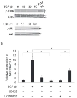
Basal activity of ERK and PI3K/Akt plays an important role in TGF- β 1-promoted NGF expression.
We evaluated how TGF- β 1-activated intracellular signals other than Smad2/3- or p38 MAPK-mediated signals affected TGF- β 1-induced expression of NGF mRNA: we examined the phosphorylation status of ERK and Akt after TGF- β 1 stimulation. TGF- β 1
(10 ng/mL) did not affect basal phosphorylation levels of ERK and Akt at 30 and 60 min after stimulation (Figure 3A, upper and lower panels, respectively). Intriguingly, the MEK inhibitor U0126 (10 μM) almost completely suppressed TGF- β 1-induced expression of
NGF mRNA (Figure 3B). In addition, the PI3K/Akt inhibitor LY294002 (10 μM) partially and significantly suppressed TGF- β 1-induced expression of NGF mRNA (Figure 3B).
LPS suppressed neurite extension from the surface of PC12 cells that were stimulated by
NGF secreted from TGF- β 1-stimulated SCDC2 cells.
ATP γ S enhanced the sensitivity of PC12 cells to NGF stimulation by activating early response kinase 1/2 and p38 MAPK mediated through the P2Y2 receptor. Then, NGF-induced neurite extension from the surface of PC12 Figure 1:LPS suppressed TGF- β 1-promoted mRNA expression of NGF in SCDC2 cells through TLR4 in a
NF- κ B-dependent manner.
Cells were seeded into type I collagen-coated 12-well tissue culture plates at a density of 7 × 10⁴ cells/
well in SCDC2 growth medium and maintained for 24 h. The growth medium was replaced with Ham’s F-12 without FBS, FGF-1, and heparin, and the cells were cultured until starvation for 24 h. (A) The starved cells were treated with TGF- β 1 (10 ng/mL) with or without LPS at various concentrations for 24 h. (B) The starved cells were treated with or without TGF- β 1 (10 ng/mL) and LPS (0.5 μ g/
mL) for 24 h. Some cells were pretreated with (B) TLR4 inhibitor TAK-242 (4 μ M) or (D) TPCA-1
(10 μ M) 30 min before LPS administration. The relative expression level of NGF was evaluated using qRT-PCR, as described in Materials and Methods. (C) The cells were seeded into type I collagen-coated tissue culture plates and maintained for 24 h. The cells were starved and subsequently cultured with or without TGF- β 1 (10 ng/mL) alone and with or without TGF- β 1 (10 ng/mL) plus LPS (0.5 μ g/mL)
for 6 days. Some cells were pre-treated with TAK-242 (4 μ M), as described above. The concentration of NGF protein secreted into the culture medium was determined using ELISA. Data represent the mean ± standard deviation (SD; n = 6). *P < 0.05 was considered significant. The cells not subjected to TAK-242 or TPCA-1 pretreatment were incubated with dimethyl sulfoxide (DMSO) as a vehicle control.
A C
B D
cells was accelerated26). We previously demonstrated that the NGF secreted from TGF- β 1-stimulated SCDC2 cells promoted neurite extension from surfaces of PC12 cells primed with ATP γ S (unpublished observation). Here, we evaluated the effect of LPS on neurite extension from ATP γ S-primed PC12 cells co-cultured with TGF- β 1-stimulated SCDC2 cells. Co-culture of TGF- β 1 (10 ng/mL)-treated SCDC2 cells and PC12 cells showed that LPS (0.5 μ g/mL)
suppressed neurite extension of PC12 cells
(Figure 4A, panel g). We also confirmed that both TGF- β type I receptor inhibitor SB- 431542 (10 μ M) (panel c) and NGF receptor
(Trk A) inhibitor GW441756 (2 nM) (panel d)
abrogated the neurite extension of PC12 cells Figure 2:LPS suppressed TGF- β 1-induced
activation of p38 MAPK.
The cells were seeded into type I collagen- coated 6-well tissue culture plates at a density of 1.4 × 10⁵ cells/well in SCDC2 growth medium and maintained for 24 h. The cells were starved and treated with or without TGF- β 1 (10 ng/mL) alone, or TGF- β 1
(10 ng/mL) plus LPS (0.5 μ g/mL) for the indicated times. The phosphorylation status of (A) p38 MAPK and (B) Smad2/3 was evaluated by western blotting.
Figure 3:Basal activity of ERK and PI3K/Akt seemed to play an important role in TGF- β 1-promoted NGF expression.
(A) The cells were seeded into type I collagen-coated 6-well tissue culture plates at a density of 1.4 × 10⁵ cells/well in the SCDC2 growth medium and maintained for 24 h. The cells were starved as described above, followed by stimulation with TGF-β1 (10 ng/mL) for the indicated times. The phosphorylation status of ERK and Akt was evaluated by western blotting, as described in Materials and Methods. Because the basal level of ERK phosphorylation in the SCDC2 cells was low, the cells were stimulated with EGF (10 ng/mL) as a positive control to confirm the positive band position of phosphorylated ERK. (B) Cells were seeded into type I collagen-coated 12-well tissue culture plates at a density of 7 × 10⁴ cells/well in the SCDC2 growth medium and maintained for 24 h. The growth medium was replaced with Ham’s F-12 without FBS, FGF-1, and heparin, and the cells cultured until starvation for 24 h.
The starved cells were treated with or without TGF-β 1 (10 ng/mL) for 24 h. Some cells were pretreated with the ERK inhibitor U0126 (10 μ M) or the PI3K/Akt inhibitor LY294002 (10 μ M) 30 min before TGF- β 1 administration. The cells without U0126 and LY294002 pretreatment were incubated with dimethyl sulfoxide
(DMSO) as a vehicle control. The relative expression level of NGF was evaluated by qRT-PCR, as described in Materials and Methods. Data represent the mean ± SD (n = 6). *P < 0.05 was considered significant.
Figure 4:LPS suppressed the neurite extension from the surface of PC12 cells which was stimulated by NGF secreted from TGF- β 1-stimulated SCDC2 cells.
(A) SCDC2 cells (2 × 10⁴ cells) and rat pheochromocytoma cells PC12 (1 × 10⁴ cells) were mixed and seeded into each well of non-coated 4-well glass culture slides (panels a to g). Cells were treated with
(panels b, c, d, f, and g) or without (panels a and e) TGF- β 1 (10 ng/mL) and incubated for 4 days. In some cases, the cells were treated with the TGF-β type I receptor inhibitor SB-431542 (10 μM) (panel c), the NGF receptor (Trk A) inhibitor GW441756 (2 nM) (panel d), or LPS (0.5 μ g/mL) (panel g)
from the start of the co-culture. In addition, ATP γ S (100 μ M) was added to all co-cultures (panels a to g). DMSO was used as a vehicle control for SB-431542 and GW441756 (panels a, and b). For the positive control experiment for neurite extension, PC12 cells alone were treated with NGF (50 ng/mL)
(panel h). The neurite extension of PC12 cells was visualized by immunostaining with the anti- neurofilament H antibody (red). The nuclei were stained with DAPI (blue). Scale bar: 50 μ m. (B)
Statistical assessment of neurite extension in the PC12 cells co-cultured with SCDC2 cells was performed. Data represent the mean ± SD (n = 8). *P < 0.05 was considered significant.
in this co-culture system. In addition, neurite extension in the PC12 cells co-cultured with SCDC2 cells was statistically analyzed. As shown in Figure 4B, LPS (0.5 μ g/mL) clearly reduced the ratio of the number of neurite- extended PC12 cells to the total number of PC12 cells, which were activated by ATP γ S
(100 μ M) treatment, and co-cultured with TGF- β 1-treated SCDC2 cells (64.8–0.8%).
Discussion
We had previously shown that 1) TGF- β 1 promoted NGF expression through Smad2/3- and p38 MAPK-mediated signal transductions in the PDL fibroblasts SCDC2, and that 2)
NGF secreted from the TGF- β 1-treated SCDC2 cells promoted neurite extension from the surface of ATP γ S-primed PC12 cells t h r o u g h t h e N G F r e c e p t o r T r k A
(unpublished observation). In this study, we evaluated how LPS affected NGF expression in SCDC2 cells. We found that the TLR4- mediated signal activated by LPS suppressed NGF synthesis in SCDC2 cells (Figure 1A, B, and C). As described above, NF- κ B is a major signal transduction molecule after TLR4 is activated by LPS. We found that IKK-2 inhibitor TPCA-1 (10 μ M), which is known to inhibit the transcriptional activity of NF- κ B by disturbing the translocation of NF- κ B into the nucleus, reversed LPS-induced suppression of the TGF- β 1-promoted NGF expression in SCDC2 cells (Figure 1D), suggesting that LPS suppressed TGF- β 1-promoted NGF expression through the activation of intra-cellular signals mediated by NF- κ B. In addition, we showed that LPS abrogated TGF- β 1-induced activation of p38 MAPK, unlike Smad2/3 (Figure 2). These results suggested that the TLR4-mediated signaling activated by LPS suppresses TGF- β
1-induced NGF expression in SCDC2 cells through the inhibition of TGF- β 1-induced p38 MAPK-mediated signal. We previously showed that the inflammatory cytokines IL-1 β and TNF- α suppressed TGF- β 1-induced phosphorylation of both Smad2/3 and p38 MAPK (unpublished observation), suggesting that TGF- β 1-induced intra-cellular signal transduction was differentially modified by inflammatory cytokines and LPS, respectively.
On the other hand, TGF- β 1 did not up- regulate the phosphorylation status of ERK and Akt (Figure 3A). However, the MEK inhibitor U0126 almost completely suppressed TGF- β 1-promoted NGF expression. In addition, the PI3K/Akt inhibitor LY294002 partially and significantly suppressed TGF- β 1-promoted NGF expression (Figure 3B).
These results suggested that the basal activities of ERK and PI3K/Akt play important roles in TGF- β 1-promoted NGF expression. In addition, we found that LPS suppressed neurite extension of PC12 cells co- cultured with TGF- β 1-treated SCDC2 cells
(Figure 4), suggesting that the LPS on the outer membrane of gram-negative bacteria in the oral cavity disturbs the regeneration of PDL neurons promoted by the NGF secreted from TGF- β 1-stimulated PDL fibroblasts.
G r a m - n e g a t i v e b a c t e r i a s u c h a s Porphyromonas gingivalis, Actinobacillus actinomycetemcomitance, Prevotella intermedia, Fusobacterium nucleatum, and Tannerella forsythensis are known to be the major pathogens involved in periodontitis27), 28), 29). Some previous studies have shown that P.
gingivalis LPS activated TLR2 rather than TLR430), 31), 32), while some other studies showed that P. gingivalis LPS activated TLR433), 34), 35). In addition, Kikuchi et al.
demonstrated that A. actinomycetemcomitance
LPS induced interleukin (IL)-12 expression in human dendritic cells through the stimulation of TLR436). Kikkert et al. also showed that A.
actinomycetemcomitance LPS promoted IL-8 expression in the human embryonic kidney cells through the stimulation of both TLR2 and TLR437). Thus, LPS on the outer membrane of the gram-negative bacteria in the oral cavity seemed to stimulate TLR2 and/or TLR4. Here, we studied how E. coli LPS affected TGF- β 1-induced NGF expression in PDL-derived fibroblasts. Actually, the chemical structure of P. gingivalis lipid A, which is the bioactive moiety of LPS, is remarkably different from that of E. coli lipid A38). Therefore, E. coli LPS is known to activate TLR4, but not TLR239). Therefore, it remains to be clarified how LPS-induced TLR2-mediated signals affect TGF- β 1-promoted NGF expression in PDL- derived fibroblasts. Therefore, further studies are required to clarify this aspect.
As described above, neurotrophic factors including BDNF, GDNF, and NGF, secreted from the MSCs in PDL, induced neurite extension of human neuronal cells10). In fact, the SCDC2 cells derived from rat PDL tissue expressed several MSC markers, including CD44, CD146, and vascular cell adhesion molecule (VCAM)-125), which may be related to the acquisition of the ability to express NGF in SCDC2 cells. In addition, SCDC2 cells such as MSCs differentiated into endothelial cell-like cells and constructed vessel-like structures in a three-dimensional culture with type I collagen gel25). In addition, it remains to be clarified whether fully differentiated PDL fibroblasts, which did not show multipotency like MSCs, retain the ability to express NGF.
Thus, taken together, we showed that LPS- a c t i v a t e d T L R 4 s u p p r e s s e d T G F - β 1-promoted NGF synthesis through abrogation
of TGF- β 1-induced activation of p38 MAPK in a NF- κ B-dependent manner. These results suggested that the gram-negative bacteria in the oral cavity are obstacles for the regeneration of PDL neurons promoted by the NGF secreted from PDL fibroblasts. Therefore, the findings of this study may help in identifying targets for treating damaged PDL neurons at the molecular level.
Acknowledgements
This study was supported in part by the JSPS KAKENHI Grant Numbers JP22592076 and JP17K11851 awarded to M.K., JP25463053 and JP16K11654 to N.C., JP26462823 to S.K., and JP26670852 and JP16H05534 to A.I., from the Ministry of Education, Culture, Sports, Science and Technology of Japan, and Grant- in-Aid for Strategic Medical Science Research Centre (to A. I.) from the Ministry of Education, Culture, Sports, Science, and Technology of Japan, 2010-2016. We would like to thank Editage (www.editage.jp) for English language editing.
Conflict of interest statement The authors declare no conflicts of interest regarding the publication of this article.
References
1) Shigenaga, Y., Mitsuhiro, Y., Shirana, Y., Tsuru, H.: Two types of jaw-muscle spindle afferents in the cat as demonstrated by intra-axonal staining with HRP. Brain Res., 514: 219-237, 1990.
2) Shigenaga, Y., Doe, K., Suemine, S., Mitsuhiro, Y., Tsuru, K., Otani, K., Shirana, Y., Hosoi, M., Yoshi- da, A., Kagawa, K.: Physiological and morphologi- cal characteristics of periodontal mesencephalic trigeminal neurons in the cat—intra-axonal stain- ing with HRP. Brain Res., 505: 91-110, 1989.
3) Maeda, T., Hayashi, S.: Morphological basis of periodontal nerve endings. Kaibogaku Zasshi, 75:
377-380, 2000.
4) Byers, M.R., Maeda, T.: Periodontal innervation:
regional specializations, ultrastructure, cytochem-
istry and tissue interactions. Acta. Med. Dent.
Helv., 2: 116-133, 1997.
5) Wakisaka, S., Atsumi, Y., Youn, T.H., Maeda, T.:
Morphological and cytochemical characteristics of periodontal Ruffini ending under normal and regeneration processes. Arch. Histol. Cytol., 63:
91-113, 2000.
6) Maeda, T., Ochi, K., Nakakuma-Ohshima, K., Youn, S.H., Wakisaka, S.: Ruffini ending as an es- sential mechanoreceptor in periodontal liga- ment–—its morphology, cytochemical feature, re- generation and development. Crit. Rev. Oral Biol.
Med., 10: 307-327, 1999.
7) Byers, M.R.: Sensory innervation of periodontal ligament of rat molars consists of unencapsulated Ruffini-like mechanoreceptors and free nerve end- ings. J. Comp. Neurol., 231: 500-518, 1985.
8) Atsumi, Y., Imai, T., Matsumoto, K., Sakuda, M., Maeda, T., Kurisu, K., Wakisaka, S. Effects of dif- ferent types of injury to the inferior alveolar nerve on the behavior of Schwann cells during the regeneration of periodontal nerve fibers of rat incisor. Arch. Histol. Cytol., 63: 43-54, 2000.
9) Terenghi, G.: Peripheral nerve regeneration and neurotrophic factors. J. Anat., 194: 1-14, 1999.
10) Kolar, M.K., Itte, V.N., Kingham, P.J., Novikov, L.N., Wiberg, M., Kelk, P.: The neurotrophic ef- fects of different human dental mesenchymal stem cells. Sci. Rep., 7: 12605, 2017.
11) Wang, W., Huang, X.R., Li, A.G., Liu, F., Li, J.H., Wang, X.L., Lan, H.Y.: Signaling mechanism of TGF-beta1 in prevention of renal inflammation:
role of Smad7. J. Am. Soc. Nephrol., 16: 1371-1383, 2005.
12) Korns, D., Frasch, S.C., Fernandes-Boyanapalli, R., Henson, P.M., Bratton, D.L.: Modulation of macrophage efferocytosis in inflammation. Front.
Immunol., 2: 57, 2011.
13) Ahmed, S., Bradshaw, A.D., Gera, S., Dewan, M.Z., Xu, R.: The TGF- β /Smad4 signaling path- way in pancreatic cartinogenesis and clinical sig- nificance. J. Clin. Med., 6: 5, 2017.
14) Heldin, C.H., Moustakas, A. Signaling receptors for TGF- β family members. Cold Spring Harb.
Perspect. Biol., 8: 8, 2016.
15) Macias, M.J., Martin-Malpartida, P, Massagué, J.:
Structural determinations of Smad function in TGF- β signaling. Trends Biochem. Sci., 40: 296- 308, 2015.
16) Mu, Y., Gudey, S.K., Landström, M.: Non-Smad signaling pathways. Cell Tissue Res., 347: 11-20, 2012.
17) Hass, S.L., Fitzner, B., Jaster, R., Wiercinska, E., Gaitantzki, R., Jesnowski, R., Löhr, J.M., Singer, M.V., Dooley, S., Breitkopf, K.: Transforming growth factor-beta induces nerve growth factor
expression in pancreatic stellate cells by activa- tion of the ALK-5 pathway. Growth Factors, 27:
289-299, 2009.
18) Shi, X., DiRenzo, D., Guo, L.W., Franco, S.R., Wang, B., Seedial, S., Kent, K.C.: TGF- β /Smad3 stimulates stem cell/developmental gene expres- sion and vascular smooth muscle cell de-differen- tiation. PLoS One, 9: e93995, 2014.
19) Beutler, B.: TLR4 as the mammalian endotoxin sensor. Curr. Top. Microbiol. Immunol., 270: 109- 120, 2002.
20) Sperandeo, P., Martorana, A.M., Polissi, A.: Lipo- polysaccharide biogenesis and transport at the outer membrane of Gram-negative bacteria. Bio- chim. Biophys. Acta., 1862: 1451-1460, 2017.
21) Kuzmich, N.N., Sivak, K.V., Chubarev, V.N., Po- rozov, Y.B., Savateeva-Lyubimova, T.N., Peri, F.:
TLR4 signaling pathway modulators as potential therapeutics in inflammation and sepsis. Vaccines, 5: 34, 2017.
22) Brown, J., Wang, H., Hajishengallis, G.N., Martin, M.: TLR-signaling networks: An integration of adaptor molecules, kinases, and cross-talk. J. Den- tal Res., 90: 417-427, 2011.
23) Bolívar, S., Santana, R., Ayala, P., Landaeta, R., Boza, P., Humeres, C., Vivar, R., Muñoz, C., Pardo, V., Fernandez, S., Anfossi, R., Diaz-Araya, G.: Li- popolysaccharide activates Toll-like receptor 4 and prevents cardiac fibroblast-to-myofibroblast differentiation. Cardiovasc. Toxicol., in press
(Epub ahead of print).
24) Kassel, K.M., Sullivan, B.P., Luyendyk, J.P.: Lipo- polysaccharide enhances transforming growth factor β 1-induced PDGF-B expression in bile duct epithelial cells. J. Gastroenterol. Hepatol., 27:
714-721, 2012.
25) Okubo, N., Ishisaki, A., Iizuka, T., Tamura, M., Kitagawa, Y.: Vascular cell-like potential of undif- ferentiated ligament fibroblasts to construct vas- cular cell-specific marker-positive blood vessel structures in a PI3K-activation-dependent man- ner. J. Vasc. Res., 47: 369-383, 2010.
26) Arthur, D.B., Akassoglou, K., Insel, P.A.: P2Y2 receptor activates nerve growth factor_TrkA signaling to enhance neuronal differentiation.
Proc. Natl. Acad. Sci. USA, 102: 19138-19143, 2005.
27) Haffajee, A.D., Socransky, S.S.: Microbial etiolog- ical agents of destructive periodontal diseases.
Periodontol. 2000, 5: 78-111, 1994.
28) Lai, C.H., Listgarten, M.A., Shirakawa, M., Slots, J.: Bacteroides forsythus in adult gingivitis and periodontitis. Oral Microbiol. Immunol., 2: 152-157, 1987.
29) van Winkelhoff, A.J., Loos, B.G., van der Reijden, W.A., van der Velden, U.: Porphyromonas gingiva- lis, Bacteroides forsythus and other putative peri-
odontal pathogens in subjects with and without periodontal destruction. J. Clin. Periodontol., 29:
1023-1028, 2002.
30) Pulendran, B., Kumar, P., Cutler, C.W., Mo- hamadzadeh, M., Van Dyke, T., Banchereau, J.: Li- popolysaccharides from distinct pathogens induce different classes of immune responses in vivo. J.
Immunol., 167: 5067-5076, 2001.
31) Re, F., Strominger, J.L.: Toll-like receptor 2
(TLR2) and TLR4 differentially activate human dendritic cells. J. Biol. Chem., 276: 37692-37699, 2001.
32) Toshchakov, V., Jones, B.W., Perera, P.Y., Thomas, K., Cody, M.J., Zhang, S., Williams, B.R., Major, J., Hamilton, T.A., Fenton, M.J., Vogel, S.N.:
TLR4, but not TLR2, mediates IFN-beta-induced STAT1alpha/beta-dependent gene expression in macrophages. Nat. Immunol., 3: 392-398, 2002.
33) Darveau, R.P., Pham, T.T., Lemley, K., Reife, R.A., Bainbridge, B.W., Coats, S.R., Howald, W.N., Way, S.S., Hajjar, A.M.: Porphyromonas gingivalis lipopolysaccharide contains multiple lipid A spe- cies that functionally interact with both toll-like receptors 2 and 4. Infect. Immun., 72: 5041-5051, 2004.
34) Hashimoto, M., Asai, Y., Ogawa, T.: Separation and structural analysis of lipoprotein in a lipo- polysaccharide preparation from Porphyromonas gingivalis. Int. Immunol., 16: 1431-1437, 2004.
35) Ogawa, T., Asai, Y., Hashimoto, M., Takeuchi, O., Kurita, T., Yoshikai, Y., Miyake, K., Akira, S.:
Cell activation by Porphyromonas gingivalis lipid A molecule through Toll-like receptor 4- and my- eloid differentiation factor 88-dependent signaling pathway. Int. Immunol., 14: 1325-1332, 2002.
36) Kikuchi, T., Hahn, C.L., Tanaka, S., Barbour, S.E., Schenkein, H.A., Tew, J.G.: Dendritic cells stimu- lated with Actinobacillus actinomycetemcomitans elicit rapid gamma interferon responses by natu- ral killer cells. Infect. Immun., 72: 5089-5096, 2004.
37) Kikkert, R., Laine, M.L., Aarden, L.A., van Win- kelhoff, A.J.: Activation of Toll-like receptors 2 and 4 by gram-negative periodontal bacteria. Oral Microbiol. Immunol., 22: 145-151, 2007.
38) Ogawa, T.: Chemical structure of lipid A from Porphyromonas (Bacteroides) gingivalis lipopoly- saccharide. FEBS Lett., 332: 197-201, 1993.
39) Vandewalle, A.: Toll-like receptors and renal bacterial infections. Chang Gung Med. J., 31: 525- 537, 2008.