ちゃお ゆーしゅ 氏名(本籍) 赵 玉雪(中国) 学位の種類 博士(薬学) 学位記番号 論博第358 号 学位授与の日付 平成31 年 3 月 18 日 学位授与の要件 学位規則第4 条第 2 項該当
学位論文題目 Characterization of the action mechanisms of arsenic disulfide against human breast cancer cells
論文審査委員 (主査)教授 平野 俊彦 教授 佐藤 隆 教授 田野中 浩一 教授 高木 教夫
論文内容の要旨
INTRODUCTIONThe medicinal application of arsenic compounds can be dated back more than 2000 years to ancient China and Europe. Over recent decades, accumulating studies have shown the efficacy of arsenics, such as arsenic trioxide (ATO), in the treatment of different types of carcinoma including breast cancer. However, these agents are limited in the clinical applications due to the serious adverse effects arise from their high toxicity. Therefore, alternative agents with similar therapeutic efficacy but fewer adverse effects are urgently required to be developed. Arsenic disulfide (As2S2), the main active ingredient of orange-red crystalline realgar, also known as
‘Xiong-Huang’ in traditional Chinese medicine, was chosen to be a candidate for its good therapeutic reputation and relatively low toxicity in the treatment of various types of malignancies. In recent years, clinical trials have shown the promising anticancer efficacies of As2S2 in treatment of refractory or relapsed acute promyelocytic leukemia (APL) and chronic
myelogenous leukemia (CML) in China. Besides, experimental researches suggested therapeutic effects of As2S2 on various solid tumors. Nevertheless, a limited number of studies
have reported the cytotoxic effect of As2S2 on human breast cancer. In addition, there are rare
cases of studies investigating the underlying mechanisms of the effects of As2S2 against breast
carcinoma.
The present study aimed to investigate: (1) the effects of As2S2 on human breast cancer cell
lines and (2) the possible molecular mechanisms underlying the action of the drug, as well as (3) the synergistic anticancer effects of As2S2 combined with L-buthionine-(S, R)-sulfoximine
(BSO) on breast cancer cells.
CHAPTER ONE Anticancer efficacies and underlying mechanism of action of arsenic disulfide on breast cancer cells in different culture systems
2
cell culture system. The effects of As2S2 on human breast cancer MCF-7 cells were compared
between 2D and 3D cultures, and the possible molecular mechanisms underlying the action of this drug were studied. The results showed that As2S2 inhibited viability of MCF-7 cells and
induced cell apoptosis in both 2D monolayers and 3D spheroids in a concentration dependent manner (Figure 1). In addition, the cytotoxic selectivity of As2S2 on MCF-7 cells in comparison
with normal human breast epithelial cells (184B5) was investigated. As2S2 exerted a cytotoxic
action in both MCF-7 and 184B5 cells in a dose-dependent manner. However, in comparison with MCF-7 cells, 184B5 cells were less sensitive to the cytotoxic effects of As2S2 (Table 1),
indicating the cytotoxic selectivity of As2S2 to human breast cancer cells as compared to normal
human breast epithelial cells. Furthermore, observations obtained from a three-dimensional (3D) cultured system provided more convincing evidence to confirm the cytotoxic selectivity of As2S2 in breast cancer cells but not in normal breast epithelial cells. This chapter thus
provided an experimental confirmation for the cytotoxicity of As2S2 against breast cancer cells
and the drug safety of As2S2 as a relatively less toxic agent to normal breast cells.
Figure 1 Effects of As2S2 on viability of
MCF-7 cells in 2D (open symbol) and 3D (closed symbol) culture systems. Cells were
cultured with As2S2 for 72h, and the cell viability was assessed by CCK-8 assay. (A) Cell viability of MCF-7 monolayers (2D) and spheroids (3D). (B) Dose response curves for the inhibitory effects of As2S2 on the cell viability of 2D-cultured (〇) and 3D-2D-cultured (●) MCF-7 cells. (C) The mean IC50 values of As2S2 on MCF-7 cell viabilities in 2D and 3D culture systems. Asterisks indicate significant differences between control and drug treatment groups (**P < 0.01 and ****P < 0.0001). Hashes indicate significant differences between 2D and 3D cultured cells (##P < 0.01 and ###P < 0.001).
Table 1 IC50 values of As2S2 on the growth of normal breast and breast cancer cells in 2D- and 3D-cultured systems
IC50 value of As2S2 (μM)
Cell line 2D 3D
184B5 12.39 ± 0.36 > 16
MCF-7 5.45 ± 0.36* 8.61 ± 3.09
Abbreviations: As2S2, arsenic disulfide; 2D, two dimensional; 3D, three dimensional. *Significantly low IC50 value (high sensitivity) was observed, as compared to the value in 184B5 cells (P = 0.0002).
3
The anticancer effects of As2S2 on two typically distinctive subtypes of breast cancer cell lines,
namely MCF-7 and MDA-MB-231, in vitro (Figure 2) and the possible underlying mechanisms were investigated. The results identified the antitumor effects of As2S2 against breast cancer
cells, which included the inhibition of cell proliferation (Figure 2), cell survival, and cell invasion. These effects of As2S2 were associated with blockade of cell cycle progression and
induction of apoptosis and autophagy. Inhibition of PI3K/Akt signals (Figure 3), decrease in Matrix metalloproteinase-9 (MMP-9) expression (Figure 4), and cellular reactive oxygen species (ROS) accumulation were also suggested to be implicated in antitumor activities of As2S2.
Figure 2 As2S2 inhibited viability of cells of the
human breast cancer cell lines MCF-7 and MDA-MB-231. MCF-7 and MDA-MB-231 cells were
treated with As2S2 for 48 h, and the cell viability was determined by CCK-8 assay. Data are presented as the mean ± SEM (n ≥ 3). *P < 0.05, **P < 0.01 vs. control group (As2S2 0 µM).
Figure 3 Effects of As2S2 on the expression of
cell survival related-proteins. MCF-7 and
MDA-MB-231 cells were treated with 0, 4, 8 and 16 µM As2S2 for 48 h, and Western blot assays were carried out to examine expression of PI3K and Akt. β-actin was used as an internal control. Asterisks indicate significant differences between the control (As2S2 0 µM) and the As2S2-treated groups (*P < 0.05, **P < 0.01).
Figure 4 Effects of As2S2 on MMP-9 protein
expression in breast cancer cells. MCF-7 and
MDA-MB-231 cells were treated with As2S2 for 48 h. Then, Western blot assays were carried out to examine the effects of As2S2 on the expression of MMP-9. β-actin was used as an internal control. Asterisks indicate significant differences between the control cells (As2S2 0 µM) and As2S2-treated cells (*P<0.05).
4
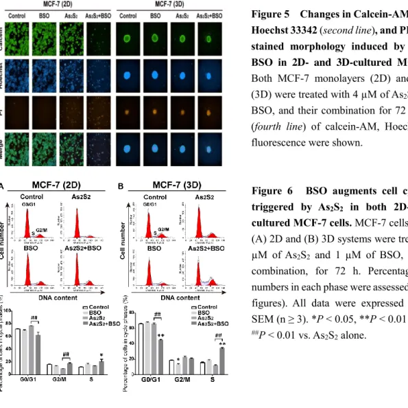
Combination chemotherapy represents an effective approach to potentiate the therapeutic efficacy, overcome drug resistance, reduce adverse effects, and minimize drug dosage of each compound alone. In this Chapter, I investigated the synergistic anticancer effects of As2S2
combined with BSO, a potent specific inhibitor of glutathione (GSH) biosynthesis, on human breast cancer MCF-7 cells cultured in both 2D monolayers and 3D spheroids (Figure 5). The results suggested that the combination of As2S2 and BSO synergistically decreased the amount
of intracellular GSH and conversely potentiated reactive oxygen toxicity in human breast cancer cells, which resulted in cell cycle arrest (Figure 6), apoptosis induction, and cell survival inhibition. Thus, the combination treatment with As2S2 and BSO might be a promising
therapeutic strategy to increase drug sensitivity of breast cancer cells to As2S2, as well as to
overcome their drug resistance.
Figure 5 Changes in Calcein-AM (first line), Hoechst 33342 (second line), and PI (third line) stained morphology induced by As2S2 and
BSO in 2D- and 3D-cultured MCF-7 cells.
Both MCF-7 monolayers (2D) and spheroids (3D) were treated with 4 µM of As2S2, 1 µM of BSO, and their combination for 72 h. Merging (fourth line) of calcein-AM, Hoechst, and PI fluorescence were shown.
Figure 6 BSO augments cell cycle arrest triggered by As2S2 in both 2D- and
3D-cultured MCF-7 cells. MCF-7 cells 3D-cultured in
(A) 2D and (B) 3D systems were treated with 4 µM of As2S2 and 1 µM of BSO, alone or in combination, for 72 h. Percentages of cell numbers in each phase were assessed (lower two figures). All data were expressed as mean ± SEM (n ≥ 3). *P < 0.05, **P < 0.01 vs. control; ##P < 0.01 vs. As
2S2 alone.
CONCLUSION
5
action in treatment of breast cancer, using 2D- and 3D-cultured human breast cancer cell lines. In addition, the therapeutic efficacy of As2S2 combined with BSO was also studied. The results
demonstrated that human breast cancer MCF-7 cells were more sensitive to As2S2 in
comparison with normal breast epithelial cells in both 2D and 3D culture systems, suggesting significant antitumor effects of As2S2 on breast carcinoma, which were further characterized by
inhibiting cell growth, inducing apoptosis, alleviating cell migration and arresting cell cycle in different breast cancer cell lines. The results also provided insights that the combination application of As2S2 with BSO could improve the therapeutic efficacy of As2S2 in treatment of
drug resistant breast carcinomas.
PUBLICATIONS
論文審査の結果の要旨 亜ヒ酸(As2O3)に代表される無機ヒ素化合物は、毒物として知られる反面、各種白血病 の治療に有効性を示す報告が多数あり、本邦では急性前骨髄球性白血病の治療薬として適応 となっている。中国ではAs2O3の他、より毒性の低いAs2S2も各種白血病の治療に臨床で用い られているが、これら無機ヒ素化合物の抗腫瘍効果には不明な点が多く、特にAs2S2に至っ ては、その作用に関する科学的解析がほとんどなされていない。またヒ素化合物が、乳癌を はじめとする種々の固形癌に有効であるとする報告がある一方、これらの癌に対するAs2S2 の効果についてはほとんど報告がない。本論文は、通常の単層培養(2次元培養)系およびス フェロイドを形成させる3次元培養系を用いて培養したヒト乳癌細胞に対する、As2S2の効果 とその作用機序を検討した結果をまとめたものであり、次の3章に分けて論じている。 第1章では、ヒト乳癌細胞株MCF-7細胞の2次元および3次元培養系を確立し、As2S2の増殖 抑制効果を比較検討した。2次元および3次元のいずれの培養系おいても、As2S2はMCF-7細 胞の生存率を用量依存的かつ有意に減少させた。さらには、これらの培養系細胞に対する As2S2のアポトーシス誘導作用や、アポトーシス関連タンパク質発現に対するAs2S2の有用な 効果を確認した。また本章では、2次元培養したMCF-7細胞に比べ3次元培養したMCF-7細胞 の方が、As2S2の作用に耐性を示すことを明らかとした。 更に本章では、MCF-7細胞および正常乳腺由来細胞株184B5細胞に対するAs2S2の効果を 比較検討した。As2S2が184B5細胞の生存率に及ぼす作用は弱かった。特に3次元培養した 184B5細胞の生存率に対しては、ほとんど影響を及ぼさなかった。これらの結果から、As2S2 はヒト乳癌細胞に特異的に作用し、正常乳腺細胞に対してはほとんど毒性を示さないものと 結論した。 第2章では、エストロゲン受容体を有するMCF-7細胞と、ホルモン受容体の無いヒト乳癌 細胞株MDA-MB-231細胞を用い、2次元および3次元培養したこれらの細胞の生存率に及ぼす As2S2の作用およびその作用機序を比較検討した。As2S2はいずれの培養系においても、両細 胞株細胞の生存率を低下させ、また細胞周期の進行を阻害してアポトーシスとオートファジ ーを誘導した。これらAs2S2の効果は、PI3K/Aktシグナルやアポトーシス関連タンパク質への 影響、および細胞内活性酸素分子種の蓄積と関連しているものと考えられた。 第3章では、第1章で述べた3次元培養MCF-7細胞のAs2S2耐性を克服する戦略として、細胞
内グルタチオンレベルを下げる作用のあるDL-buthionine-(S, R)-sulfoximine (BSO)をAs2S2