Investigations of the quality medicines
distributed in Myanmar and Cambodia, through different surveys
著者 ムハンマド ラフィクル イスラム
著者別表示 MD Rafiqul Islam journal or
publication title
博士論文本文Full 学位授与番号 13301甲第4602号
学位名 博士(学術)
学位授与年月日 2017‑09‑26
URL http://doi.org/10.24517/00049707
Creative Commons : 表示 ‑ 非営利 ‑ 改変禁止 http://creativecommons.org/licenses/by‑nc‑nd/3.0/deed.ja
INVESTIGATIONS OF THE QUALITY MEDICINES DISTRIBUTED IN MYANMAR AND CAMBODIA, THROUGH DIFFERENT
SURVEYS
A DISSERTATION
SUBMITED TO THE DIVITION OF PHARMACEUTICAL SCIENCE IN PARTIAL FULFILLMENT OF THE REQUIRMENTS FOR THE
DEGREE OF DOCTORATE IN PHILOSOPHY
by
MD. RAFIQUL ISLAM
REGISTRATION NO.: 1429012012 ADVISOR: DR. HIROHITO TSUBOI
GRADUATE SCHOOL OF MEDICAL SCIENCE & TECHNOLOGY KANAZAWA UNIVERSITY
KAKUMA, KANAZAWA, JAPAN
JULY, 2017
2
3
INVESTIGATIONS OF THE QUALITY MEDICINES DISTRIBUTED IN MYANMAR AND CAMBODIA, THROUGH DIFFERENT
SURVEYS
Ph.D. THESIS
by
MD. RAFIQUL ISLAM
GRADUATE SCHOOL OF MEDICAL SCIENCES & TECHNOLOGY KANAZAWA UNIVERSITY
KAKUMA, KANAZAWA, JAPAN
JULY, 2017
4
5
Dissertation
Investigations of the quality medicines distributed in Myanmar and Cambodia, through different surveys
Graduate School of Medical Science & Technology Kanazawa University
Major Subject: Pharmaceutical Science
School Registration No.: 142912012 Name: Md. Rafiqul Islam
Chief Advisor: Dr. Hirohito Tsuboi
6
7
Abstract
Falsified or substandard medicines can present a health hazard to us. We have been attempting to clarify how often we might encounter such medicines and also to identify the specific features of defects to find clues for improvement. Despite of our results, much remains to be studied. Therefore, we reviewed the quality of medicines for lifestyle diseases in Cambodia for three years, and the quality of antimicrobial medicines in Cambodia for four years. In addition, we surveyed counterfeit or substandard medicines in Yangon, Myanmar survey in 2014 for collecting more data.
We conducted a four-year and three-year study to evaluate the quality of selected
antimicrobials and lifesaving medicines and to examine the prevalence of falsified or
substandard antimicrobial and lifesaving medicines in Cambodia, aiming to promote
efforts to improve the quality of medicines. We collected samples of clarithromycin,
sulfamethoxazole/trimethoprim, ceftriaxone, cefuroxime, levofloxacin, gentamicin,
ciprofloxacin, fluconazole, nalidixic acid, ofloxacin, phenoxymethyl penicillin and
roxithromycin medicines as well as cimetidine, amlodipine, esomeprazole, rabeprazole,
glibenclamide and metformin from pharmacies, Depot-A, Depot-B, wholesalers and
non-licenced drug outlets in five provinces (rural areas) and Phnom Penh (an urban
area), during 2011 to 2014 (antimicrobial) and 2011 to 2013 (lifesaving). The
authenticity of the collected medicines was investigated, and the medicines were
8
analyzed to determine whether they met the appropriate pharmacopoeial standards. We collected 647 samples, produced by 179 manufacturers, from 353 outlets. Only 51 (15%) of the outlets were air-conditioned. We found different-coloured packaging of the same brand (different lots) of products from some manufacturers. The insert information of one sample was different from the package information. Twelve (1.9%) samples were not officially registered with DDF. In authenticity investigation, 43 of 179 manufacturers replied and confirmed the authenticity of 154 samples (out of 647);
also, 18 out of 54 MRAs replied to enquiries about whether products were licensed or not (one was not). Among the samples, 84 (16.5%), 58 (12.5%) and 47 (8.1%) failed in dissolution, content uniformity and quality tests, respectively. Samples of cefuroxime and roxithromycin that failed were significantly cheaper than those that passed. Poor- quality antimicrobial medicines were found in Cambodian markets, though no falsified medicines were detected. Manufacturers should be encouraged to improve GMP implementation. Storage conditions in the distribution chain may also need to be improved. Continuous efforts by MRAs are needed to ensure that medicines are properly licensed.
In the case of three-year survey, we found 342 samples (223 from Phnom Penh)
were collected from 263 outlets; among them, 32 (9.4%) had no inserts, and 14 (4.1%)
were not registered with DDF. 38 (11.1%) were domestically produced. The containers
9
of one amlodipine and three cimetidine samples were different from those of authentic samples. Nonstandard inserts were found in two samples (amlodipine and metformin).
Only 21/81 manufacturers and 16/35 MRAs replied during authenticity investigation.
In quality evaluation, 38 (11.1%), 52 (15.2%) and 48 (14%) samples failed dissolution, content uniformity and quantity tests, respectively. The failure rate in quality tests was significantly associated with the results of visual analysis of samples. Poor-quality medicines were prevalent in Cambodia in 2011-2013. Further surveys should be conducted to monitor the situation. Measures are desirable to improve the quality of domestically manufactured products.
We also investigate the current situation of substandard or counterfeit medicines in Myanmar. Samples of oral medicines, cefuroxime axetil (CXM), donepezil hydrochloride (DN) and omeprazole (OM), and injections, ceftriaxone sodium (CTRX) and gentamicin sulfate (GM), were collected from pharmacies, hospitals and wholesalers in Yangon, Myanmar in 2014. Authenticity and registration were verified.
Quality tests of samples were performed according to the pharmacopeia indicated on
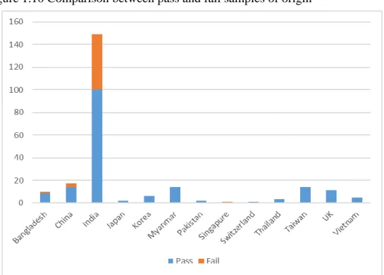
the label. There were 221 (94%) foreign medicines among 235 samples collected from
75 locations. Five samples of GM and 1DN sample were not registered with Myanmar
Food and Drug Administration (MFDA). In quality analysis, 36 samples out of 177
(20.3%) did not pass quantity tests, 27 samples out of 176 (15.3%) did not pass content
10
uniformity tests, and 23 out of 128 samples (18.0%) did not pass dissolution tests. Three of the unregistered GM samples failed in both identification and microbial assay tests.
Counterfeit GM is being sold in Yangon. Also, the quality of OM is a matter of concern, and requires follow-up. Poor-quality medicines were frequently found among the products of a few manufacturers. Regular surveys to monitor counterfeit and substandard medicines in Myanmar are recommended.
We found that poor-quality medicines are the urgent problems in Cambodia and
Myanmar, even though the medicines were not counterfeit. Serious dissolution failure
is the dominant problem in these countries. It is necessary to collect more information
of such medicines, and to analyze the characteristics of the data for preventing health
hazards caused by falsified or substandard medicines.
11
Acknowledgements
I wish to express my indebtedness, sincere appreciation and deepest sense of gratitude to my respected supervisor Dr. Kazuko Kimura, Professor, Institute of Medical, Pharmaceutical & Health Sciences, Kanazawa University, Japan for her highly valued supervision, solemn instruction, valuable suggestions and constant encouragement during the entire period of this research work and in the preparation of this dissertation.
I accord my heartily reverence to Dr. Hirohito Tsuboi, Associate Professor, Institute of Medical, Pharmaceutical & Health Sciences, Kanazawa University, Japan for his whole-hearted co-operation, encouragement and enthusiastic suggestion throughout my research work.
I am very much grateful to Dr. Naoko Yoshida, Assistant Professor, Institute of Medical, Pharmaceutical & Health Sciences, Kanazawa University, Japan for her cordial help with suggestion in my experiment.
I am very much grateful to Dr. Nobuko Tuno, Associate Professor, Department of Ecology, Kanazawa University, Japan for using her laboratory equipment.
I am very much thankful to Dr. Tsv Yashi and others Myanmar FDA staff for their great support during our survey.
I am very much beholden to Heng Bun Kiet and Eav Dararath DDF in Cambodia for their excellent support and providing the idea about other rural in Cambodia
In fine, all praises go to Almighty Allah, the omniscient and the most merciful.
The Author
12
13
Dedicated to
the memories of
2011 Tōhoku earthquake and tsunami victims
14
15
Table of Content
Abstract………. 7
Acknowledgement………. 11
Table of content………. 15
General Introduction……….. 1
Chapter One: An investigation into the quality of medicines in Yangon, Myanmar… 2 1.1 Introduction………... 3
1.2 Aim of This Work………. 4
1.3 Sample Collection………. 5
1.3.1 Observation Analysis ……… 6
1.3.2 Sample Authenticity Investigation………. 6
1.3.3 Samples for Chemically Analysis………... 7
1.3.4 Samples for Biological Analysis………. 25
1.4 Results………... 37
1.4.1 Sample collection ………... 37
1.4.2 Drug outlets and registration status in Myanmar FDA ……….. 37
1.4.3 Observations………... 39
1.4.4 Authenticity………. 41
1.4.5 Quality evaluate of samples……… 44
1.4.6 Factors influencing the outcome of the quality test………... 52
1.4.7 Effect of air-conditioning……….
521.4.8 To observe again of the unacceptable samples by using new judge which is wider than original (pharmacopeial criteria) ……… 53
1.4.9 Result of fluorescence spectrophotometer analysis……….. 55
1.5 Discussion ……….. 59
1.6 Conclusion ………. 63
Chapter two Four-year survey of the quality of antimicrobials in Cambodia……… 64
2.1 Introduction……… 65
2.2 Objective………. 65
2.3 Materials and Methods……… 66
2.3.1 Selection of sampling areas ………. 66
2.3.2 Samples collected……… 66
2.3.3 Observation………. 66
2.3.4 Authenticity………. 67
2.3.5 Sample chemically analysis………. 67
2.3.6 Statistical analysis……… 69
2.4 Results ……… 69
2.4.1 Drug outlets ……… 69
2.4.2 Observations ………... 72
2.4.3 Authenticity ……… 74
2.4.4 Quality investigation of samples……….. 74
2.5 Discussion ……….. 79
2.6 Conclusion………... 81
Chapter three Quality survey of selected medicines in Cambodia, 201 1- 2013……….. 82
3.1 Introduction……… 83
3.2 Methods………... 84
3.2.1 Sample collection………. 84
16
3.2.2 Observation……….. 84
3.2.3 Authenticity………. 85
3.2.4 Quality analysis……… 85
3.2.5 Statistical analysis………... 85
3.3 Results……… 88
3.3.1 Drug Outlets……… 88
3.3.2 Observations……… 89
3.3.3 Authenticity………. 89
3.3.4 Quality evaluation………... 92
3.4 Discussion………... 94
3.5 Conclusion ………. 95
Chapter four Comparatively study between Myanmar and Cambodia……… 96
Comparatively study between two-countries ……… 97
Conclusion of these surveys………... 99
References……….. 101
Annex 1.1………... 111
Annex 1.2………... 112
Annex 1.3………... 113
Annex 1.4………... 116
Annex 1.5………... 117
Annex 1.6………... 119
Annex 1.7………... 121
Annex 1.8………... 140
Annex 1.9………... 154
Annex 2.1………... 169
17
List of Tables
Table 1.1 Preparation of the different concentration of endotoxin
solution……….. 28
Table 1.2 Outline of samples collection………... 38
Table 1.3a Reply from manufacturers with their number of samples……….. 42
Table 1.3b Reply from manufacturers with their number of samples……….. 43
Table 1.4 Reply from MRAs……… 44
Table 1.5 Summary of quality test of samples ……… 46
Table 1.6 Association between price and medical quality (CXM, GM, OM and CTRX) ………. 52
Table 1.7 Association between air conditioning and temperature /humidity... 52
Table 1.8 Showing of the comparisons of the pharmacopeial quality test between original and newly considered value………... 53
Table 1.9 are showing the comparisons between original all and new all tests……… 53
Table 1.10 Compare the results between pharmacopeial guideline and considered new judge……… 55
Table 2.1 HPLC conditions for pharmacopoeial tests………. 68
Table 2.2 Outline of sample collection in Cambodia……… 70
Table 2.3 Number of sample collected which were produced domestically (Cambodia) foreign samples……… 71
Table 2.4 Significance association among the drug outlets in quantity test………… 72
Table 2.5 Number of abnormal samples were found during observation analysis….. 73
Table 2.6 Number of unregistered samples in DDF………. 73
Table 2.7 MRAs and manufacturers replied during the authenticity investigation….. 75
Table 2.8 Summary of quality test of samples ……… 76
Table 2.9 Comparison between price and result of the quality test in samples……… 77
Table 2.10 Factors association with quality test found in roxithromycin samples which were originated from Cambodia and other countries……… 78
Table 3.1 HPLC conditions for pharmacopoeial tests ………. 87
Table 3.2 Number of samples collected from different outlets ……… 88
Table 3.3 Samples without registration or insert ………. 91
Table 3.4 Summary of quality test of samples ……… 92
Table 3.5 Statistical analysis ………... 93
18
19
List of Figures and Illustrations
Figure 1.1 Chromatogram of cefuroxime standard……….. 9
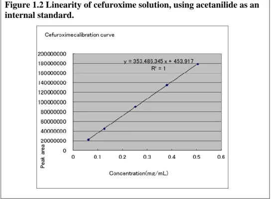
Figure 1.2 Linearity of cefuroxime solution, using acetanilide as an internal standard………... 10
Figure 1.3 Outline of endotoxin gel test………... 29
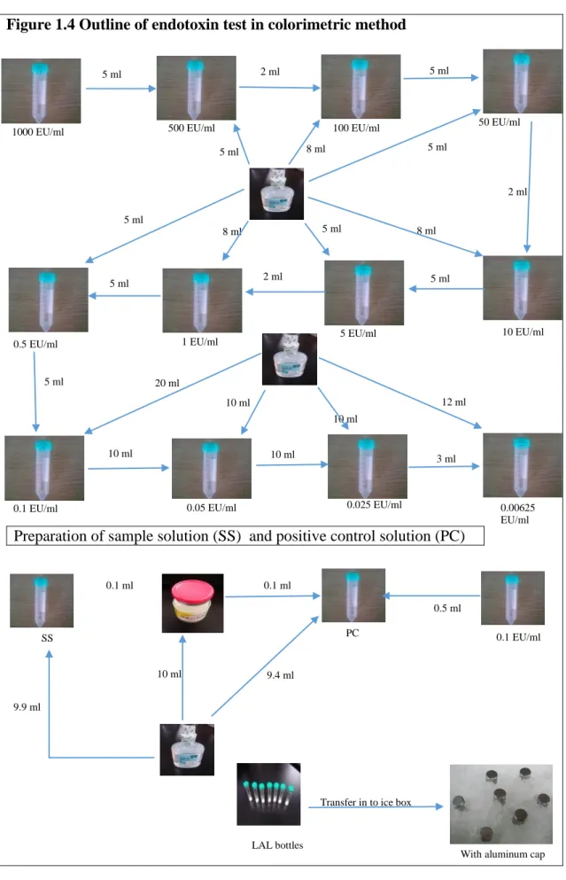
Figure 1.4 Outline of endotoxin test in colorimetric method ……….. 33
Figure 1.5 outline of sterility test………. 36
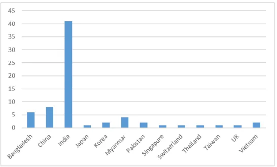
Figure 1.6 Number of Manufacturers found in the program ……….. 40
Figure 1.7 Number of samples collected from that origins……….. 40
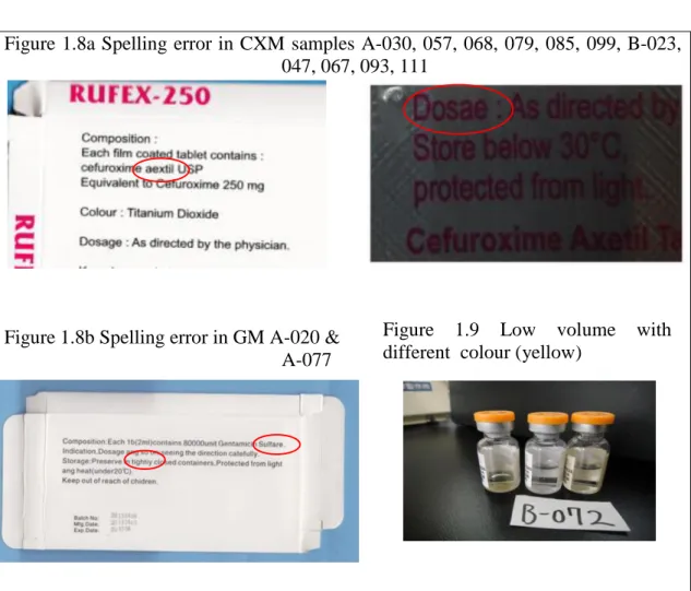
Figure 1.8a Spelling error in CXM samples A-030, 057, 068, 079, 085, 099, B-023, 047, 067, 093, 111………. 41
Figure 1.8b Spelling error in GM A-020 & A-077……….. 41
Figure 1.9 Low volume with different colour (yellow)……….. 41
Figure 1.10 Comparison between pass and fail samples from different countries………….. 47
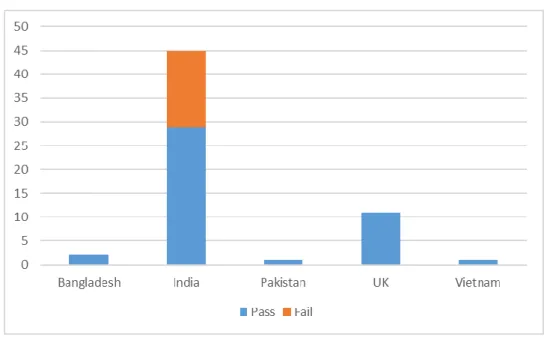
Figure 1.11 Comparison between CXM pass and fail samples from different countries …… 48
Figure 1.12 Comparison between OM pass and fail samples from different countries…….. 48
Figure 1.13 Comparison between GM pass and fail samples from different countries…….. 49
Figure 1.14 Comparison between CTRX pass and fail samples from different countries……… 49
Figure 1.15 Comparison between DN pass and fail samples from different countries……… 50
Figure 1.16 Chromatogram of GM standard………... 50
Figure 1.17 Chromatogram of counterfeit GM samples………... 50
Figure 1.18
Counterfeit gentamicin samples………
51Figure 1.19 Zone of inhibition (microbial assay) are showing between standard concentration and counterfeit GM samples………... 51
Figure 1.20 Image of fluorescence spectrophotometer of counterfeit samples A-020 (China)………... 55
Figure 1.21 Image of fluorescence spectrophotometer of counterfeit sample A-069 (China)………... 55
Figure 1.22 Image of fluorescence spectrophotometer of counterfeit sample A-077 (China)………... 56
Figure 1.23 Image of fluorescence spectrophotometer of pass sample B-09 (Bangladesh)………... 56
Figure 1.24 Image of fluorescence spectrophotometer of pass sample of B-072 but colour change white to yellow before expiration (India)………... 57
Figure 1.25 Image of fluorescence spectrophotometer of pass sample A-024 (Myanmar)………... 57
Figure 1.26 Image of fluorescence spectrophotometer of pass sample A-040 (Taiwan)………. 58
Figure 1.27 Image of fluorescence spectrophotometer of pass sample A-090 (Vietnam)………... 58
Figure 3.1a Different boxes or containers of cimetidine………. 90
Figure 3.1b Different boxes of amlodipine ………. 90
Figure 3.1c Different tablets of amlodipine………... 91
20
General Introduction:
Medicine is one of the most essential elements especially for human being to survive in the in the world. People used different types of plants for their treatments before 5000 years [1] however, now in modern world patients are using biotech medicines like as insulin, interferon, interleukin and so on [2]. Sir Alexander Fleming discovered the
benzylpenicillin (Penicillin G) from the mould Penicillium notatum in 1928 [3], sincethen patients in the world wants to use particular elements (active ingredient) for their
treatments. From the historical reason and requirement in the world manufacturers are
producing lot of medicines and supplying to the markets. Some manufacturers are taking
a chance and preparing counterfeit or falsified or poor quality medicines and supply to
the markets. These types of medicines are accessed in both developed and developing
countries [4-6]. One investigation was occurred and found around 1% and 10% in
developed and developing countries, respectively [7]. People are suffering and even died
due to effect of counterfeit / falsified / substandard / poor quality medicines which
evidences were already established in the world [8-10]. Perception from the above of
story the governments of Myanmar and Cambodian were started more than one
collaborative projects with Kanazawa University investigated to observe their own
situations and evaluate the quality of selected medicines through different surveys.
Chapter One:
An investigation into the quality of medicines in Yangon,
Myanmar survey in 2014
3 1.1 Introduction
Medicines are the most essential elements especially for human beings for surviving their lives in this world. It is almost impossible to imagine the remedy of human body from various diseases without taking good quality of medicines. Deliberately, many pharmaceuticals have been producing counterfeit medicines and supply to the patients as well as they are taking a chance to earn more money by producing such detrimental counterfeit medicines and even extending their imposture day by day. This is also happening in both developed and developing countries [4-6]. In this vast sector it is very difficult to optimize the counterfeit medicines. Depending on geographic region the range of counterfeit drugs supply to the developed countries as well as the rising countries are about 1% and 10%, respectively [7]. Another investigation from the World Health Organization (WHO) about 20%-90% falsified medicines were found in several Africa countries [11, 12]. The
incidence due tocounterfeit medicines were estimated in
Cambodia with the range of 4%- 90% from 2001 to 2010 [13-16]. Furthermore, owing tofake medicines
around 200 children were died in Bangladesh in 1990–1993 ingesting counterfeit paracetamol that contained diethylene glycol [17].
In Myanmar, a massive investigation occurred by World Health Organization
(WHO) in 1999 and caught counterfeit medicines [18]. It is very difficult to identify such
4
counterfeit medicines however it is also possible to buy good quality medicines. The problem is that the sellers demand extra money for good quality medicines which is illegal and unethical. For this reason, an emergency cases, people suffer or may die for the prevalence of counterfeit drugs [17, 19]. In most cases the patients from developing counties do not want or cannot fulfill antibiotic courses due to their economic crisis. Thus the misuses or inadequate doses of antibiotics may guide them to the advance of resistance [20] while support the extra food demands of the rising population of the world antibiotics are using in husbandry sectors specially in poultry industry as a growth promoter and transmitted to the human that is occurred resistance by several types of microorganisms particularly in bacteria [21, 22].
Counterfeiting or poor quality antibiotic is Worldwide spreading that is one of the biggest and vital factors and is making sub-inhibitory concentrations naturally and enhance the selection of resistant strains from various types of microorganisms [23, 24].
1.2 Aim of This Work
People in low income countries are suffering in counterfeit or poor quality
medicines in their daily life. People in this type of country are almost depending on
foreign country medicines. In 1999, a massive investigation occurred by WHO to evaluate
the quality of medicines and found counterfeit medicines in Myanmar. Since then there
5
were no systematic survey conducted on medicines in Myanmar. We want to investigate the quality of medicines which associate outlet condition, outlet types, price of medicine, type of medicines (domestically produced or not) and medicines entered in to Myanmar that is needed to fulfil Myanmar government policies. Finally, we suggested to the government of Myanmar how to remove counterfeit or poor quality medicines from Myanmar markets.
1.3 Sample Collection
From the suggestions of Myanmar FDA (MFDA), we selected a populated region as well as from the MFDA essential drugs list, we selected five types of medicines.
Samples of oral medicines, cefuroxime axetil (CXM) [25], donepezil hydrochloride (DN)
and omeprazole (OM) [26] and injections, ceftriaxone sodium (CTRX) and gentamicin
sulfate (GM) [27] were purchased during 27 September- 4 October 2014 by two teams
from Yangon, Myanmar (Annex 1.1). Each team consisted of one supervisor from MFDA,
one local assistant and one or two Japanese researcher (s). All team members received
training before starting the sampling work. During the sampling period, we maintain a
sampling form for each number of samples (Annex 1.2). We collected samples from the
governmental hospital and private hospital as well as community pharmacies, clinical
6
pharmacies and wholesalers. Obtained samples were stored at 20-25ºC before analyzed in Kanazawa University, Japan.
1.3.1 Observation Analysis
During the sampling we observed room conditions like as temperature, humidity and also observed in both internal and external environmental conditions around the surrounding of the retail shops. After sampling, the obtained samples were checked physical shape, size of sample volume or shape, uniform colour, insert, spelling, registration number from MFDA, manufacturing date, expire date, lot no., name of active ingredient, doses form etc. that were utilized in the form of the
“Tool for Visual Inspection of Medicines” (Annex 1.3) [28]. For establishing the evidences, photographs were takenfor each samples with scanned insert and the sample box.
1.3.2 Sample Authenticity Investigation
The authenticity investigation and registration verification was performed
according to the modification method of World Health Organization (WHO) [16]. We
asked some questions to the responsible manufacturers by using a form (annex 1.4) with
sent scan copy of the samples box, samples photographs and insert of the sample by E-
mail (annex 1.5). At the same time, we asked to the Medicine Regulatory Authority7
(MRA) of each country regarding the medicines were registered or not (annex 1.6). While,
we asked to the MFDA about obtaining medicines were registered or not.
1.3.3 Samples for Chemical Analysis
Samples were evaluated according to the pharmacopeia that mentioned on the
package of the samples. In the following method we used and evaluated our collected
samples. Our collected cefuroxime samples 250 mg tablets were performed dissolution,
content uniformity and quantity test. To determine the amount of cefuroxime
(C
16H
16N
4O
8S) dissolved by employing UV absorption at the wavelength of maximum
absorbance at 278 nm on filtered portions of the solution under test, suitably diluted with
dissolution medium 0.07N Hydrochloric Acid; 900 ml, if necessary, in comparison with
a standard solution having a known concentration of UPS cefuroxime axetil RS,
equivalent to about 0.01 to 0.02 mg of cefuroxime (C
16H
16N
4O
8S) per ml, in the same
medium. 55 rpm (for test 1) and 100 rpm (for test 2) were used during the dissolution test,
while samples considered at 15 and 45 minutes not less than 60%, 75% for 1
ststage and
50% ,70% for 2
ndstage gradually. 0.2 M monobasic ammonium phosphate (purchased
from Nakalai Tesque Kyoto, Japan) dissolve 23.0 gm of monobasic ammonium
phosphate in water to preparer 1000 ml of solution. 620 ml solution were taken from 1000
ml and added 380 ml methanol (Wako, Japan) to make 1000 ml mobile phase. 5.4 mg
8
acetanilide dissolved in per ml methanol to preparer internal standard solution. For
Resolution Solution, mix 10.0 ml of a solution of USP cefuroxime axetil RS in methanol
containing 1.2 mg per ml then transfer in a 50 ml volumetric flask and including of 5.0
ml of internal standard solution with 3.8 ml of a solution of cefuroxime axetil Delta-3
Isomers RS in a methanol containing 0.16 mg per ml. Finally, to fill with dilute with 0.2M
monobasic ammonium phosphate to make the target the volume, and well mix. For
standard preparation, transfer 30 mg of USP cefuroxime axetil RS to a 25 ml volumetric
flask dissolve dilute to make the volume. Promptly transferred 10.0 ml of this solution to
another 50 ml volumetric flask then added 5.0 ml of internal standard solution and 3.8 ml
of methanol, finally added dilute to make the volume. In assay preparation, fine powder
not fewer than 10 tablets were accurately counted. Transfer the powder, with the aid of
methanol, to a volumetric flask of such capacity that when filled to volume, the solution
will contain the equivalent of about 2 mg of cefuroxime (C
16H
16N
4O
8S) per ml. Added
methanol to fill the volumetric flask to about half of its capacity, and shake by mechanical
means for about 10 min. Dilute with methanol to volume, and mix. Filter a portion of this
stock mixture, and transfer 5.0 ml of the filtrate to a 50 ml volumetric flask. Add 5.0 ml
of internal standard solution and 8.8 ml of methanol dilute with 0.2 M monobasic
ammonium phosphate to the volume, and mix. The HPLC system from JASCO (Tokyo,
9
Japan) were maintained 278 nm UV detector, 4.6 mm x 25 cm; 5µm packing L13 column, 40
0C column oven temperature, flow rate 1.2 ml/min and injection volume 10 µl. In quantity analysis 90.0≦mean≦110.0 and content uniformity AV≦15.0 were followed [29],
cefuroxime peak observed (Figure 1.1).The linearity of the standard cefuroxime/diluent solution was maintained and analyzed between 0.025 and 0.5 mg/ml and the 0.6 to 0.5 (Figure 1.2).
Figure 1.1 Chromatogram of cefuroxime (standard)
10
Figure 1.2 Linearity of cefuroxime solution, using acetanilide as an internal standard.
For content uniformity test of ceftriaxone for injection (1 gm vial) at first we prepared P
H7.0 buffer, dissolve 17.415gm of dibasic potassium phosphate in 500 ml of water and dissolve 13.605 gm of monobasic potassium phosphate in 1000 ml of water.
Control the p
Hof dibasic potassium phosphate solution to p
H7.0 by using monobasic
potassium phosphate solution. p
H5.0 buffer, dissolved 12.9 gm of sodium citrate in 250
ml of water and dissolved 9.6 gm of citric acid in 500 ml of water. Control the p
Hof
sodium citrate solution to p
H5.0 by using citric acid solution. Dissolved 3.2 gm of
tetraheptyl ammonium bromide were taken in 400 ml of acetonitrile, added 44 ml of p
H7.0 buffer and 4 ml of p
H5.0 buffer, and added water to make 1000 ml to make the mobile
phase. 0.5 µm membrane filter was used then allow to degas. 450 ml Acetonitrile were
taken into the 1000 ml volumetric flask then added water up to the volume for diluents as
11
well as IS preparation 5 mg of diethyl terephthalate were taken to dissolve in diluents and
make up to the volume 50 ml. Regarding the standard solution preparation, 5 mg of
ceftriaxone sodium RS were transferred to 50 ml volumetric flask, and dilute with diluents
to the volume (0.1 mg/ml) then 2 ml of these solutions were transferred into 10 ml
volumetric flask (20µg/ml) to make 200%. Mixed 3 ml of 20µg/ml solution and 1ml of
diluents (15 µg/ml) to make150%. Mixed 2 ml of 20µg/ml solution with solution 2 ml
diluents (10 µg/ml) for 100%. Again mixed 2 ml of 10 µg/ml solution with 2 ml of
diluents (5 µg/ml) to make 50% of the solution. While, mixed 2 ml of 5 µg/ml solution
and 2 ml of diluents then prepared (2.5 µg/ml) 25% of the samples. Mixed 1 ml of each
(200%, 150%, 100%, 50% & 25%) of this solution with 1 ml of IS solution for linier
carve. During the assay preparation, 1 gm ceftriaxone sodium were transferred to a 100
ml volumetric flask then added diluents to the volume. Transferred 1 ml of this solution
to 50 ml volumetric flask and added with diluents to the volume. Mix 0.5 ml of this
solution and 2.5 ml of diluents. Mix 1 ml of this solution and 1 ml of IS solution. In
chromatographic system 270 nm detector, 4.0 x 10 cm column, 2.0 ml/min flow rate,
injection volume and 40
0C oven temperature were maintained. In quantity analysis
90.0≦mean≦115.0 and content uniformity AV≦15.0 were followed [30].
12
Donepezil samples were investigated according to the Japanese Pharmacopoeia 16th edition (JP 16). 2.5 gm of sodium-1 decane sulfonate were dissolved in 650 ml of water, and added 350 ml of acetonitrile, 1 ml of per chloric acid to make mobile phase, then to prepare standard solution and weighed accurately about 50 mg of JP donepezil hydrochloride RS, and dissolved in diluent-1 (Methanol and 0.1 mol/L hydrochloride 3:1) to make exactly 25 ml. Transfer 5 ml of this solution to a suitable test tube, added diluent- 1 to make exactly 50 ml. For assay preparation, one tablet of donepezil hydrochloride with added diluent-1 so that it contained the concentration about 0.2 mg per ml then sonicate and properly mix until a tablet is disintegrated for 10 min. After sonicate then centrifuge for 4000 rpm for 15 min with continued 25℃ and supernatant solution were taken. For dissolution test was performed at 50 revolutions per minute according to the puddling method as directed under the dissolution test in JP16, using 900 ml of the dissolution medium. After the dissolution, performed the test with 50 µl each of the sample solution and standard solution as directed under Liquid Chromatography in JP16 followed and calculate the ratios of AT and AS, of the peak area of donepezil hydrochloride. 3.4 gm Potassium dihydrogen phosphate and 3.55 gm of sodium dihydrogen phosphate were taken in 1000 ml water to make the phosphate buffer.
Phosphate buffer (pH 6.8) and water (1:1) were used as a dissolution medium. Mightysil
13
RP 18GP 150×4.6 mm (5 µm) column, wavelength 271 nm, 35℃ in column temperature,
1.0 ml/min flow rate, 50 µl injection volume, mobile phase: water, acetonitrile and per chloric acid (650:350:1) were used in this test. To make the standard solution we weighed accurately about 20 mg of JP donepezil hydrochloride RS with dissolve in diluent-1 to make exactly 20 ml. 1 ml of this solution were transferred to a suitable test tube then added with the dissolution medium to make exactly 100 ml. In addition, transferred 5 ml of this solution to a suitable test tube and added the dissolution medium to make exactly 10 ml solution. The sample solution was withdrawal not less than 20 ml of the medium at 15 min, after starting the test and filtered with a membrane filter which contained the pore size of 0.45 µm. Discard the first 10 ml filtrate of the sample then transfer the subsequent filtrate to a suitable test tube. Not less than 80 % of the labeled amount of donepezil hydrochloride (C24H29NO3・HCl) is dissolved in 15 min were considered [31].
Identification was performed on the gentamicin samples. 1gm of o- phthalaldehyde in 5 ml of methanol and added 95ml of 0.4 M boric acid that previously adjusted with 8 N potassium hydroxide to a p
Hof 10.4 and 2 ml of thioglycolic acid.
Adjust, obtained of the solution with 8 N potassium hydroxide to a pH of 10.4. To prepare
the mobile phase and maintained the ratio of methanol, water and glacial acetic acid
14
(68:27:5) as well as 5gm of sodium 1-heptanesulfonate were added per liter to this solution. Then standard solution was prepared to use of USP gentamicin sulfate RS in water to make the concentration of 0.65mg per ml. 10 ml of this solution were transferred to a suitable test tube and added of 5ml of isopropyl alcohol with 4 ml of o-phthalaldehyde solution then properly mix and finally isopropyl alcohol were added to obtain 25 ml of solution. At 60°C tempereture were maintained in a water bath for 15 minutes then cool.
In the case of sample solution preparation, 1ml of the sample were taken and mix with 60.5 ml of water. Taken 10 ml from the mixture transfer to a suitable test tube with added 5ml of isopropyl alcohol as well as 4 ml of o-phthalaldehyde solution then properly mix and finally added isopropyl alcohol to obtain 25 ml of solution. At 60°C tempereture were maintained in a water bath for 15 minutes then cool. Phenomenex Luna C18 L1 150×4.6 (mm) column, 1.0 ml/min flow rate, 330 nm UV detector and 20 μl injection volume were used in the chromatographic system. In analysis part, we compared the peak of the sample with that of the RS to quantitate GM, and determine whether any impurity peaks appear in the chromatogram [32].
Content uniformity test in omeprazole at first to make for the solution A (1L)
10.454 gm tri-sodium phosphate 12-water and 15.616 gm disodium hydrogen phosphate
were taken in a 1000 ml volumetric flask. Suitable amount of distill water were added
15
and sonicate to dissolve it. Adjust the volume with distill water then adjust the p
Hto 11.0
± 0.05 with 10 M sodium hydroxide or orthophosphoric acid were used. For solution B (500 ml) 5 ml of 10 M NaOH were taken in 500 ml volumetric flask. To make the volume of 500 ml with 0.05 M phosphate buffer solution p
H4.5 and well mix. In 1 L phosphate 6.8 gm potassium dihydrogen were taken in a 1000 ml volumetric flask and added suitable amount of distill water to dissolve and used a sonicate, adjust the volume and then filter by a vacuum filter and degas it. 210 ml of 0.05 M phosphate buffer solution (p
H4.5) with 60 ml of solution B were mixed, from it 200 ml solution were taken in a 1000 ml volumetric flask and make volume with solution A. Finally, this solution used for diluent.
Regarding the mobile phase, 1.17 gm of sodium dihydrogen phosphate dihydrate
(NaH
2PO
4.2H
2O) were taken in a 500 ml volumetric flask, allow to the sonicator for
dissolved and added with dilute to the volume. In another 500 ml volumetric flask was
taken and transferred 1.06 gm of disodium hydrogen phosphate (Na
2HPO
4) dissolve in
diluent and make sure the volume. Transferred the NaH
2PO
4.2H
2O solution to a 1000 ml
beaker and adjust the p
Hto 7.6 ± 0.05 with Na
2HPO
4solution in a p
Hmeter. For 1 Liter
of mobile phase 600 ml of this solution were taken in a 1000 ml volumetric flask and
added 400 ml of acetonitrile (60:40 ratio). Filtered the solution in a suction filter and then
degas the mixture in a sonicator for 30 minute. For standard solution preparation 10 mg
16
of omeprazole RS were weighted and dissolve to a 50 ml volumetric flask with medium in a sonicator than sure to make the final volume and properly mix which was 200%
(solution concentration 0.2mg / ml). From this concentration of the solution with medium to make 150%, 100%, 50% and 25% of solution. while weighted accurately about 10 mg of lansoprazole were dissolved to a 100 ml volumetric flask with medium in a sonicator, make final volume and mix for IS preparation (0.1 mg/ml). Transferred 1 ml of 200% ~ 25% to each test tube with 1 ml IS added to each mix then allow to filter and put it in 1 ml vial. For assay preparation in content uniformity, 10 capsules granules were transferred in 50 ml volumetric flask. Dissolved the capsules with diluent in a sonicator and continue sonication until it dissolves. Transferred 1 ml from each to 10 test tube and added 3 ml of diluent and mix then filtered of the sample by 0.45 μm filter paper, 1 ml of this solution was taken and added 1 ml of IS solution with well mix and transferred to 1 ml vial. 4.6 mm × 150 mm column (C18) column, 302 nm wavelength, 30
0C column oven temperature, 0.5 ml/min flow rate and 10 μl injection volume were maintained in chromatographic system. In quantity analysis 95.0≦mean≦105.0 for BP as well as 90.0≦mean≦110.0 for USP and content uniformity were followed in AV≦15.0 [33-35].
In dissolution test for omeprazole samples were performed according to BP and USP that
were mentioned on the package label. Regarding the dissolution test in BP, solution A
17
and solution B were used as well as to prepare solution C with 1.170 gm Sodium
dihydrogen phosphate dihydrate and 1.061 gm disodium hydrogen phosphate were taken
in a separate 500 ml volumetric flask. Suitable amount of distil water were used then
allow for sonicate to dissolve and then adjust volume. Added 400 ml of disodium
hydrogen phosphate to 500 ml of sodium dihydrogen phosphate dihydrate and adjust p
Hto 7.6±0.05. We prepared the mobile phase and 400 ml acetonitrile with 600 ml of
solution C were properly mixed (p
H7.6±0.05) then filter it in a vacuum filter and degas
it for 30 minute in a sonicator. At that time, we prepared this solvent, 13.6 gm potassium
dihydrogen phosphate were taken in a 2000 ml volumetric flask with added distil water
to dissolve in a sonicator (about 10 min), adjust the volume and allow for filter by a
vacuum filter then degas it. For the first stage, 0.05 M phosphate buffer solution p
H4.5
and solution A (1:4, v/v) as well as in the final stage, 0.05 M phosphate buffer solution
p
H7.6 and solution A (1:4, v/v) were mixed to make the diluent. In the dissolution tester
water were transferred before test, to keep warmed to 37 ± 0.5 ℃. Measure the degassed
solvent 700 ml in a graduated cylinder and put it in the vessel. Filled all the six vessels
following the first one. Mount the paddle up, then lower it to the original position when
temperature reaches to the desired level, set rotational speed to 100 rpm. Put the weighed
samples, in each vessel in 1 min interval. In acid stage (p
H4.5), after 45-minute elution,
18
5ml medium were withdrawal and filter the aliquot of dilute to 25 ml with solution A in a 25 ml volumetric flask then transferred 1 ml of this test solution to a test tube with added 1 ml IS solution to it and properly mixed. Proceed immediately to the final stage.
Preparation of standard we used, 1) 40 ml of 0.05 M phosphate buffer solution (p
H4.5)
were taken in a 200 ml volumetric flask and fill it up to the mark with solution A (Solution
D). 2) Accurately weigh 5 mg of lansoprazole IS were put in 50 ml a volumetric flask,
added a suitable amount of Solution D, sonicate for 10 minutes to dissolve and then make
it up to 50 ml with solution D (0.1mg/ml). Taken 1 ml of this solution to place it in a
volumetric flask of 10 ml and filled up with Solution D (IS solution with 10 μg / ml). 3)
Accurately weigh 5 mg of Omeprazole RS and put in a volumetric flask of 50ml, add an
appropriate amount of Solution D, sonicate for 10 minutes to dissolve, and make volume
(0.1mg/ml). 2 ml of this solution were transferred to 10 ml volumetric flask and dilute it
up to the mark with solution D 200% solution (concentration of 20 μg/ml). From this
concentration of this solution with diluent to make 150%, 100%, 50% and 25% of the
solution. Transferred 1 ml from solution 200% ~ 25% to each test tube and added 1 mL
IS to each with mix then filter and put it in 1ml vial. For the buffer stage (pH 6.8), within
5 minutes of the 200 ml of solution B at 37±0.5
0C were added to each vessel. The rotation
speed at 100 revolutions per minute was controlled and continue to operate the apparatus
19
for 45 minutes as well as again 5 ml of the dissolution medium were withdrawn 45 minutes, after starting the test and transferred to a 25 ml volumetric flask, make sure to the volume with dilute then 1 ml of above test solution were transferred to a test tube and added 1 ml of IS solution with well mix. Regarding the buffer stage again we prepared for standard, 1) 21 ml of 0.05M phosphate buffer solution (p
H4.5) were mixed with 6 ml of Solution B, from this solution 20 ml were transferred in a 100 ml volumetric flask and make volume with solution A (Solution E). 2) 5 mg Lansoprazole were transferred in a 50 ml volumetric flask with added the suitable amount of solution E and sonicate for 10 minutes to dissolve then actual make the volume (0.1mg/ml). Taken 1 ml from it to a 10 ml volumetric flask then make volume with solution E that was IS solution. 3) 5 mg omeprazole RS were put in a 50 ml volumetric flask, added a suitable amount of solution E and sonicate for 10 minutes to dissolve then make volume (0.1mg/ml). From this volume 2 ml were placed in a 10 ml volumetric flask and filled it up with Solution E (20μg / ml) and obtained 200% solution. From 200% solution with diluent used to make 150%, 100%, 50% and 25% of the solution. Transferred 1 ml from solution 200% ~ 25%
to each test tube and added 1 ml of IS to each with mix then allow to filter and put it in
1ml vial. In chromatographic system 302 nm detector, Gemini-NX column, 0.5 ml/min
flow rate, 30℃ temperature and 10μl injection were used. In acid stage, no individual
20
unit exceeds 10% dissolved and buffer stage no unit is less than Q+5% (Q= 65%) were considered. According to the USP dissolution method, for the mobile phase we used 340 ml of Acetonitrile to a 1000 ml volumetric flask, dilute with p
H7.6 phosphate buffer to the volume then allow for filtration through membrane filter then degas for 30 minutes.
1) For Acid Resistance Stage, 40 ml of 5N HCl were measured exactly and placed it in a 2000 ml volumetric flask dilute were used to make the volume (0.1NHCl). 2) pH 10.4, 0.235M disodium hydrogen phosphate (For 1L) 2.4 L for Buffer Stage 33.36 g of disodium hydrogen phosphate were dissolved in 1000 ml of water and adjust with 2N sodium hydroxide for p
Hof 10.4 ± 0.1. 3) pH 6.8 phosphate buffer (900mL), 500 ml of 0.1N hydrochloric acid were added with 400 ml of disodium hydrogen phosphate p
H10.4.
0.235 M dibasic sodium phosphate (Na
2HPO
4.7H
2O) were used to adjust with 2N hydrochloric acid or 2N sodium hydroxide, if necessary to the contain of p
Hof 6.8 ± 0.05.
4) p
H7.6 phosphate buffer (1L) for the mobile phase, 0.178 gm sodium dihydrogen
phosphate and 1.12 gm disodium hydrogen phosphate were transferred in a 250 ml
volumetric flask and dissolve it with distilled water. If necessary, adjust to p
H7.6 ± 0.1
with utilized 2N sodium hydroxide or 2N hydrochloric acid. Total solution was
transferred to 1000 ml volumetric flask and make the volume with dilute. 5) For 0.01M
sodium borate solution (1L), 3.8137 gm of Sodium tetra-borate decahydrate (Borax) were
21
taken in a 1000 ml volumetric flask and make the volume with distilled water. Regarding the dissolution of the Sample, water was pre-fill to the dissolution tester and to keep warm to 37.0 ± 0.5 ℃. 500 ml of media (0.1 N HCl) were placed in each of the six dissolution
vessels. The apparatus was assembled and warm the media to 37
o± 0.5
oC. Weigh and place pellets equivalent to 20 mg omeprazole were maintaining one-minute interval in each vessel and immerse paddle in media to a distance of 2.5 ± 0.2 cm between the paddle and bottom of the vessel. Analyze the sample by the following HPLC method. For the acid resistance stage in standard solution, 40 ml methanol were transferred in 200 ml volumetric flask than added with 160 ml of 0.01 M sodium borate solution for diluent preparation. To prepare the IS solution, 5 mg Lansoprazole RS were used and put it in a 50 ml volumetric flask as well as added a suitable amount of diluent allow to sonicate for 10 minutes for dissolving and then make it up to 50 ml with diluent (0.1mg/ml). From this solution 1 ml were taken and placed in a10 ml volumetric flask with filled up with solution D. To make the WS solution, put 5 mg of accurately weighed omeprazole RS into 50 ml volumetric flask with added an appropriate amount of diluent to allow sonicate for 10 minutes then filled the volume with diluent. 4 ml solution were put in a10 ml volumetric flask of and make up to volume with diluent for 200%) Solution. From 200%
solution with diluent to make 150%, 100%, 50% and 25% of the solution. Transferred 1
22
ml from solution 200% ~ 25% to each test tube and added 1 ml IS to each as well as mix to allow filter and put it in 1ml vial. In the case of test solution, after 2 hours filtered the dissolution medium which were containing the pellets through a sieve with an aperture not more than 0.2 mm. Collected the pellets on the sieve and rinse them with water and were using approximately 60 ml of 0.01M sodium borate solutions with carefully transfer the pellets quantitatively to a 100 ml volumetric flask then sonicate for about 20 minute until the pellets are broken up. Added 20 ml of methanol to the flask with dilute of 0.01M sodium borate solution to volume and properly mix. Dilute an appropriate amount of this solution with 0.01M sodium borate solutions were obtained a solution which having a concentration of about 0.02 mg per ml. Filter the solution through Whatman No. 42 or equivalent omeprazole filter paper were used. Then filter the filtrate again done through syringe filter of 0.20 micron. During the buffer stage, proceed as directed for Acid resistance stage with accurately weighed fresh pellets from the same batch. After 2 hours 400 ml of 0.235M dibasic sodium phosphate were added to the 500 ml of 0.1N hydrochloric acid medium in the vessel as well as of adjust, if necessary, with 2N hydrochloric acid or 2N sodium hydroxide to a p
Hof 6.8 ± 0.05 were used. At the end of 30 minutes, determine the amount of omeprazole dissolved in p
H6.8-phosphate buffer.
Regarding the test solution (for 20 mg display of capsule), after dissolution for 30
23
minutes, immediately transferred 5 ml of the solution under test to a test tube which containing 1 ml of 0.25 M sodium hydroxide, well mix well and filter the solution through Whatman No. 42 or equivalent filter paper. Then filter the filtrate again through syringe filter of 0.20 micron. To prepare the standard solution, 200 ml p
H6.8 phosphate buffer with 40 ml 0.25 M sodium hydroxide were used for diluent as well as in the IS solution, accurately 5 mg Lansoprazole RS were weighed and put it in a volumetric flask of 50 ml with added a suitable amount of the diluent then sonicate for 10 minutes to dissolve with the making for 50 ml which containing of the diluent, from this solution taken for 1 ml and placed it in a volumetric flask of 10 ml fill up with solution D. Then we were making the WS solution and put 5 mg of accurately weighed omeprazole RS into 50 mL volumetric flask with added an appropriate amount of diluent then allow for sonicate for 10 minutes finally to prepare the volume with diluent. Transferred 4 ml solution in a volumetric flask of 10ml and filled the volume with diluent which was 200% solution.
From this solution (200%) with diluent to make 150%, 100%, 50% and 25% of the solution. Transferred 1 ml from the solution of 200%~ 25% to each test tube and were added 1 ml of the IS solution to each and well mix allow to filter and put it in 1ml vial.
In chromatographic system 280 nm detector, 4.0 mm×12.5 cm including packing L7 of
5μm of column, 1.0 ml/min flow rate and 10 μl injection volume were used. In acid
24
resistance stage tolerance, level L1 individual data will not exceed 15% of the omeprazole dissolved, for the level L2 of 12 average units within 20% dissolved omeprazole in individual data will not exceed 35% omeprazole dissolved. Regarding the level L3of 24 within 20% of the average dissolution omeprazole units, greater than 35% of the maximum in also dissolved within 2 units is omeprazole, individual units is not greater than 45% of omeprazole dissolved. While in the case of buffer stage, level B1 Each unit is not less than Q+5% (Q=75%) and the level B2 average of 12 units was equal to or greater than Q and no individual unit were less than Q-15%, finally the level B3 average of 24 units is equal to or greater than Q and not more than 2 units were less than Q-15%
and no unit was then Q-25%.
In our investigation, we caught counterfeit gentamicin samples. For this reason,
we further investigation in this way to use fluorescence spectrophotometer and observed
and compare in pass and counterfeit samples.
In fluorescence spectrometry both anexcitation spectrum (the light that is absorbed by the sample) and/or an emission spectrum
(the light emitted by the sample) can be measured. The concentration of the analyte is
directly proportional with the intensity of the emission with excitation of wavelength.
25 1.3.4 Samples for Biological Analysis
Our collected gentamicin samples (injection) which were performed in microbial
assay according to the analysis of USP. Regarding this test, Staphylococcus epidermidis
ATCC 12228 strain were performed during this test. We used Base layer media which
consists of peptone, pancreatic digest of casein, yeast extract, beef extract, dextrose, agar
and water (12:8:6:3:2:32:2000) and controlled the p
H6.6±0.1. culture organisms were
transferred in this media. 16.73 gm/l of diabasic potassium phosphate and 0.523g/L of
monobasic potassium phosphate were mixed to make 0.1 M buffer with adjust the pH to
8.0±0.1 with 18 N phosphoric acid or 10 N potassium hydroxide. Microorganisms were
suspend in 10 ml saline and adjust the solution to give a transmittance of around 1.0% at
580 nm as a solution. For the standard solution, we weighted 10 mg of gentamicin RS
and dissolve in 10 ml of the buffer solution. From the serial dilution we prepared the
standard solution 5 (4.0 gm/ml), solution 4 (3.0 gm/ml), solution 3 (2.286 gm/ml),
solution 2 (1.0 gm/ml) and solution 1 (0.5 gm/ml). Also, prepared a control solution which
were containing 2.0 gm/ml (
=590 ug/mg as potency) of gentamicin RS. To make the
sample solution,1ml of the solution from the ampoule (sample) were taken and added to
a flask with adjust to 17576 fold dilution of buffer (= 2.2758 gm/ml) then transferred the
26
solution to a clean bench, allow for filter and place it a 2 ml tube. In this method we maintain the following procedure, at first we injected 100 ml of microrganism solution on the base layer, and spread with a turn table and spreader. At least five test plates are needed to make the standard curve. Second, place four cylinder-cups on each plate. Third, injected 250 ml of one of the standard solutions 1 to 5 and control solution on each plate.
Put control solution in one cylinder on each plate and fill the remaining cylinders as follows. 1) Plate 1 has one control and three cylinders of solution1. 2) Plate 2 has one control and three cylinders of solution 2. 3) Plate 3 has one control and three cylinders of solution 3. 4) Plate4 has one control and three cylinders of solution 4. 5) Plate 5 has one control and three cylinders of solution 5. 6) Plate 6 has one control and three cylinders of sample (1). 7) Plate 7 has one control and three cylinders of sample (2). Fourth, place all the test plates were in an incubator at 35°C and cultivate for twenty hours [36].
According to the USP (our collected samples) the endotoxin and sterility tests were applicable in both cefteriaxone (for injection) and gentamicin (injection) samples.
For endotoxin test were performed in two ways one for gel-clot thecnique and another
was chromogenic technique. In gel-clot technique, at first 5 ml pure water were injected
into the Limulus Ambocyte Lysate (LAL) vial. For another 10 ml pure water were
injected into the standard endotoxin which concentration 1000 EU/ml and then vortex.
27
From this concentration to prepare 100, 10, 1, 0.1, 0.6 (2λ), 0.03 (λ), 0.015 (0.5λ) and 0.0075 (0.25λ) (Table 1.1). Each step was done for vortex in one minute and solutions
were keep into an ice box. 10 ml pure water were used with sample and vortex for 1 minute, to make the sample solution. 0.6 ml were taken from the stoke solution with 5.4 ml pure water then vortex for 1 minute to make for dilute stoke solution. For the positive control, 1 ml from stoke solution with 1 ml from 100 EU/ml solution and then added 8 ml pure water to allow for vortex to make 10 EU/ml solution 1. 1 ml from the solution 1 with 9 ml pure water were used for 1 EU/ml solution 2. 0.3 ml solution 2 were taken and added 4.7 ml pure water for 2 λ solution 3. 1 ml from solution 3 with 7 ml of pure water were used to make 0.25 λ solution 4. A total 44 bottles were taken and transferred 0.1 ml of LAL reagent. Three bottles were used in each of the sample solution, positive control 10 EU/ml, standard endotoxin concentration 10 EU/ml and pure water as well as 1 battle was used for normal water. For an another case four bottle were used in each of the sample solution, positive control 10 EU/ml, 1 EU/ml, 2 λ and 0.25 λ. While two battles were used in each of the standard concentration 10 EU/ml, 1 EU/ml, 2 λ, 0.25 λ and pure water.
After one an hour incubation and we observed about the positive control of 10 EU/ml and
1 EU/ ml, standard concentration 10 EU/ml and 1 EU/ml solution with normal water
28
containing were solid in the bottles and rest of the bottles were liquid (Fig 1.3a & 1.3b) [37].
Table 1.1 Preparation of the different concentration of endotoxin solution
Concentration taken from the amount Pure water Concentration
From 1000 to 2 ml 18 ml 100
From 100 to 1 ml 9 ml 10
From 10 to 1 ml 9 ml 1
From 1 to 1 ml 9 ml 0.1
From 0.1 to 9 ml 6 ml 0.06 (2λ)
From 2 λ to 4 ml 4 ml 0.03 (λ)
From λ to 4 ml 4 ml 0.015 (0.5λ)
From 0.5 λ to 5 ml 5 ml 0.0075 (0.25λ)
29
0.6 ml Sample solution 1 ml
Figure 1.3 Outline of endotoxin gel test
5 ml PW
+
5 ml
5 ml 1 ml
2 ml LAL
10 ml
+
1000 EU/ ml
10 ml 4 ml
1 ml
10 EU/ml 1 EU/ml
0.25 λ EU/ml
Dilute Sample
5 ml
18 ml 100 EU/ml
+
Preparation the solution for positive control
4 ml
0.5 λ EU/ml
6 ml 9 ml 9 ml 9 ml
λ EU/ml
4 ml 4 ml 9 ml
0.1 EU/ml 2λ EU/ml
30
1 ml DS + 1 ml 100
EU/ml 1 ml (1)
8 ml PW 9 ml PW
Dilute Sample 10 EU/ml (1) 1 EU/ml (2)
1 ml from (3)
4.7 ml PW 0.3 ml from (2)
7 ml PW Vortex were done
each step for 1 min.
0.25 λ EU/ ml 2 λ EU/ml (3)
0.1 ml of LAL Transfer
A total 44 bottles were taken. Three bottles were used in each of the sample solution, positive control 10 EU/ml, standard endotoxin concentration 10 EU/ml and pure water as well as 1 battle was used for normal water. For an another case four bottle were used in each of the sample solution, positive control 10 EU/ml, 1 EU/ml, 2 λ and 0.25 λ. While two battles were used in each of the standard concentration 10 EU/ml, 1 EU/ml, 2 λ, 0.25 λ and pure water.
PW= Pure Water, DS= Dilute Sample
Incubation for 1 h
31
In colorimetric methods, 5 ml were taken from 1000 EU/ml and 5 ml PW were
added to make the concentration 500 EU/ml then to prepare 100 EU/ml, 50 EU/ml, 10
EU/ml, 5 EU/ml, 1 EU/ml, 0.5 EU/ml, 0.1 EU/ml, 0.05 EU/ml, 0.025 EU/ml and 0.00625
EU/ml of the solution for calibration curve. 10 ml PW were injected into the sample for
the sample stock solution (SS). For the making of sample solution, 0.1 ml were taken
from the sample stock solution and added 9.9 ml PW were added as well as for the positive
control of the solution, 0.1 ml taken from the stock solution and 0.5 ml were from standard
concentration 0.1 EU/ml solution then 9.4 ml PW were added in a test tube. While PW
were used as a negative control. 7 LAL bottles were taken and keep into the ice box with
aluminum cap then marking for sample, positive control, negative control, standard
concentration 0.1 EU/ml, 0.05 EU/ml, 0.025 EU/ml and 0.0065 EU/ml. 0.2 ml of buffer
solution were added into each LAL battle (pipetting with no bubble). Each of 0.2 ml of
the solutions sample, positive control, negative control, standard concentration 0.1 EU/ml,
0.05 EU/ml, 0.025 EU/ml and 0.0065 EU/ml were transferred into the representative LAL
bottles and keep into the water bath for 30 minutes. During the bath preparation we were
taken Pyrocolour MP which were containing 1, 1A, 2, 3, 3A kits. Just, solutions of the kit
1A were transferred into the kit 1 (sodium nitrite) as well as solutions of the kit 3A (N-
Methyl- 2- pyrrolidone) were transferred into the kit 3 (N- (1-Napthyl) ethylenediamine
32
dihydrochloride). 4 ml Water were added into the kit 2 which containing Ammonium sulfamate. 7 LAL bottle were picked up from the water bath and put into the ice box.
Each 0.5 ml solutions were taken from the prepared solutions 1, 2 and 3 and added of the
seven bottles then allow for measuring spectrophotometer with 545 nm wavelength. After
the measurement calculates the average concentration of endotoxin based on the
calibration curve. Expected the absolute value of the correlation coefficient of the
calibration curve is 0.98 or more (Figure 1.4a & 1.4b). Whether the measurement results
of the water for injection (negative control) does not exceed the limit of the blank test,
which is set in the lysate reagent, bellow the detection limit of endotoxin. For positive
control and is based on the difference between the endotoxin concentration of the sample
solution that, the recovery rate is calculated and it is in the range of 50% to 200% (Figure
1.4b). Based on the average endotoxin concentration of the sample solution to determine
the endotoxin concentration of the sample, when meeting the endotoxin standards that
value is defined, and pass the endotoxin test [38].
33
8 ml
Figure 1.4 Outline of endotoxin test in colorimetric method
2 ml 5 ml
5 ml
500 EU/ml
1000 EU/ml 100 EU/ml 50 EU/ml
8 ml 5 ml
8 ml 5 ml
5 ml
5 ml
2 ml
2 ml 5 ml 5 ml
10 EU/ml
0.5 EU/ml 1 EU/ml
20 ml
5 ml
10 ml
9.4 ml
10 ml 10 ml 3 ml
10 ml
12 ml
0.025 EU/ml 0.00625
EU/ml 5 EU/ml
Preparation of sample solution (SS) and positive control solution (PC)
0.1 EU/ml 0.05 EU/ml
With aluminum cap
0.1 ml 0.1 ml
0.1 EU/ml
9.9 ml
LAL bottles
Transfer in to ice box
0.5 ml SS PC
10 ml
34
PC, among 0.2 ml
Each of the 7 bottle 0.2 ml buffer transfer
SS, among 0.2 ml among of 0.2 ml transfer
0.00625 EU/ml among of 0.2 ml transfer
0.025 EU/ml among of 0.2 ml transfer
0.05 EU/ml among of 0.2 ml transfer
NC, among of 0.2 ml transfer to LAL
0.1 EU/ml, among of 0.2 ml transfer
Transfer to water bath for 30 minutes, 37˚C
1A 1
3A 3
2
4 ml
0.5 ml for each LAL bottle 0.5 ml for each
LAL bottle 0.5 ml for each LAL bottle