Baculovirus ‑ inducing fast ‑ acting innate immunity kills Plasmodium liver stages
著者 タルハ ビン エムラン
著者別表示 Talha Bin Emran journal or
publication title
博士論文本文Full 学位授与番号 13301甲第4793号
学位名 博士(学術)
学位授与年月日 2018‑09‑26
URL http://hdl.handle.net/2297/00053153
doi: 10.4049/jimmunol.1800908
バキュロウイルスが誘導する即効性自然免疫応答による 肝臓期マラリア原虫の殺傷効果
Baculovirus-inducing fast-acting innate immunity kills Plasmodium liver stages
Talha Bin Emran
Dissertation
Baculovirus-inducing fast-acting innate immunity kills Plasmodium liver stages
Graduate School of Medical Sciences Kanazawa University
Division: Pharmaceutical Sciences
Laboratory: Vaccinology & Applied Immunology
School Registration No. : 1529012008
Name : Talha Bin Emran
Primary Supervisor Name : Prof. Shigeto Yoshida
SUMMARY
Malaria is a life-threatening disease cause 216 million cases worldwide and almost 445,000 deaths every year. Plasmodium sporozoite parasites injected by a female Anopheles mosquito, travel to the liver and infect hepatocytes, afterward the clinical phase of malaria infection occurred in blood stage. The liver-stage parasites asymptomatically produce thousands of merozoites that will subsequently infect erythrocytes.
Therefore, an effective innate immune response raised before or during the liver-stage infection could prevent the onset of disease. Autographa californica nucleopolyhedrosis virus (AcNPV), a type of Baculovirus (BV), is an enveloped insect virus with a circular double- stranded DNA genome, possesses unique characteristics to induce strong innate immune responses in various mammalian cells and in mice. The aim of my study was to assess the capability of the BV-induced innate immune responses for protecting mice against malaria pre-erythrocytic stage along with its adjuvant properties.
Here I show that the innate immune responses induced by BV not only eliminate Plasmodium liver-stage parasites but also elicit sterile protection against Plasmodium sporozoite infection through type I interferon (IFN) signaling pathway. Mice had infected with liver-stage parasites before 24 h completely prevented blood-stage parasites following single dose of BV intramuscular administration, which was much superior to primaquine (PQ), the only drug approved to eradicate liver-stage parasites indicating an excellent therapeutic effect as well as the short-term prophylactic effect. This BV-mediated liver-stage parasite elimination was also observed in TLR9-/- mice. In addition to the therapeutic effect, BV intramuscular administration sterilely protects mice for at least seven days from subsequence sporozoite infection, indicating the prophylactic effect. At 6 h post-BV administration, IFN-α and IFN-γ were robustly produced in the sera, and RNA transcripts of interferon-stimulated genes were drastically upregulated in the liver compared with control mice. The in vivo passive transfer with sera from mice intramuscularly administered with BV effectively eliminated liver-stage parasites and this effect was canceled by neutralization of IFN-α but not IFN-γ in the sera, indicating a killing mechanism downstream of type I IFN signaling pathway. In conclusion, my results provide a great potential of BV for development of BV-based vaccine and anti-hypnozoite drug as a new stand-alone therapeutic and prophylactic immunostimulatory agent with a killing mechanism downstream of type I IFN signaling pathway, which is applicable not only for malaria but also for other serious infectious diseases such as viral hepatitis and liver cancers.
LIST OF CONTENTS
SUMMARY ... i
LIST OF CONTENTS ... ii
ABBREVIATIONS ... iii
INTRODUCTION ... 1
AIMS OF THE STUDY ... 3
MATERIALS AND METHODS ... 4
RESULTS ... 11
DISCUSSION ... 39
REFERENCES ... 45
ABBREVIATIONS
Ab antibody
AcNPV Autographa californica nucleopolyhedrosis virus AdHu5 human adenovirus type 5
AdHu5-luc recombinant AdHu5 expressing luciferase
Ag antigen
ALT alanine aminotransferase / alanine transaminase ANOVA analysis of variance
AST aspartate aminotransferase / aspartate transaminase BDES baculovirus dual-expression system
BES baculovirus expression system
BV baculovirus
CpG a cytosine-guanine dinucleotide, p indicates the phosphate bond CSP cirucumsporozoite protein
DC dendritic cell
DAF decay-accelerating factor
DAI DNA-dependent activator of IFN-regulatory factors dsDNA double stranded deoxyribonucleic acid
EGFP enhanced green fluorescent protein ELISA enzyme-linked immunosorbent assay
G6PD glucose-6-phosphate-dehydrogenase enzyme IFNAR interferon-α/β receptor
IFNGR interferon-gamma receptor IFN interferon
IFITs IFN-induced proteins with tetratricopeptide repeat ILs interleukins
i.m. intramuscular i.n. intranasal i.p. intraperitoneal
IRF interferon-regulatory factor ISGs interferon stimulated genes IVIS in vivo imaging system i.v. intravenous
KO knockout
NK natural killer cell NKT natural killer T-cell
OAS 2′-5′ oligoadenylate-synthetase ODN oligodeoxynucleotides
PAMPs pathogen-associated molecular patterns PCR polymerase chain reaction
PBS phosphate-buffered saline
Pb-conGFP transgenic P. berghei ANKA parasite line that constitutively express GFP Pb-luc transgenic P. berghei ANKA parasite line that constitutively express luciferase pCMV cytomegalovirus immediate early promoter
PfCSP-Tc/Pb transgenic P. berghei ANKA parasite line that express P. falciparum CSP under the control of the P. berghei CSP promoter
pfu plaque-forming unit p.i. post-infection PKR protein kinase R pPolh polyhedron promoter pRBC parasitized red blood cell PRRs pattern recognition receptors
PQ primaquine
qRT-PCR real-time quantitative reverse transcription polymerase chain reaction RNaseL ribonuclease L
RLU relative luminescence units SEM standard error mean
Spz sporozoites
TLR toll-like receptor TNF tumor necrosis factor VLP virus like particle
VSV-G vesicular stomatitis virus G glycoprotein WHO World Health Organization
WT wild type
INTRODUCTION
Malaria remains a severe public health problem and causes significant economic losses worldwide. In 2016, there were approximately 216 million malaria cases and an estimated 445,000 malaria deaths, mainly in children under five years age (1). Malaria infection is initiated following injection of Plasmodium sporozoites injected into the skin during the taking of a blood meal by Anopheles mosquitoes. The sporozoites then migrate to the liver and invade hepatocytes.
Before clinical symptoms of malaria occur during the blood stage of infection, Plasmodium falciparum in the liver develops into exoerythrocytic schizonts for 5 to 6 days. Plasmodium vivax and Plasmodium ovale can develop dormant liver-stage forms, known as hypnozoites, which cause relapsing blood-stage infections months or years after the primary infection. The clinically silent pre-erythrocytic stages of mammalian Plasmodium spp, composed of both the sporozoite and liver stages represent the first opportunity the mammalian host has to mount a defense against Plasmodium infection (2). Moreover, the liver-stage development compensates for the low numbers of transmitted sporozoites, a major bottleneck of the Plasmodium life cycle.
Therefore, the pre-erythrocytic stages are deal targets for both the vaccine interventions and prophylactic drug discovery (2).
Currently, the only licensed drug for the radical cure of P. vivax hypnozoites is artemisinin-based combination therapies and primaquine (PQ) which are recommended by the World Health Organization (WHO) as the first-line treatment for blood-stage P. falciparum malaria. Artemisinin-based combination therapies (ACTs) are recommended by WHO as the first-line treatment for uncomplicated Plasmodium falciparum malaria. However, artemisinin resistance has emerged in Southeast Asia, (3) and possible ACT-resistant P. falciparum malaria cases have been reported in Sub-Saharan Africa (4). Therefore, the WHO advocates increased monitoring and surveillance to identify and contain artemisinin resistance (5). For drug development against the liver-stage parasite, currently, the only licensed drug for the radical cure of P. vivax malaria is primaquine (PQ), which kills liver stage parasites, including hypnozoites (6). But, PQ has a high associated risk of life-threatening hemolytic anemia in people with glucose-6-phosphate-dehydrogenase enzyme (G6PD) deficiency (8). For future malaria eradication strategies, safer radical curative compounds that efficiently kill hypnozoites are required. The absence of a fully efficacious vaccine and continuous emergence of drug-resistant parasites pose a serious threat of malaria resurgence. Hence, to control the spread of malaria, development of a safe and fully efficacious control strategies is an urgent priority (5, 7).
A series of studies performed by Nussenzweig and colleagues in 1986-1987 revealed that exogenously administered IFN-γ effectively inhibits the development of liver-stage parasites in vitro and in vivo (8-11). Recently, Boonhok et al. reported that IFN-γ-mediated inhibition occurs at least partially in an autophagy-related protein-dependent manner in infected hepatocytes (12). Additionally, Liehl et al. reported that hepatocytes infected with wild type (WT) liver-stage parasites induce type I IFN secretion via host cell sensing of Plasmodium RNA, resulting in a reduction of the liver-stage burden (13). These findings suggest that IFN-mediated immunotherapy against liver-stage parasites might be effective. However, new anti-hypnozoite drugs (e.g. rIFNs or appropriate IFN inducers) have not been developed yet.
Autographa californica nucleopolyhedrosis virus (AcNPV), a type of baculovirus (BV), is an enveloped, double-stranded DNA virus that naturally infects insects. BVs possess unique characteristics that activate dendritic cell (DC)-mediated innate immunity through both MyD88/Toll-like receptor 9 (TLR9)-dependent and -independent pathways (14). Takaku and colleagues reported that BV also directly activates murine natural killer (NK)-cells through the TLR9 signalling pathway (15, 16), which leads to the induction of NK cell-dependent anti- tumour immunity. With the outstanding adjuvant properties on maturation of DC and activation of NK cells, BV has recently emerged as a new vaccine vector with several other attractive attributes, including (i) low cytotoxicity, (ii) an inability to replicate in mammalian cells, and (iii) an absence of preexisting antibodies. Our lab has developed BV-based malaria vaccines effective for all three parasite stages, the pre-erythrocytic stage (17-19), asexual blood stage (20, 21), and sexual stage (22, 23).
Here, I investigated BV-mediated innate immunity against the pre-erythrocytic stage parasites. My results clearly demonstrate that BV intramuscular administration not only elicits short-term sterile protection against Plasmodium sporozoite infection but also eliminates liver- stage parasites completely through the type I IFN signalling pathway. I propose that, due to its potent IFN-inducing characteristics, BV has the potential to be developed not only as a new malaria vaccine additive capable of protecting vaccine recipients for a short period before and after malaria infection but also as a new non-haemolytic single-dose drug.
AIMS OF THE STUDY
BV is an enveloped insect virus with a circular double-stranded DNA genome, possesses unique characteristics to induce strong innate immune responses in various mammalian cells and in mice. Stimulation of innate immune system is a prerequisite for generating robust and long-lasting adaptive immune responses. Immune potentiators (e.g., TLR agonists or cytokines) function by activating innate immune responses, leading to enhanced antigen presentation. Therefore, an effective innate immune response raised before or during the liver-stage infection could prevent the onset of disease.
The overall aim of this thesis was to examine the characteristics features of BV with activation of innate immunity. In the long run, I was chasing against the malaria pre- erythrocytic stage by exploiting the adjuvant properties of baculoviral vector.
In addition, the specific aims of the study were:
- To investigate whether BV-mediated innate immunity could protect animals against malaria challenge infection.
- To study whether BV-mediated innate immunity could eliminate the existing liver- stage parasites.
- To assess whether BV possessed more prophylactic effectiveness than CpG.
- To evaluate whether BV possessed better therapeutic effectiveness than PQ.
- To identify the role of TLR9 in BV-mediated liver-stage parasite elimination.
- To check the interferon’s (IFNs) dependent killing mechanism.
- To determine the association of interferon stimulated genes (ISGs) in the killing of liver-stage parasites.
MATERIALS AND METHODS
Animals and cell lines
Female inbred BALB/c (H-2d) mice were obtained from Japan SLC (Hamamatsu, Shizuoka, Japan) and used in all experiments at 7-8 weeks of age. TLR9-deficient (TLR9-/-) mice on a BALB/c background were kindly provided by Shizuo Akira (University of Osaka, Suita, Japan). Spodoptera frugiperda (Sf9) and HepG2 cells were maintained as according to the standard protocol (18). Mice were anesthetized with ketamine (100 mg/kg; intramuscular;
Daiichi Sankyo, Tokyo, Japan) and xylazine (10 mg/kg; intramuscular; Bayer, Tokyo, Japan) when necessary.
Parasites and mosquitoes
The Anopheles stephensi mosquito strain SDA 500 was maintained at the Kanazawa University according to a standard protocol (17, 24). Three transgenic P. berghei ANKA parasites were used in this my study: green fluorescent protein (GFP)-P. berghei (Pb- conGFP) (25), luciferase-P. berghei (Pb-Luc) (26), and P. falciparum cirucumsporozoite (PfCSP)-P. berghei (PfCSP-Tc/Pb) (27). Transgenic PfCSP-Tcell (Tc)/Pb parasites were generated to replace the PbCSP gene with PfCSPA361E using the same method as described previously (27, 28). These transgenic parasites were maintained by cyclical passaging through BALB/c mice and A. stephensi (SDA 500 strain) at the Kanazawa University and Jichi Medical University according to a standard protocol (17, 24).
Parasite production and sporozoite collection
Transgenic parasites used to challenge mice were produced at the insectary of Laboratory of Vaccinology & Applied Immunology, Kanazawa University. A. stephensi mosquitoes (SDA 500 strain) were infected either with Pb-conGFP, Pb-Luc, or PfCSP-Tc/Pb by allowing them to feed on parasite-infected mice. The condition of the parasites (in terms of their infectivity) to recipient mice was checked by exflagellation tests before blood feeding commenced. Mosquitoes were exposed to anaesthetized infected mice for 10-30 minutes.
Mosquitoes were then maintained for 21 days in a humidified incubator at a temperature of 19–21 °C on a 12-hour day-night cycle and fed with a fructose/PABA solution. On day 18 to 21 after infection, the salivary glands of the mosquitoes were collected by hand-dissection.
The mosquitoes used for challenge had salivary gland sporozoite infection rates of 60 to 90%
at 18 to 21 days after the infectious blood meal. Salivary glands were collected in DMEM (Thermo Fisher Scientific K.K., Tokyo, Japan) or RPMI-1640 media (Gibco, Life technologies) and homogenized by a plastic homogenizer. The free sporozoites were counted in haemocytometer (C-Chip, NanoEnTek) counting chamber using phase-contrast microscopy.
Recombinant viruses
The recombinant BVs BES-GL3 and BDES-sPfCSP2-WPRE-Spider have been described previously (29). Recombinant baculovirus, BES-GL3-Spider was designed to express the luciferase gene under the control of the pCMV single promoter, which also contains a gene cassette for hDAF display as described previously (17, 20). Purification of viral particles was performed as described previously (17). The purified BV particles were free of endotoxin (<0.01 endotoxin units/109 PFU), as determined by the Endospecy®
endotoxin measurement kit (Seikagaku Co., Tokyo, Japan). The recombinant adenovirus, AdHu5-Luc was designed to express the luciferase gene under the control of the CAG promoter, as described earlier (18). To generate the human type 5 adenoviral vector AdHu5- sPfCSP2, the gene cassette encoding the GPI-anchor lacking PfCSP (Leu19-Val367) fused to VSV-G protein membrane anchor sequence with followed by a wpre sequence was excised from pFast-sPfCSP2-WPRE-Spider (30) by digestion with EcoRI and XhoI and then inserted into the EcoRI and XhoI sites of pAd/PL-DEST (Invitrogen, Carlsbad, CA, USA) under the control of CAG promoter sequences. Purification and titration of adenovirus were carried out using fast-trap adenovirus purification and concentration kits (Millipore, Temecula, CA, USA) and Adeno-X™ Rapid Titer Kit (Clontech, Palo Alto, CA), respectively, according to the manufacturer's protocols. In this thesis, BDES-sPfCSP2-WPRE-Spider and AdHu5- sPfCSP2 are described as BDES-PfCSP and AdHu5-PfCSP, respectively.
Analysis of protective effects against sporozoite parasites
BALB/c mice were intravenously, intramuscularly, or intranasally administered 104– 108 plaque forming units (pfu) of BES-GL3. Alternatively, instead of BES-GL3, BALB/c mice were intramuscularly injected with 50 µg of CpG ODN 1826 (TCCATgACgTTCCTgACgTT, Fasmac Inc., Tokyo, Japan). The mice were intravenously challenged with 1,000 Pb-conGFP sporozoites or 1,000 blood-stage parasite-infected red blood cells at various time intervals (6 h–14 days). The mice were checked for P. berghei blood-stage infection by microscopic examination of Giemsa-stained thin smears of their tail
blood, prepared on days 5, 6, 7, 8, 11, and 14 post-challenge. The time required to reach 1%
parasitaemia was determined as described previously (31). A minimum of 20 fields (magnification: 1,000×) were examined before a mouse was deemed to be negative for infection. The percentage of parasitaemia was calculated as follows: parasitaemia (%) = [(number of infected erythrocytes)/(total number of erythrocytes counted)] × 100. Protection was defined as the complete absence of blood-stage parasitaemia on day 14 post-challenge.
Analysis of elimination effects on liver-stage parasites
BALB/c mice were intravenously injected with 1,000 Pb-conGFP sporozoites and then intravenously (107 pfu) or intramuscularly (108 pfu) injected with BES-GL3 at various time intervals (6, 24, or 42 h post-infection). Alternatively, instead of BV, a single high (2 mg) or low (0.1 mg) dose of PQ (primaquine diphosphate 98%, Sigma-Aldrich, St. Louis, MO, USA), with corresponding concentrations of roughly 100 mg/kg body weight and 5 mg/kg body weight respectively, was intraperitoneally administered 24 h after the injection of 1,000 Pb-conGFP sporozoites. The mice were checked for P. berghei blood-stage infection and evaluated for 1% parasitaemia as described above.
In vivo bioluminescent imaging
Luciferase activity in mice was visualized through imaging of whole bodies using in vivo imaging system, IVIS (PerkinElmer, Waltham, MA, USA) as described previously (32- 34). BALB/c mice were intravenously or intramuscularly injected with BES-GL3 on day 0, and D-luciferin (15 mg/ml; OZ Biosciences, Marseille, France) was then intraperitoneally administered (150 µl/mouse) to these mice at various timepoints. The animals were anesthetized with a ketamine (100 mg/kg)/xylazine (10 mg/kg) mixture 10 min later, and the luciferase expression was detected with an in vivo imaging system, IVIS® Lumina LT in vivo imaging system (PerkinElmer, Waltham, MA, USA). Alternatively, BALB/c mice were intravenously injected with 1,000 Pb-Luc sporozoites followed 24 or 42 h later by intramuscular administration of BES-GL3 (108 pfu) into the left thigh muscle. At 72 h after the sporozoite injection, the luciferase expression was detected as described above. At days 5-14 post-infection, the same mice were analysed for blood-stage infections by determination of the course of parasitaemia in Giemsa-stained thin blood films of tail blood.
Cytokine ELISA
BALB/c mice were intravenously or intramuscularly injected with BV, and serum samples were subsequently harvested from whole blood obtained by cardiopuncture at various times and stored at −20 °C until analysis. The concentrations of cytokines in the sera were determined by sandwich ELISA using a Mouse IFN-γ ELISA MAX™ standard kit (Biolegend Inc., San Diego, CA, USA), mouse IL-12/IL-23 (p40) ELISA MAX™ standard kit (Biolegend Inc.), or mouse TNF-α ELISA MAX™ deluxe kit (Biolegend Inc.) according to the manufacturer’s instructions. Briefly, ninety-six-well plates (Corning Inc.; Corning, NY, USA) was coated with the capture Ab (100 µl, 1:200), diluted in coating buffer (8.4 g NaHCO3, 3.56 g Na2CO3 in 1L DI H2O, pH 9.5) for overnight at 4 °C. After washing four times with PBS containing 0.05% Tween 20 (200 µl, washing buffer) and blocking with PBS containing 1% BSA (200 µl, assay diluent) for 1 h at room temperature. After washing the plates four times in ELISA washing buffer, diluted samples (in assay diluent) of either sera or cytokine standards were dispensed in duplicate (100 µl/well) and the plates incubated for at 37 °C for 2 h. Standards were serially diluted with assay diluent according to manufacturer’s protocol. The plates were then washed as before and added detector mouse biotinylated cytokine (IFN-γ, TNF-α, and IL-12) at dilutions of 1:200 (100 µl/well). This was followed by incubation at 37 °C for 1 h, and washed as before. Streptavidin horseradish peroxidase (SA- HRP; Sigma, USA) diluted 1:1000 in assay diluent was added 100 µl/well and incubated at 37 °C for 30 min before a further six washes. Color development was achieved by adding 100 µl/well of tetramethylbenzidene (TMB) substrate solution A : B in equal proportion (1:1). After incubating for 30 min at room temperature, the optical density was measured at 450 and 570 nm on a multiskan bichromatic microplate reader (Thermo Lab systems, Vantaa, Finland) following the addition of 100 µl of stop solution (1 N H2SO4). IFN-γ, TNF-α, and IL-12 concentration was calculated using a standard curve according to kit protocol.
The IFN-α concentration was determined by sandwich ELISA as described previously (13). In brief, rat monoclonal antibody against mouse IFN-α (clone RMMA-1; PBL Biomedical Laboratories Piscataway, NJ, USA) was used as the capture antibody (2 µg/ml for coating), rabbit polyclonal antibody against mouse IFN-α (PBL Biomedical Laboratories) was used at 80 neutralizing units per ml for detection, and HRP-conjugated goat anti-rabbit IgG (Bio-Rad Laboratories, Hercules, CA, USA) was used as the secondary reagent.
Recombinant mouse IFN-α (PBL Biomedical Laboratories) was used as the standard. The lower detection limits for the IFN-γ and IFN-α immunoassays were each <20 pg/ml, whereas those for the IL-12 and TNF-α immunoassays were each <10 pg/ml.
Aspartate transaminase (AST) and alanine transaminase (ALT) assays
Serum samples from intravenously or intramuscularly injected mice with BV were collected at various times by cardiopuncture method and stored at −20 °C until analysis. The levels of alanine transaminase (ALT/GPT) and aspartate transaminase (AST/GOT) from the sera were determined by using a GPT/GOT assay kit (Transaminase CII-test; Wako Pure Chemical Industries, Ltd., Japan), according to manufacturer’s instruction. The manufacturer’s instructions were precisely followed.
Serum transfer and IFN administration analysis
Pooled sera were obtained from blood harvested by cardiopuncture from five BALB/c mice that had been intramuscularly injected 6 h previously (at −6 h) with BES-GL3, and the concentrations of IFN-α and IFN-γ were measured immediately. On the same day, the IFN-α and IFN-γ in 100 µl aliquots of the pooled sera were neutralized by incubation on ice for 6 h with anti-IFN-α (anti-mouse interferon alpha, rabbit serum; PBL Biomedical Laboratories) and anti-IFN-γ (Ultra-LEAFTM purified anti-mouse IFN-γ antibody; Biolegend Inc.) antibodies, respectively, according to the manufacturer’s instructions. At 24 h after being intravenously injected with 1,000 Pb-conGFP sporozoites, BALB/c mice were subsequently intravenously injected with 100 µl of the sera that had been treated with either anti-IFN-α or anti-IFN-γ. For the IFN administration experiment, BALB/c mice that had been intravenously injected with 1,000 Pb-conGFP sporozoites 24 h before were then intravenously administered either 8,619 pg of IFN-α or 4,705 pg of IFN-γ. For each experiment, the mice were checked for P. berghei blood-stage infection and evaluated for 1% parasitaemia as described above.
RNA isolation from livers and qRT-PCR quantification
BALB/c (WT or TLR9-/-) mice were intramuscularly injected with 108 pfu of BES- GL3. Alternatively, 50 µg of CpG ODN 1826 were administered intramuscularly. Six hours later, whole livers were obtained by dissection of the treated mice. Each whole liver was placed in a 5-ml plastic tube with a cap containing 4 ml of buffer RLT (Qiagen, Valencia, CA, USA) containing 1% 2-mercaptoethanol. Two stainless steel beads (5-mm external diameter) were added to the mixture. Once the tube was capped, it was attached to a µT-12 Beads Crusher (TAITEC, Saitama Japan), and vigorously shaken at 2,500 rpm for 3.5 min.
Total RNA was isolated from 100-µl aliquots of the homogenates by using an RNeasy kit (Qiagen). cDNA was synthesized by using random hexamers and Multiscribe reverse transcriptase (Applied Biosystems, Foster City, CA, USA). Quantitative analysis of RNA
transcripts was performed by Real-time PCR with SYBR® Green Premix Ex Taq™ (Takara, Tokyo, Japan) and the following primer sets:
gapdh (F: TGCCCCCATGTTTGTGATG, R: TGTGGTCATGAGCCCTTCC), mx1 (F: AACCCTGCTACCTTTCAA, R: AAGCATCGTTTTCTCTATTTC), oas1a (F: ACTCCTTTGTGGCTCAGTGG, R: ACCAGCTCCACGTCTGTAGTG), oas1b (F: TTCTACGCCAATCTCATCAGTG, R: GGTCCCCCAGCTTCTCCTTAC), oasl1 (F: ATGTTAATACTTCCAGCAAGC, R: GCAAAGACAGTGAGCAACTCT), pkr (F: GATGGAAAATCCCGAACAAGGAG, R: AGGCCCAAAGCAAAGATGTCCAC), ifit1 (F: CCTTTACAGCAACCATGGGAGA, R: GCAGCTTCCATGTGAAGTGAC), ifit3 (F: CTGAACTGCTCAGCCCACAC, R: TGGACATACTTCCTTCCCTGA), ifit44 (F: TCGATTCCATGAAACCAATCAC, R: CAAATGCAGAATGCCATGTTTT), irf3 (F: GATGGCTGACTTTGGCATCT, R: ACCGGAAATTCCTCTTCCAG),
irf7 (F: CTTCAGCACTTTCTTCCGAGA, R: TGTAGTGTGGTGACCCTTGC).
Amplification of gapdh was performed in each experiment. The Ct value of each sample was standardized based on the gapdh Ct value (∆Ct), and each ∆Ct value was normalized to that of the ∆Ct value from PBS-treated control WT mice (∆∆Ct). Results are shown as the relative expression (1/2∆∆Ct).
Protective efficacy of heterologous human adenovirus serotype 5 (AdHu5)-prime/BV dual expression system (BDES)-boost immunization against sporozoite challenge
Recombinant viruses expressing PfCSP, AdHu5-PfCSP, or BDES-PfCSP have been described previously (19, 29). BALB/c mice were first intramuscularly immunized with AdHu5-PfCSP (5 × 10⁷ pfu). After 3 weeks, the mice were intravenously challenged with 1,000 PfCSP-Tc/Pb sporozoites, and, after 24 h, the mice were then intramuscularly immunized either with BDES-PfCSP (1 × 10⁸ pfu) or PBS. The mice were checked for P.
berghei blood-stage infection and evaluated for 1% parasitaemia as described above.
Protected mice were intravenously re-challenged with 1,000 PfCSP-Tc/Pb sporozoites, and protection was defined as described above.
Statistical analysis
Details concerning the study outline, sample size, and statistical analysis are shown in the main text, figures, and figure legends. A two-tailed Fisher’s exact probability test was performed to determine the significance of differences in the protective efficacies of the vaccines, using SPSS software (version 19, Chicago, IL, USA). In all other experiments,
statistical differences between the experimental groups were analysed by the methods described in the individual figure legends; p values of <0.05 were considered statistically significant. Statistical analyses were performed with either Prism version 7.0a (GraphPad Software Inc., La Jolla, CA, USA) or Microsoft® Excel (Redmond, WA, USA).
RESULTS
Construction of recombinant BV
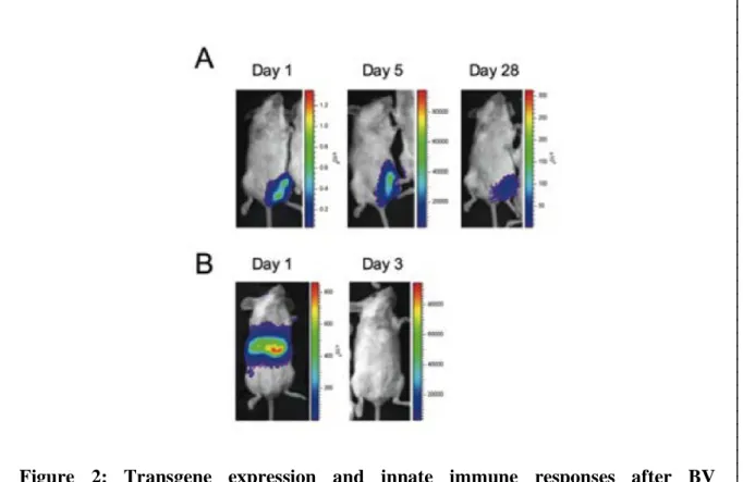
Construction of BES-GL3 Spider (BV) is shown in Figure 1. I used the BV called BES-GL3, which harbours two gene cassettes consisting of the luciferase gene under the control of the CMV immediate-early enhancer-promoter (pCMViep) promoter and the decay accelerating factor (DAF) gene under the control of the p10 promoter. The gene encoding EGFP was driven invertedly by the polyhedrin promoter (pPolh). BES-GL3 was designed to express luciferase as a transducing marker and to display DAF as protection for BV from complement attack, respectively (29).
BV administration induces transgene expression and innate immune responses
BV is a promising gene therapy vector capable of transducing broad range of mammalian cells in vitro with significant efficiency leading to stable gene expression (35, 36). BES-GL3 intramuscular administration into the left thigh muscle of mice initially increased the luciferase expression levels robustly, but these levels gradually decreased to 2%
on day 28 (Figure 2A), which is consistent with findings from previous studies (29, 37).
Among the various cell types tested in vitro, hepatocytes were found to be the most effective at taking up BV (38), suggesting a potential use for BV as a vector for liver-directed gene transfer. However, direct evidence of in vivo liver-directed gene transfer has not been
Figure 1: Schematic representation of the luciferase-expressing BV. BES-GL3 Spider vectors express firefly luciferase GL3 under the pCMV promoter and contain a gene cassette for DAF display. S, gp64 signal sequence; and EGFP, enhanced green fluorescent protein.
reported previously because BV-mediated gene transfer into hepatocytes via intravenous injection is severely hampered by serum complement (39). Here, use of our complement- resistant DAF-shielded BES-GL3 revealed for the first time that intravenously administered BV effectively transduces hepatocytes in vivo (Figure 2B).
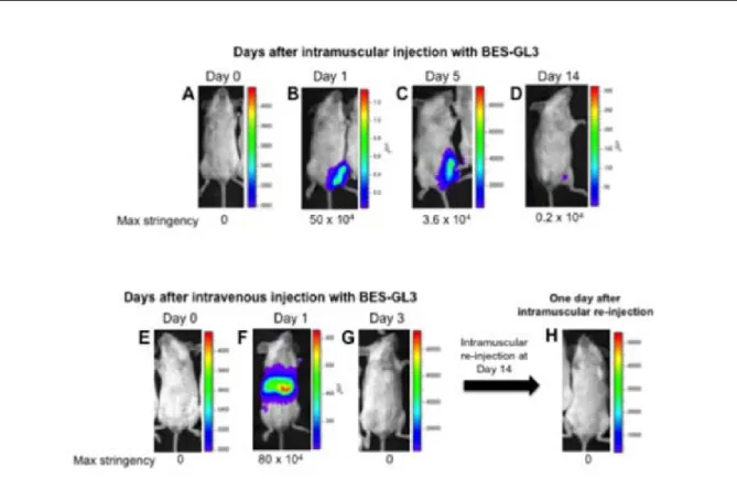
BV-mediated innate immune responses reject re-administration of BV
To examine antigen expression in vivo, I monitored luciferase expression in the mice by bioluminescence imaging on days 0, 1, 5, and 14 post-intramuscular injections of BV into the left anterior muscle of BALB/c mice (Figure 3A-D). We have previously shown that luciferase expression levels in intramuscular route were gradually reduced over time but detection of luminescence is achievable up to day 11 (29). BV might induce innate immune
Figure 2: Transgene expression and innate immune responses after BV administration via intravenous or intramuscular routes. (A, B) Luciferase expression at different timepoints after intramuscular (108 pfu) (A) or intravenous (107 pfu) (B) administration of luciferase-expressing BES-GL3 (described as BV), detected by using the IVIS® Lumina LT in vivo imaging system. The heatmap visible in each mouse image represents the total flux of photons (p/sec/cm2) in that area. Rainbow scales are expressed in radiance (p/s/cm2/sr).
responses at the injection site or systemic immunity for more than 10 days, which is consistent with findings from previous studies (29). Moreover, intravenous administration of BES-GL3 initially increased the luciferase expression levels robustly, but the levels gradually decreased to 0% on the day 3 (Figure 3E-G), which is also stable with previous studies (33).
Interestingly, when BV-Luc was intramuscularly re-administrated into the left tibialis anterior muscle at day 14, the luciferase expression was not detectable (Figure 3H). The result indicates that BV intravenous administration strongly induces innate immune responses which systemically clear upon the re-administration of BV.
Figure 3: Effects of BV administration. Luciferase expression at different time points after intramuscular (108 pfu) (A-D) or intravenous (107 pfu) (E-G) administration of BES-GL3 (BV) detected by using the IVIS® Lumina LT in vivo imaging system. After 14 days intravenous BV administration mice were re-administered with BES-Luc (108 pfu) by intramuscular route. The expression of luciferase was monitored on day 15 (14+1) post-BV administration (H). (A-H) The heat map images visible in the mice represent the total flux of photons (p/s/cm2) in that area.
Recombinant BV induces inflammatory cytokines in both WT and TLR9-/- mice
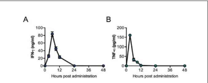
Baculovirus virions displaying PbCSP administration was reported to elicit specific antibodies and gamma interferon (IFN-γ) and conferred protection against P. berghei challenge infection (40). Since IFN-γ is a critical component of antimalarial liver stage protective response (41), I examined the serum kinetics of proinflammatory cytokines following BES-GL3 intravenous administration. To explore the cellular immune responses, I first determined the Th1-type (IFN-γ and TNF-α) cytokine levels at various times (0, 3, 6, 9, 12, 24, and 48 h) after BES-GL3 intravenous administration. IFN-γ and TNF-α levels rapidly reached their peaks at 6 h and decreased to baseline by 24 h (Figure 4).
I also compared different cytokines (IFN-γ and TNF-α) production at 6 h after post- BES-GL3 administration by intravenous and intramuscular routes (Figure 5A, B).
Intravenous routes robustly produced both Th1-type cytokines levels compare with intramuscular route. In particular, IFN-γ and TNF-α level detected from sera of mice immunized with BES-GL3 by intravenous route was highly significant compared with control (p < 0.0001). On the other hand, intravenously administered mice with Ad-Luc and PBS did not induce any significant proinflammatory cytokines.
Figure 4: Kinetics of proinflammatory cytokines (IFN-γ and TNF-α). Kinetics of proinflammatory cytokines, IFN-γ (A) and TNF-α (B) in the sera at different timepoints post-BV intravenous administration (107 pfu) (n = 6). Line graph shows the increment of the cytokines level with time.
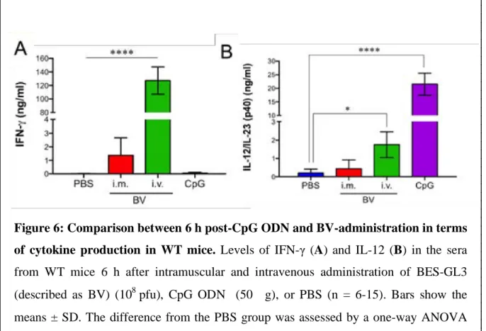
CpG ODNs are known to induce IFN-γ secretion in previous studies (42). Gramzinski et al., reported that, IFN-γ might be involved in the CpG-induced protection against sporozoite challenge (43). As a positive control, less amount of IFN-γ was detected from 6 h intramuscular CpG ODN 1826 (50 µg) sera compare with BV (Figure 6A). The possible reason might be the B-type CpG ODNs which has a complete phosphorothioate backbone and induce the production of modest levels of IFN-α, with much weaker NK cell activation thus lower IFN-γ secretion (44).
BV is known to activate bone marrow-derived dendritic cells to induce the expression of activated cell-surface markers and to produce various kinds of cytokines (IL-6, IL-12p70, and TNF-α) (45). Since IL-12 has been implicated with a role in CpG ODN-induced immunity in other systems (46), so I compared the serum IL-12 levels 6 h post-BV intramuscular administration in WT mice (Figure 6B). Consistent with previous findings (43), CpG intramuscular administration induced a robust IL-12 response than BV administered sera. Results suggested that a strong inflammatory response with a complex pattern of inflammatory cytokines might contribute to the intrinsic immunogenic properties of the administered BV.
Figure 5: Comparison between different routes of BV-administration in terms of cytokine production. Levels of IFN-γ (A) and TNF-α (B) in the sera from WT mice 6 h after intramuscular and intravenous administration of BES-GL3 (described as BV) (108 pfu), Ad-Luc (1010 pfu), or PBS (n = 6). Bars show the means ± SD. The difference from the PBS group was assessed by a one-way ANOVA with Dunn’s correction. ****p <
0.0001. i.m., intramuscular; i.v., intravenous.
To further investigate, I proceeded in TLR9-/- mice model for detection of different cytokines (IFN-γ and IL-12) production at 6h after intramuscular administration of BES-GL3 (Figure 7A, B). Significant amount IFN-γ was detected in both the TLR9-/- and WT mice. In particular, IFN-γ level detected from sera of TLR9-/- mice immunized with BES-GL3-Spider by intramuscular route was highly significant compared with TLR9-/- control group mice (p <
0.0001). In contrast, less amount of IL-12 was detected in both TLR9-/- and WT group which is not statistically significant with control.
Figure 6: Comparison between 6 h post-CpG ODN and BV-administration in terms of cytokine production in WT mice. Levels of IFN-γ (A) and IL-12 (B) in the sera from WT mice 6 h after intramuscular and intravenous administration of BES-GL3 (described as BV) (108 pfu), CpG ODN (50 µg), or PBS (n = 6-15). Bars show the means ± SD. The difference from the PBS group was assessed by a one-way ANOVA with Dunn’s correction. *p < 0.05, ****p < 0.0001. i.m., intramuscular; i.v., intravenous.
Intravenous BV-administration cause liver injury
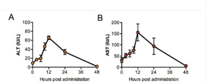
Since previous study suggest that CD8+ T cell-mediated immune pathology occurs in the brain but not the liver, while parasite-dependent pathology occurs in both organs during P. berghei ANKA infection (47). Although BES-GL3 can completely eliminate the liver- stage parasites, I was interested in liver damages caused by the BES-GL3 administration through different routes. To evaluate the liver damage, I first examined the serum kinetics of ALT and AST following BES-GL3 intravenous administration. The ALT and AST levels each rapidly reached their peaks at 12 h and decreased to baseline by 48 h (Figure 8).
Figure 7: Recombinant baculovirus induces inflammatory cytokines in TLR9-/- mice. Levels of IFN-γ (A) and IL-12 (B) in the sera from TLR9-/- mice 6 h after intramuscular administration of BES-GL3 (described as BV) (108 pfu) or PBS (n = 6- 15). Bars show the means ± SD. The difference from the PBS group was assessed by a one-way ANOVA with Dunn’s correction. ***p < 0.001, ****p < 0.0001, ns, not significant.
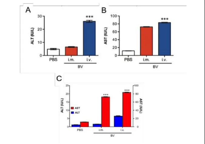
I also measured liver damage markers levels after 6 h of BES-GL3 administration through different routes. Compared with intravenous administration, intramuscular administration did not affect the ALT levels; although the AST level trended higher following intramuscular administration, this difference did not reach statistical significance (Figure 9A-C). ALT is a sensitive indicator of liver damage (48), so these results suggest that, for BV, intramuscular administration may be less destructive than intravenous administration. Alternatively, if desired, intravenous administration can induce stronger systemic innate immune responses than intramuscular, depending upon target diseases such as cancers.
Figure 8: Kinetics of liver damage markers (ALT and AST). Kinetics of liver damage markers, ALT (A) and AST (B), in the sera at different timepoints post-BV intravenous administration (107 pfu) (n = 6). Line graph shows the increment of the enzymes level with time.
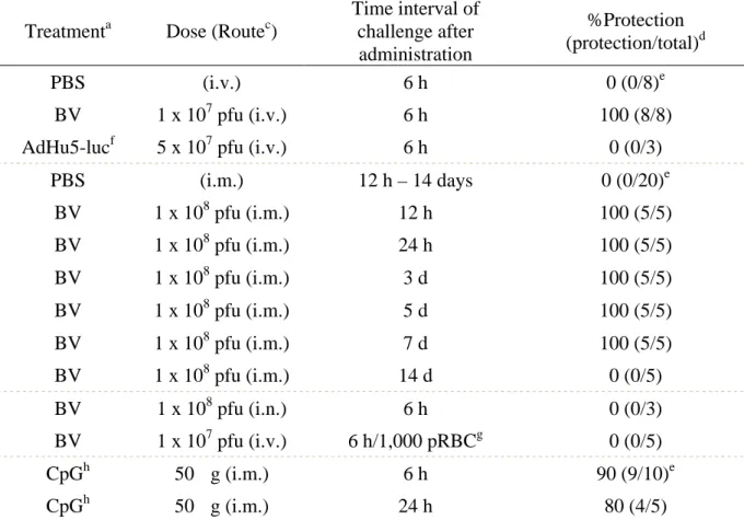
BV administration elicits sterile protection against sporozoite
Table 1 summarizes the protective efficacy results for BV administration against malaria sporozoite challenge. First, to examine the effects of BV intravenous administration, mice were intravenously administered 107 pfu of BES-GL3. At 6 h post-BV injection, which coincides with peak IFN-γ production, the mice were intravenously challenged by 1,000 Pb- conGFP sporozoites, which are transgenic P. berghei constitutively expressing GFP. All BV- injected mice were protected, whereas all PBS- and AdHu5-injected mice treated similarly became infected. Next, I investigated the effects of BES-GL3 intramuscular administration (108 pfu) followed by sporozoite challenge at various intervals post-BV injection. After intramuscular administration of BES-GL3, all mice were protected for at least 7 days.
However, there was a complete loss of protection by 14 days post-BES-GL3 intramuscular Figure 9: Comparison between different routes of BV-administration in terms of liver enzymes. Comparison of ALT (A), AST (B), and both (C) values in the sera at 6 h after intramuscular (108 pfu) or intravenous (107 pfu) administration of BV (n = 6). Bars or points show the mean ± SD. The difference from the PBS group was assessed by a Kruskal-Wallis test with Dunn’s correction. ***p < 0.001. i.m., intramuscular; i.v., intravenous.
administration and no delay of parasitaemia was observed in these mice. Additionally, no protection was observed in mice treated intranasally with BES-GL3.
BES-GL3 intravenous administration failed to provide protection against challenge with 1,000 parasitized red blood cells (pRBCs) at 6 h post-BV injection, indicating that BV has no residual effect on blood-stage parasites. CpG intramuscular administration at 6 or 24 h prior to challenge conferred protection against sporozoite challenge in 90% or 80% of mice, respectively. This is consistent with previous work showing short-term (2-days) protection induced by CpG intramuscular administration (50 µg) against challenge with 100 P. yoelii sporozoites (43), although only partial protection (50%) was observed when the challenge occurred at 7 days post-CpG intramuscular injection. Thus, the protective efficacy induced by BES-GL3 intramuscular administration is more effective and longer-lasting (7-days) compared with that induced by CpG. All PBS-treated control mice developed blood-stage infection within 6 days following an intravenous injection of 1,000 Pb-conGFP sporozoites.
Table 1. Protective efficacy of BV injection against P. berghei sporozoite challengea, b
Treatmenta Dose (Routec)
Time interval of challenge after administration
%Protection (protection/total)d
PBS (i.v.) 6 h 0 (0/8)e
BV 1 x 10⁷ pfu (i.v.) 6 h 100 (8/8)
AdHu5-lucf 5 x 10⁷ pfu (i.v.) 6 h 0 (0/3)
PBS (i.m.) 12 h – 14 days 0 (0/20)e
BV 1 x 10⁸ pfu (i.m.) 12 h 100 (5/5)
BV 1 x 10⁸ pfu (i.m.) 24 h 100 (5/5)
BV 1 x 10⁸ pfu (i.m.) 3 d 100 (5/5)
BV 1 x 10⁸ pfu (i.m.) 5 d 100 (5/5)
BV 1 x 10⁸ pfu (i.m.) 7 d 100 (5/5)
BV 1 x 10⁸ pfu (i.m.) 14 d 0 (0/5)
BV 1 x 10⁸ pfu (i.n.) 6 h 0 (0/3)
BV 1 x 10⁷ pfu (i.v.) 6 h/1,000 pRBCg 0 (0/5)
CpGh 50 µg (i.m.) 6 h 90 (9/10)e
CpGh 50 µg (i.m.) 24 h 80 (4/5)
aBALB/c mice were injected with BES-GL3 (described as BV) by the indicated route. After the indicated interval, mice were intravenously challenged with 1,000 Pb-conGFP
sporozoites. Parasitaemia was monitored on days 5-8, 11, and 14 after sporozoite challenge.
Once parasites appeared in the blood, all mice died.
bScheme of the experimental design is shown in Figure 19A.
ci.v., intravenous; i.n., intranasal; i.m., intramuscular.
dProtection is defined as the complete absence of blood-stage parasitaemia on day 14 post- challenge.
eCumulative data from two or four experiments.
fBALB/c mice were intravenously injected with AdHu5-luc.
gBALB/c mice were intravenously challenged with 1,000 Pb-conGFP-pRBC.
hBALB/c mice were intramuscularly administrated with 50 µg of CpG ODN 1826 (described as CpG).
BV administration completely eliminates of liver-stage parasites
Pathways stimulated by type I and II IFNs can lead to the killing of hepatocytes infected with liver-stage parasites (8-13). Because BV is a potent inducer of type I and II IFNs (45, 49), and I observed BV-mediated protection in Table 1, so in my next experiment I investigated whether BV-induced IFNs could kill liver-stage parasites in vivo. To examine the elimination effects on the trophozoite and exoerythrocytic (mature) schizont stages, I administered BES-GL3 intravenously or intramuscularly at two different intervals following sporozoite challenge, 24 and 42 h, respectively. Table 2 summarizes these results on the elimination efficacy of BES-GL3 administration against liver-stage parasites. Blood-stage parasites were completely prevented in all mice that had been intravenously injected with BES-GL3 at 24 h post-infection; in contrast, the protective effectiveness of BES-GL3 intravenous administration was diminished when mice received it at 42 h post-infection instead. The same results were obtained when mice were intramuscularly injected with BES- GL3.
As PQ is the only licensed drug for the radical cure of P. vivax hypnozoites, I also compared the elimination effects of BV with those of PQ. Two different doses of PQ, high dose (2 mg/mouse) and low dose (0.1 mg/mouse), were intraperitoneally administered. A single administration of high dose of PQ completely eliminated the liver-stage parasites (Table 2), whereas a single low dose of PQ was unable to reduce liver parasite burden but caused a significant delay of parasitaemia (Figure 11). The WHO-recommended treatment schedule for PQ is 15 mg/day for 14 days, but because high doses of PQ often cause side
effects like nausea, vomiting, and stomach cramps, these side effects can limit patient compliance, potentially resulting in PQ resistance (50, 51). Thus, BV intramuscular administration may have important advantages of over PQ.
Table 2. Elimination of liver-stage parasites by BV administrationa, b
Treatmenta Dose (Routec)
Time interval of administration after
challenge
%Elimination (uninfected/total)
PBS (i.v.) 24 h 0 (0/12)e
BV 1 x 10⁷ pfu (i.v.) 24 h 100 (13/13)e
BV 1 x 10⁷ pfu (i.v.) 42 h 0 (0/3)
PBS (i.m.) 24 h 0 (0/9)e
BV 1 x 10⁸ pfu (i.m.) 24 h 100 (7/7)
BV 1 x 10⁶ pfu (i.m.) 24 h 0 (0/5)f
BV 1 x 10⁴ pfu (i.m.) 24 h 0 (0/5)f
BV 1 x 10⁸ pfu (i.m.) 42 h 0 (0/3)f
PQ (High)d 2 mg (i.p.) 24 h 100 (5/5)
PQ (Low)d 0.1 mg (i.p.) 24 h 0 (0/5)f
aBALB/c mice were intravenously injected with 1,000 Pb-conGFP sporozoites. After the indicated interval, mice were administrated either with BES-GL3 (described as BV) or PQ.
Parasitaemia was monitored on days 5-8, 11, and 14 after sporozoite injection. Once parasites appeared in the blood, all mice died.
bScheme of the experimental design is shown in Figure 19B.
ci.v., intravenous; i.m., intramuscular; i.p., intraperitoneal.
dThe two different doses of PQ as High (2 mg/100 µl) and Low (0.1 mg/100 µl) were administrated to eliminate liver-stage parasites.
eCumulative data from three experiments.
fSignificant delay of parasitaemia was observed in infected mice, compared with the PBS group as showing in Figure 11 and 12.
Detection of liver stage parasites elimination by in vivo imaging system
Our group generated a transgenic rodent malaria parasite (P. berghei) that consist of the luciferase gene under a promoter region of elongation factor-1α (33). The transgenic parasites designated Pb-Luc expressed luciferase in all stages of their life cycle. To visualize the parasite elimination by BV, mice were infected with Pb-Luc, which are transgenic P.
berghei constitutively expressing luciferase, and then examined via IVIS; this is a highly sensitive method for detecting liver- and blood-stage parasites. Parasites were observed in the liver at both 24 h and 42 h post-infection (Figure 10A and B, respectively; left panels). BES- GL3 intramuscular administration into the left thigh muscle at 24 h post-infection completely eliminated the liver-stage parasites completely at 72 h post-infection, whereas the PBS control treatment failed to prevent the development of blood-stage parasites (Figure 10A;
right panel). Although BES-GL3 intramuscular administration into the right thigh muscle at 42 h post-infection also failed to prevent the development of blood-stage parasites (Figure 10B; right panel), it caused a significant delay of parasitaemia (Figure 11). The exoerythrocytic merozoites of P. berghei are released from infected hepatocytes into the blood stream at 44–48 h after the liver stage (52) Therefore, this result indicates that even for exoerythrocytic schizonts (42 h post-infection), the elimination effect of BV intramuscular administration was invoked in the liver within 2–6 h.
BV possesses great prophylactic efficiency than CpG and primaquine
Next, I investigated whether BV possessed high prophylactic and therapeutic effectiveness than CpG and primaquine, respectively. Intramuscular administration of BES- GL3 into the left thigh muscle at 24 h post-infection completely eliminated the liver-stage parasites at 72 h post-infection as mentioned before. While 42 h post-infection failed to prevent the development of blood-stage parasites but still caused a significant delay of parasitaemia (Figure 11). Results suggested that 42 h post-infection is too late to eliminate the liver-stage parasites and innate immunity induced by BES-GL3-Spider is not effective for
Figure 10: Effect of BV intramuscular administration on liver-stage parasites (A, B). Mice were challenged by infection with Pb-Luc sporozoites at 0 h, followed by intramuscular administration of BES-GL3 (108 pfu; described as BV) at the indicated timepoints. Luminescence in the liver indicates parasite growth, whereas that in the thigh shows the transgene expression by intramuscular BV injection. The heatmap visible in each mouse image represents the total flux of photons (p/s/cm2) in that area.
Rainbow scales are expressed in radiance (p/s/cm2/sr).
blood-stage parasites. A single administration of high dose PQ completely eliminated the liver-stage parasites whereas a single low dose of PQ was suboptimal, producing only a reduction in parasite burden in the liver and caused a significant delay of parasitaemia (Figure 11). It was conventional that, pretreatment of mice with CpG ODN 1826 provided complete protection from infection when the CpG ODN 1826 was administered 1 or 2 days prior to P. yoelii sporozoites challenge (43). Consistent with the previous findings, CpG has a little effect on mature schizonts (24 h post-infection) as it caused a significant delay of parasitaemia (Figure 11). Results implicated that BV has an important potential for anti-liver stage drug.
Liver stage parasites elimination efficacy is dependent on dose of BV
Lower doses (104 and 106 pfu) of BES-GL3 administered at 24 h post-infection failed to prevent blood-stage parasites. However, a significant delay of parasitaemia was observed for the dose of 106 pfu of BES-GL3 (Figure 12), indicating that the elimination effect is
Figure 11: Comparison among BV, CpG, and primaquine in the elimination of liver stage parasites. Delay of parasitaemia in infected mice. Parasitaemia of the groups of infected mice shown in Table 2 (108 pfu of BV injected intramuscularly at 42 h post- infection, and PQ low dose administered at 24 h post-infection), and Table 3 (CpG administered 24 h post-infection). Bars or points indicate the mean ± SD. The difference from the PBS group was assessed by a two-way ANOVA. *p < 0.05, **p < 0.01, ****p
< 0.0001.
dependent on the amount of BV that is intramuscularly administered. From the dose dependent study, I can conclude that 108 pfu BES-GL3-Spider dose is the best for the complete elimination of malaria parasites.
BV-mediated liver-stage parasite killing occurs through TLR9-independent pathways CpG intramuscular administration completely eliminated early liver-stage parasites completely at 6 h post-infection (Table 3); however, although this treatment caused a significant delay of parasitaemia, it had little effect on mature schizonts (24 h post-infection) as mentioned in Figure 11. BV possesses unique characteristics that activate DC-mediated innate immunity through MyD88/TLR9-dependent and -independent pathways (14).
Therefore, next I investigated whether TLR9 plays an important role in BV-mediated parasite killing in the liver. A single dose of intramuscularly administered BES-GL3 completely prevented blood-stage parasites in all TLR9-/- mice that had been previously infected with liver-stage parasites. In contrast, no elimination effect or parasitaemia delay was observed in
Figure 12: Dose dependency of BV on liver stage parasite elimination. Delay of parasitaemia in infected mice. Parasitaemia of groups of infected mice shown in Table 2 (106 or 104 pfu of BV injected intramuscularly 24 h post-infection). Bars or points indicate the mean ± SD. The difference from the PBS group was assessed by a two-way ANOVA. **p < 0.01.
TLR9-/- mice following intramuscular administration of CpG (50 µg) (Table 3). These results clearly demonstrate that BV-mediated parasite killing occurs via TLR9-independent pathways.
Table 3. Elimination of liver-stage parasites by BV injection in TLR9-/- micea
Treatmenta Mouse
strain Dose
Time interval of administration after
challenge
%Elimination (uninfected/total)
PBS TLR9-/- - 24 h 0 (0/7)
BV TLR9-/- 1 x 10⁸ pfu 24 h 100 (7/7)
BV WT 1 x 10⁸ pfu 24 h 100 (5/5)
CpG WT 50 µg 6 h 100 (5/5)
CpG WT 50 µg 24 h 0 (0/4)b
CpG TLR9-/- 50 µg 24 h 0 (0/5)
aTLR9-/- (BALB/c background) or WT mice were intravenously injected with 1,000 Pb- conGFP sporozoites. After 24 h, mice were intramuscularly administrated either with BES- GL3 (described as BV) or CpG ODN 1826 (described as CpG). Parasitaemia was monitored on days 5-8, 11, and 14 after sporozoite injection. Once parasites appeared in the blood, all mice died.
bSignificant delay of parasitaemia was observed in infected mice, compared with the PBS group as shown in Figure 11.
Intramuscular BV administration rapidly induces type I and II IFNs in sera
BV intravenous administration was reported to produce type I IFNs through TLR- independent and IRF3-dependent pathways in mice (14). To further investigate IFN production following BV intramuscular administration, the IFN serum levels were measured in WT and TLR9-/- mice at 6 h after BES-GL3 intramuscular administration. As with intravenous administration, intramuscular administration of BES-GL3 produced IFN-α in not only WT mice (6,311 ± 2,363 pg/ml) but also TLR9-/- mice (1,590 ± 737 pg/ml), whilst mice intramuscularly injected with PBS or CpG did not produce detectable IFN-α (< 20.0 pg/ml) (Figure 13A). IFN-γ, a type II IFN was also produced in both WT mice (1,367 ± 1,303 pg/ml) and TLR9-/- mice (488 ± 132 pg/ml) following intramuscular administration of BES-GL3 (Figure 13B). Compared with BV, CpG intramuscular administration in WT mice induced much less IFN-γ but much more IL-12 (Figure 13C). Notably, CpG intravenous administration induced a high level of IFN-γ with considerable systemic side effects (53, 54).
These results indicate that BES-GL3 intramuscular administration induces production of both type I and II IFN via TLR9-independent pathways.
Liver-stage parasites are killed by IFN-mediated immunity
To determine whether the serum cytokines act as effectors against liver-stage parasites, a serum transfer assay was performed. Pooled sera were collected from donor mice at 6 h after they had been intramuscularly injected with BES-GL3 or PBS. An aliquot of the pooled sera (100 µl/animal) was transferred to each recipient mouse at 24 h after their intravenous injection with 1,000 sporozoites. One of the five recipient mice effectively eliminated the liver-stage parasites, and the other four infected recipient mice showed a significant delay in
Figure 13: BV intramuscular administration BV administration rapidly induces type I and II IFNs in sera. (A-C) Levels of IFN-α (A), IFN-γ (B), and IL-12 (C) in sera from WT or TLR9-/- mice at 6 h after intramuscular administration of BES-GL3 (described as BV) (108 pfu), CpG, or PBS (n = 9-10). Bars show means ± SD. The difference from the PBS group was assessed by a Kruskal-Wallis test with Dunn’s correction. **p < 0.01, ****p < 0.0001.
the time to 1% parasitaemia (mean delay of 3.54 days; p = 0.0008, compared with the PBS sera group) (Figure 14A).
Next I examined whether neutralization of IFN-α or IFN-γ in the sera altered the effect of the sera on liver-stage parasites. Either anti-IFN-α or anti-IFN-γ antibody was incubated with 100 µl of the sera, which contained 8,619 pg/ml of IFN-α and 4,705 pg/ml of IFN-γ.
Complete neutralization of IFN-α was confirmed by ELISA (Figure 14B). The IFN-α- or IFN-γ-neutralized sera (100 µl) were intravenously administered to recipient mice that had been intravenously 24 h previously injected with 1,000 sporozoites. The anti-IFN-α antibody treatment completely abrogated the serum-induced delay of parasitaemia, whereas the anti- IFN-γ antibody treatment only partially impaired the serum-induced elimination effect (Figure 14A). These data suggest that the IFN-γ-mediated killing may be mediated via an effector mechanism distinct from that activated by IFN-α. It is possible that the effector mechanism induced by IFN-α and IFN-γ might still have been synergistically operative but that an alternate protective mechanism(s) may be activated with BV.
Figure 14: IFN-α induced by BV intramuscular administration contributes to elimination of liver-stage parasites. Results of a serum transfer assay to determine the role of IFN-α and IFN-γ in the elimination of liver-stage parasites (A). Sera collected from mice at 6 h after BV administration was neutralized by either anti-IFN-α or anti- IFN-γ antibody. Passive transfer of antibody-treated sera, non-treated sera, or PBS was conducted at 24 h after sporozoite infection (n = 5). The difference from the PBS group was assessed by a Kruskal-Wallis test with Dunn’s correction. *p < 0.05, ***p < 0.001.
Neutralization of IFN-α in sera from BV-administered mice (B). Aliquots of pooled sera from five BV-administered mice were neutralized by anti-IFN-α or anti-IFN-γ antibody.
Complete neutralization of IFN-α was assessed by ELISA. Ab, antibody.
Role of exogenous IFNs in the elimination of liver-stage parasites
To assess the effects of exogenous IFN-α and IFN-γ on the elimination of liver-stage parasites, recombinant IFN-α (8,619 pg/mouse) or recombinant IFN-γ (4,705 pg/mouse) was intravenously administered to mice that had been intravenously injected 24 h before with 1,000 sporozoites. IFN-α administration eliminated the liver-stage parasites completely, whereas IFN-γ administration only partially eliminated them but caused a significant delay in the time to 1% parasitaemia (mean delay of 3.82 days; p = 0.0082, compared with the PBS group) (Figure 15). The IFN-α-mediated parasite elimination may be occur via an effector mechanism distinct from that activated by IFN-γ. It is also possible that the effector mechanisms induced by IFN-α and IFN-γ may still be synergistically operative but that an alternate protective mechanism may be activated by BV. Miller et al. similarly showed that IFN-γ produced by NKT cells following type I IFN signalling from infected hepatocytes play an important role in the elimination of liver-stage parasites (41). Result suggests that, the IFN-α-mediated parasite elimination may be mediated via an effector mechanism distinct from that activated by IFN-γ. Table 4 summarizes the results on the elimination efficacy against liver-stage parasites of serum transfer and IFN administration.
Figure 15: Effect of exogenous IFN-α or IFN-γ on the elimination of liver-stage parasites. Recombinant mouse IFN-α or IFN-γ was intravenously administered at 24 h after sporozoite infection (n = 5). The difference from the PBS group was assessed by a Kruskal-Wallis test with Dunn’s correction. **p < 0.01.
Table 4. Elimination of liver-stage parasites by serum transfer and IFNsa,b
Treatmenta Dose %Elimination
(uninfected/total)
Days to 1% parasitaemia
(p value compared with PBS control group) Data from Fig. 14Ab
PBS sera 100 µl 0 (0/5) 6.03 ± 0.08
BV sera 100 µl 20 (1/5) 9.57 ± 0.38 (p = 0.0008)c
BV sera treated with
anti-IFN-α antibody 100 µl 0 (0/5) 6.18 ± 0.16 (p > 0.999) BV sera treated with
anti-IFN-γ antibody 100 µl 0 (0/5) 7.59 ± 0.20 (p = 0.0288)c Data from Fig. 15b
PBS 100 µl 0 (0/5) 6.46 ± 0.13
BV sera 100 µl 20 (1/5) 10.02 ± 0.14 (p = 0.0075)c
Recombinant IFN-α 8,619 pg 100 (5/5) -
Recombinant IFN-γ 4,705 pg 20 (1/5) 10.28 ± 0.41 (p = 0.0082)c
aBALB/c mice were intravenously injected with 1,000 Pb-conGFP sporozoites. After 24 h, mice were treated as indicated. Parasitaemia was monitored on days 5-11, and 14 after sporozoite injection. Once parasites appeared in the blood, all mice died.
bSchemes of the experimental designs are shown in Figure 19, C and D.
cSignificant delay of parasitaemia was observed in infected mice, compared with the PBS control group by Kruskal-Wallis test.
IFN-stimulated genes (ISGs) are upregulated in the liver after BV intramuscular administration
Signal transduction of type I IFNs results in the induction of numerous ISGs (55). Some ISGs participate in direct antimicrobial activities, such as apoptosis induction and post- transcriptional event regulation of microbial killing, mainly acting as antiviral responses.
Gene-targeting studies have distinguished four effector pathways of the IFN-mediated antiviral response: the Mx GTPase pathway, 2′-5′ oligoadenylate-synthetase (OAS)-directed ribonuclease L pathway, protein kinase R (PKR) pathway, and ISG15 ubiquitin-like pathway (56). Additionally, several ISGs, such as IFN-induced proteins with tetratricopeptide repeats
(IFITs), as well as the transcription factors IRF3 and IRF7 are responsible for sensing the liver-infection by Plasmodium sporozoites (13). To confirm the involvement of ISGs, the gene expression levels in the livers of mice that had been intramuscularly injected with BES- GL3 were measured by quantitative RT-PCR (qRT-PCR). BES-GL3 significantly induced the gene expression of several antiviral proteins (Isg15, Mx1, Oas1a/b, Oasl1, and Pkr) in WT mice (Figure 16). All these genes, except Oas1a/b, possibly due to the gene locus, were also upregulated by BV in TLR9-/- mice.
Recent study demonstrated that similar subsets of ISGs such as Ifit1, Ifit3, and Ifit44 were upregulated by Plasmodium liver-stage infection as a type I IFN response (13). In my study, the expression levels of such genes were significantly induced by the immunization of BES- GL3 with much higher extent than that by Plasmodium infection (Figure 17). Consistent with the profile of other ISGs, Ifit1, Ifit3, and Ifit44 were upregulated in TLR9-/- mice immunized with BES-GL3 (Figure 17).
Figure 16: ISG induction in the liver after BV intramuscular administration. Gene expression of antiviral proteins in the livers of WT and TLR9-/- mice at 6 h after intramuscular administration of BES-GL3 (described as BV) (108 pfu) was measured by real-time RT-PCR (n = 5-7). Bars show means ± SEM. The difference from the PBS group was assessed by a Mann-Whitney’s U test. *p < 0.05, **p < 0.01.