Evaluation of the effects of gastrectomy on the development of metabolic bone disease
著者 東 勇気
著者別表示 Higashi Yuki journal or
publication title
博士論文本文Full 学位授与番号 13301甲第4588号
学位名 博士(医学)
学位授与年月日 2017‑06‑30
URL http://hdl.handle.net/2297/00049302
doi: 10.1016/j.jss.2016.11.057.
Creative Commons : 表示 ‑ 非営利 ‑ 改変禁止 http://creativecommons.org/licenses/by‑nc‑nd/3.0/deed.ja
Evaluation of the effects of gastrectomy on the development of metabolic bone disease
Yuki Higashi, BM,
aTakashi Fujimura, MD,
bKatsunobu Oyama, MD,
a,* Jun Kinoshita, MD,
aTomoharu Miyashita, MD,
aHidehiro Tajima, MD,
aSachio Fushida, MD,
aand Tetsuo Ohta, MD
aaDepartment of Gastroenterological Surgery, Kanazawa University, Kanazawa, Ishikawa, Japan
bDepartment of Surgery, Toyama City Hospital, Toyama, Toyama, Japan
a r t i c l e i n f o
Article history:
Received 6 June 2016 Received in revised form 27 November 2016
Accepted 30 November 2016 Available online 12 December 2016
Keywords:
Metabolic bone disease Gastrectomy
Rat surgical model Reconstruction method Stomach-preserving surgery
a b s t r a c t
Background:Metabolic bone disease after gastrectomy is one of the complications leading to deterioration in quality of life. The exact mechanism of the metabolic bone disease re- mains unclear. To clarify the cause of metabolic bone disease after gastrectomy, we evaluated the associations between the method of gastrectomy and the development of metabolic bone disease in a rat model.
Methods:Rats were assigned to four groups as follows: (1) sham operation (control group);
(2) resection of the glandular stomach with Billroth I reconstruction (RGBI group); (3) Roux- en-Y anastomosis preserving the secretory function of the whole stomach (PSRY group);
and (4) total gastrectomy with Roux-en-Y reconstruction (TGRY group). In all groups, body weight, serum biochemistry (total protein, albumin, calcium, phosphorus, tartrate- resistant acid phosphatase, and bone alkaline phosphatase), bone density, and bone breaking strength were measured.
Results: Body weights and serum calcium levels were significantly lower in the three operation groups compared with the control group. Bone density was significantly lower in the PSRY and TGRY groups compared with the control group. Bone breaking strength was significantly lower in the three operation groups compared with the control group.
Conclusions:Surgical methods led to metabolic bone disease. However, exclusion of the duodenum from food passage had major influence to reduction in bone density and breaking strength. A stomach-preserving procedure and physiological reconstruction which enable food passage through duodenum and proximal jejunum contribute to miti- gation of metabolic bone disease.
ª2016 Elsevier Inc. All rights reserved.
Introduction
Although the incidence of gastric cancer has declined, it re- mains one of the most common causes of cancer-related mortality worldwide. Surgical resection is the primary treat- ment for gastric cancer. The proportion of patients with early
gastric cancer who are capable of long-term survival con- tinues to increase due to the recent advances in diagnostic techniques. Therefore, some of the surviving patients suffer from sequelae caused by their surgical procedure. Major long- term complications after gastrectomy, such as dumping syn- drome, reflux esophagitis, weight loss, malnutrition, and
* Corresponding author.Department of Gastroenterological Surgery, Kanazawa University, 13-1, Takara-machi, Kanazawa, Ishikawa 920-8641, Japan. Tel.:þ81 76-265-2362; fax:þ81 76-234-4260.
E-mail address:[email protected](K. Oyama).
Available online at www.sciencedirect.com
ScienceDirect
journal home page: www.Journa lofSurgicalResea rch.com
0022-4804/$esee front matterª2016 Elsevier Inc. All rights reserved.
http://dx.doi.org/10.1016/j.jss.2016.11.057
anemia caused by deficiency of iron and/or vitamin B12, are well known.1,2 Metabolic bone disease after gastrectomy is one of the complications leading to deterioration in quality of life because decreased bone quality results in pain and frac- tures. This disorder proceeds slowly and asymptomatically, becoming apparent 5-15 y after gastrectomy.3Metabolic bone disease reported to be observed in 20%-50% of patients after gastrectomy.4 The proportion of elderly patients receiving gastrectomy has recently increased, and consequently, the risk of metabolic bone disease after gastrectomy is likely to have increased. In textbooks for orthopedics and endocri- nology, the main cause of postgastrectomy metabolic bone disease is described as deficiency of vitamin D and calcium (Ca), leading to osteoporosis and osteomalacia due to a decrease in bone quality. It is speculated that the loss of gastric juice and gastric reservoir function, exclusion of food passage through the duodenum, and pancreatocibal asyn- chrony can lead to malabsorption of Ca and vitamins and malnutrition. Although various types of gastrectomy and reconstruction methods are performed clinically, the influ- ence of these surgical procedures on bone metabolism re- mains unclear. The advantages and demerits of several surgical procedures have been discussed, but there is little available information on the surgical procedures from the viewpoint of bone metabolism. In fact, analysis of metabolic bone disease after gastrectomy is difficult in clinical cases, because the characteristics of the patients vary widely.
Therefore, we planned our experiments using rat gastric surgery models, which have uniform backgrounds. To eval- uate the influence of gastric juice and food passage route, we created several gastrectomized rat models similar to clinical gastric surgery. In this study, we focused on the association between the type of surgical intervention and the develop- ment of metabolic bone disease.
Materials and methods
Animals and surgical proceduresTwelve-wk-old Wistar male rats (Charles River Japan, Kana- gawa, Japan) weighing approximately 400 g were used for the experiments. They were housed at three rats per cage and maintained under conditions of 223C room temperature and 555% humidity with a 12-h/12-h light/dark cycle. They were fed a standard solid chow (CRF-1; Charles River Japan).
Body weights were measured at the beginning of the study and then weekly thereafter. We followed the ARRIVE Guide- lines (Animal Research: Reporting In Vivo Experiments) 18,5 and the Animal Welfare Committee of Kanazawa University approved the experiments before the start of the study.
For evaluation of the association between the types of gastrectomy/reconstruction and postoperative metabolic bone disease, we created several surgical models. Before sur- gery, the rats were anesthetized by intraperitoneal injection of medetomidine, midazolam, and butorphanol. All operations involved a ventral midline celiotomy. Y.H. performed all sur- gical procedures. The stomach of the rat can be distinguished anatomically into a proximal part (forestomach) and a distal part (glandular stomach). The forestomach corresponds to the
esophagus in humans, lacking the ability to secrete digestive juice, whereas the glandular portion most closely resembles the human stomach.
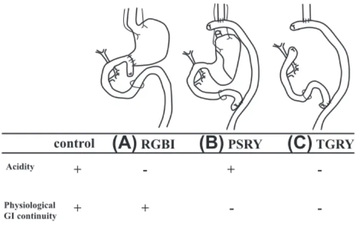
The rat with sham operation was defined as simple lapa- rotomy with blunt manipulation of the viscera. In the rats with resection of the glandular stomach with Billroth I (B-I) reconstruction (RGBI; Fig. 1A), the glandular stomach was removed. The forestomach stump was then anastomosed to the duodenum near its cut end in an end-to-side fashion. The vagal nerve was preserved. These rats secrete no gastric juice, but their intestinal continuity is physiologically maintained including the duodenum and proximal jejunum. In the rats Roux-en-Y (RY) anastomosis preserving the stomach (PSRY;
Fig. 1B), the total stomach and vagal nerve were preserved.
The esophagus was cut, and the end remaining attached to the stomach was ligated. The jejunum was cut approximately 4-cm distal to the Treitz ligament, and the end closest to the stomach was ligated with suture. The oral esophagus was then anastomosed to the distal jejunum near its suture cut end in an end-to-side fashion. The proximal jejunal cut end was anastomosed to the jejunum approximately 5 cm distal from the esophagojejunal anastomosis in a side-to-side fashion. These rats secrete gastric juice, but the food also bypasses the stomach. In the rats with total gastrectomy with RY reconstruction (TGRY;Fig. 1C), both the glandular stomach and the forestomach were removed, and the duodenal stump was closed with sutures. The jejunum was cut approximately 4 cm distal to the Treitz ligament, and both ends were closed with sutures. The esophageal stump was anastomosed to the distal jejunum near its cut end in an end-to-side fashion. The proximal jejunal cut end was anastomosed to the jejunum at approximately 5 cm distal from the esophagojejunal anasto- mosis in a side-to-side fashion. The vagal nerve was not pre- served. These rats secrete no gastric juice, and their food passage bypasses the duodenum and proximal jejunum. In all gastrectomy groups, anastomosis was carried out with inter- rupted full thickness stitches using 8-0 monofilament absorbable surgical sutures (Surgipro II Medtoronic, Dublin, Ireland). The rats received water for 3 days after the surgery,
(B) PSRY (C) TGRY
(A) RGBI control
Acidity
Physiological GI continuity
+
+ +
+ -
-
- -
Fig. 1eDiagrams of the surgical procedures. (A) RGBI, (B) Roux-en-Y anastomosis with preserving the secretory function of the whole stomach, and (C) total gastrectomy with Roux-en-Y reconstruction (TGRY).
2
j o u r n a l o f s u r g i c a l r e s e a r c h1 5 m a y 2 0 1 7 ( 2 1 2 ) 1e7and then received the standard diet from 4 days after the surgery. We assigned 15 rats in each group.
The influence of gastric juice can be evaluated by com- parison between control and RGBI. The comparison between control and PSRY clarify the influence of the food passage route. TGRY had deficiency both of gastric juice and food passage route.
The rats were euthanized by exsanguination (cardiac puncture) under isoflurane anesthesia at 22 wk after sur- gery. Blood samples were collected, and serum samples were immediately stored at 80C until analysis of total protein (TP), albumin (Alb), Ca, phosphorus (P), tartrate- resistant acid phosphatase (TRACP-5b), and bone alkaline phosphatase (BAP). The femurs were dissected out, cleaned of soft tissue, wrapped in saline-soaked gauze, and stored at 80C until determination of bone density and breaking strength.
Measurement of serum biochemical parameters
The value of TP was measured by the Biuret test (Total Protein- HR II; Wako Pure Chemical Industries, Osaka, Japan), Alb by the bromcresol green test (Albumin II Test Wako; Wako Pure Chemical Industries), Ca by the o-cresolphthalein complexone test (Nescauto Ca-V2; Alfresa Pharma Corporation, Osaka, Japan), and P by an enzyme test (DETERMINER L IP II; Kyowa Medex Co Ltd, Tokyo, Japan). The value of TRACP-5b was measured using a rat tartrate-resistant acid phosphatase 5b ELISA Kit (Cusabio, Wuhan, China). TRACP-5b is a reliable bone resorption marker that is not affected by feeding and renal dysfunction. The value of BAP was measured using a rat bone alkaline phosphatase ELISA Kit (Cusabio, Wuhan, China).
BAP is a bone formation marker produced by osteoblasts.
Measurement of bone density and strength
The right femurs were fixed in 10% neutral-buffered formalin, degreased in 100% ethanol, and fixed in cyanu- ric chloride (Wako Pure Chemical Industries). The fixed femurs were decalcified in 10% formic acid, neutralized in 5% sodium sulfate, and embedded in paraffin. Thin sections of the femoral metaphysis were cut with a microtome (LEICA SM 2000R; Leica Geosystems, Tokyo, Japan) along its length. A section of each femur was stained with hema- toxylin and eosin. The morphologic changes were evalu- ated microscopically. The pathologic images of the distal femur were captured using a bio-imaging navigator (Bio- revo BZ-9000, Keyence, Osaka, Japan). The proportions of calcified bone area in the epiphysis of distal femur were calculated using analytical software (Hybrid Cell Count Soft, Keyence, Osaka, Japan). We used these values as an index of bone density.
The bone breaking strength (N) of the left femur was measured by a three-point bending test (AG-100KNplus; Shi- madzu, Kyoto Japan). Briefly, each specimen was placed hor- izontally in the loading part of the testing machine, and the center of the diaphysis was pressed by 50 kgf (distance be- tween support points: 15 mm). We evaluated the bone breaking strength as the bone strength.
Statistical analysis
Serum biochemistry values, bone density, and bone breaking strength were analyzed by the KruskaleWallis H-test and ManneWhitney U-test with the Bonferroni correction. The values ofP<0.05 were considered statistically significant. All statistical analyses were performed using SPSS statistical software, version 11.0 (International Business Machines Cor- poration, Armonk, NY).
Results
Outcomes of surgical procedures and changes in body weights
None of the rats in the control and the RGBI groups died. In the TGRY and PSRY groups, three and four rats died, respectively.
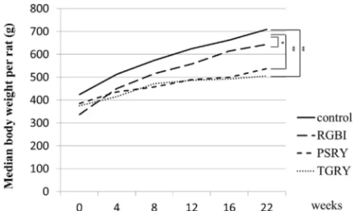
All animals died of ileus, which was diagnosed by post- operative vomiting and inspection at autopsy. Body weight in all surgical groups increased gradually after surgery. The body weight in the control group; 709 (619.5-744.5) was significantly greater than those in the other three groups at the 22nd wk after surgery (RGBI: 644 [616.5-668.5]; PSRY: 527.5 [496-599];
and TGRY: 501 [474.5-533.5],P<0.0;Fig. 2).
Serum biochemistry
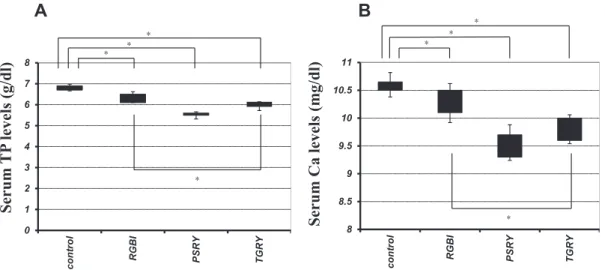
The serum TP (RGBI: 6.5 [6.1-6.5]; PSRY: 5.5 [5.5-5.6]; and TGRY: 5.9 [5.9-5.9]) and Ca (RGBI: 10.4 [10.1-10.5]; PSRY: 9.4 [9.3-9.7]; and TGRY: 9.7 [9.6-10]) levels were significantly reduced in the three gastric surgery groups compared with the control group (TP: 6.8 [6.7-6.9]; Ca: 10.6 [10.5-10.65], P< 0.05). The serum TP and Ca levels in the TGRY group were significantly lower than those in the RGBI group (P<0.05;Fig. 3A and B). There were no significant differences in the serum P, Alb, BAP, and TRACP-5b levels in the three gastric surgery groups compared with the control group (date not shown).
Fig. 2eChanges in body weights in the four groups from 0 to 22 wk after surgery. The body weight in the surgical groups was significantly lower than the control group at the 22nd wk after surgery (the KruskaleWallis H-test and ManneWhitneyU-test with the Bonferroni correction). *P
<0.05, **P<0.01.
Bone morphology
The trabecular bone was thicker and more numerous in the control and RGBI groups compared with PSRY and TGRY groups (Fig. 4).
Bone density
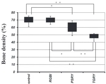
The median bone density values in the TGRY; 49 (45.8-51) and in PSRY; 65 (54.5-65) groups were significantly lower than those in the control group; 69 (67-74,P<0.05). There
Serum TP levels (g/dl)
8 8.5 9 9.5 10 10.5 11
control RGBI PSRY TGRY
0 1 2 3 4 5 6 7 8
control RGBI PSRY TGRY
A B
Serum Ca levels (mg/dl)
Fig. 3eSerum biochemistry. (A) Serum TP levels at the 22nd wk after surgery. The serum TP levels are significantly reduced in the three gastric surgery groups compared with the control group. The serum TP levels in the TGRY group were significantly lower than those in the RGBI group (P<0.05). (B) Serum Ca levels at the 22nd wk after surgery. The serum Ca levels are significantly reduced in the three gastric surgery groups compared with the control group. The serum Ca levels in the TGRY group were significantly lower than those in the RGBI group (P<0.05) (the KruskaleWallis H-test and
ManneWhitneyU-test with the Bonferroni correction). *P<0.05.
Fig. 4eThe trabecular bone was thicker and more numerous in the control and RGBI groups compared with the PSRY and TGRY. (Color version of figure is available online.)
4
j o u r n a l o f s u r g i c a l r e s e a r c h1 5 m a y 2 0 1 7 ( 2 1 2 ) 1e7was no significant difference between the control and RGBI groups; 70 (67-73, P¼ 0.55). The bone density in the TGRY group was significantly lower than that in the PSRY (P<0.01) and RGBI (P <0.01) groups. The bone density in the PSRY
group was significantly lower than that in the RGBI groups (P<0.05;Fig. 5).
Bone-breaking strength
The median bone-breaking strength in the control group; 252 (237.5-271) was significantly higher than those in the three gastric surgery groups (RGBI: 234 [180-248.5];
PSRY: 134 [120.9-159]; TGRY: 153 [136.5-160.8], P < 0.05).
There was no significant difference in the breaking strength between the TGRY and PSRY groups (P ¼ 0.27).
The breaking strength in the TGRY and PSRY groups was significantly lower than that in the RGBI group (P<0.01; Fig. 6).
Discussion
Surgery plays a vital role in the treatment of gastric cancer, but it is important to prevent postoperative complications.
Metabolic bone disease is one of the major long-term complications after gastrectomy, which can lead to frac- tures and decreased quality of life. Metabolic bone disease arising after gastrectomy was first identified more than 60 y ago.
When considering the mechanism for metabolic bone disease after gastrectomy, the process of Ca absorption is important. Most of the Ca in food is present in complexes with other dietary constituents, and therefore, the food must be broken down to release Ca in a soluble and ionized form before absorption.6 Ionized Ca is absorbed from the duodenum and the proximal part of the jejunum. In our study, the serum Ca levels were significantly reduced in the RGBI compared with the control group. Gastric juice breaks down food, ionizes calcium, and increases the solubility of Ca complexes.7Since Ca tends to precipitate from solutions with pH values greater than 6.1,8achlorhydria caused by loss of the stomach results in Ca malabsorption. In the present study, the serum Ca levels were also significantly reduced in the PSRY and TGRY groups compared with the control group. Therefore, it is also important that ionized Ca pass through the duodenum and proximal jejunum. The serum TP levels were significantly reduced in the surgical groups compared with the control group. It is possible that the surgical groups had malnutrition under the influence of surgical intervention. It is undeniable that clinically, malnutrition is one of the causes of metabolic bone disease.
However, TP levels were not different between the RGBI and PSRY groups. Therefore, the changes in bone quality were not caused by malnutrition alone.
In a clinical study, Fukudaet al.9reported that patients treated with the Billroth II procedure were predisposed to osteopenia after gastrectomy through a disorder in the process of Ca absorption. On the other hand, several reports showed that there were no differences in the development of metabolic bone disease after gastrectomy using the Bill- roth I and Billroth II procedures.10,11In the present study, the bone breaking strength and bone density with PSRY were significantly lower than control, suggesting that exclusion of the duodenum from food passage was the major cause of
Bone breaking strength (N)
0 50 100 150 200 250 300
control RGBI PSRY TGRY
Fig. 6eBone strengths at the 22nd wk after surgery. The bone breaking strength in the surgical groups is
significantly lower than that in the control group. The breaking strength in the TGRY and PSRY groups was significantly lower than that in the RGBI group (P<0.01) (the KruskaleWallis H-test and ManneWhitneyU-test with the Bonferroni correction). **P<0.01.
Bone density (%)
0 10 20 30 40 50 60 70 80
control RGBI PSRY TGRY
Fig. 5eBone densities at the 22nd wk after surgery. There is no significant difference between the control and RGBI groups. The bone densities in the TGRY and PSRY groups were significantly lower than those in the control group.
The bone density in the TGRY group was significantly lower than that in the PSRY (P<0.01) and RGBI (P<0.01) groups. The bone density in the PSRY group was
significantly lower than that in the RGBI groups (P<0.05) (the KruskaleWallis H-test and ManneWhitneyU-test with the Bonferroni correction). *P<0.05, **P<0.01.
the metabolic bone disease after gastrectomy. The bone breaking strength was suppressed in RGBI group. However, the bone density was uncharged. The loss of bone density was severest in the TGRY group among all models. Both the loss of gastric juice and the change of food passage route affected the bone quality after gastric surgery. Therefore, the change of food passage route had a major impact on the development of metabolic bone disease because the change of bone quality was greater in the model with rout modifi- cation; PSRY and compared to the model without rout change; RGBI.
In bariatric surgery, metabolic bone disease after surgery is also receiving a lot of attention. Jammah AA12reviewed complications of bariatric procedures and demonstrated metabolic bone disease was seen frequently as late compli- cation. They recommended preoperative assessment and postoperative regular follow-up for metabolic bone disease.
Stemmeret al.13reported that bone volume was significantly reduced after RY gastric bypass surgery but not vertical sleeve gastrectomy in rats. It is possible that the physiolog- ical passage of food prevented metabolic bone disease. The result of our study is consistent with this data after bariatric surgery.
There were no significant differences in the serum biochemical markers, such as TRACP-5b, and BAP levels among the groups receiving the different surgical proced- ures. Serum biomarkers of bone metabolism were evaluated in several reports, but the assessments of their utility were varied. Bernsteinet al.11reported that biochemical markers reflect only bone turnover, they are not useful in predicting bone mineral content. Baueret al.14reported that there was no significant association between values of BAP and hip bone loss. Changes in the levels of bone markers are pre- dominantly associated with rapid changes in bone meta- bolism and may not reflect chronic changes associated with feedback mechanisms maintaining biological homeostasis.
In our study, these serum biochemical markers were measured at a single time point after a prolonged period of time after surgery. The blood samples in our study may reflect the condition after receiving feedback mechanisms. It is important for gastrectomized patients to measure bone density after surgery, but the need of preventive medication is not clear. Further study disclosed to the prevention and therapy of metabolic bone disease after gastrectomy are needed.
Currently, the efficacy of function-preserving surgery is being discussed. The present study clarified the efficacy of stomach preservation for the mitigation of metabolic bone disease after gastrectomy. The Billroth I and RY methods are the most commonly used reconstruction techniques after distal gastrectomy in clinical cases. There is a lack of consensus with regard to the choice of reconstructive procedure after distal or subtotal gastrectomy. From the results of the present study, the B-I method preserving the rout of food passage through the duodenum and prox- imal jejunum appears preferable in an attempt to prevent metabolic bone disease.
Conclusions
A stomach-preserving procedure and physiological recon- struction allowing food to pass through the duodenum contribute to the mitigation of metabolic bone disease after gastrectomy.
Acknowledgment
The authors particularly thank Dr Seiji Naganuma, Depart- ment of Pathology, Kochi University, for advice on the path- ological measurement method for bone density.
Authors’ contributions: Y.H. drafted the article and pro- duced animal models. T.F. contributed in conception and design of the study. K.O. performed the critical revision of the article for important intellectual content. J.K. assessed the biochemical findings. T.M. contributed for surgical instruction and animal assessment. H.T. contributed for assessment of pathologic findings. S.F. contributed in analysis and inter- pretation of data. T.O. participated in final approval of the article.
Disclosure
The authors reported no proprietary or commercial interest in any product mentioned or concept discussed in the article.
r e f e r e n c e s
1. Liedman B. Symptoms after total gastrectomy on food intake, body composition, bone metabolism, and quality of life in gastric cancer patientseis reconstruction with a reservoir worthwhile?Nutrition.
1999;15:677e682.
2. Tovey FI, Godfrey JE, Lewin MR. A gastrectomy population: 25- 30 years on.Postgrad Med J. 1990;66:450e456.
3. Tovey FI, Karamanolis DG, Godfrey J, Clark J. Post- gastrectomy nutrition: methods of outpatient screening for early osteomalacia. Hum Nutr Clin Nutr.
1985;39:439e446.
4. Vestergaard P. Bone loss associated with gastrointestinal disease and pathogenesis.Eur J Gastroenterol Hepatol.
2003;15:851e856.
5. Kilkenny C, Browne WJ, Cuthill IC, Emerson M, Altman DG.
Improving bioscience research reporting: the ARRIVE guidelines for reporting animal research.PLoS Biol.
2010;8:e1000412.
6. Schachter D, Dowdle E, Schenker H. Active transport of calcium by the small intestine of the rat.Am J Physiol.
1960;198:263e268.
7. Mahoney AW, Holbrook RS, Hendricks DG. Effects of calcium solubility on adsorption by rats with induced achlorhydria.
Nutr Metab. 1975;18:310e317.
8. Allen LH. Calcium bioavailability and absorption. a review.
Am J Clin Nutr. 1982;35:783e808.
6
j o u r n a l o f s u r g i c a l r e s e a r c h1 5 m a y 2 0 1 7 ( 2 1 2 ) 1e79. Fukuda M, Shibata H, Hatakeyama K, et al. Difference in calcium metabolism following Billroth-I and Billroth-II procedures for gastric and duodenal ulcers.Jpn J Surg.
1979;9:295e303.
10. Higgins PM, Pridie RB. Postgastrectomy osteomalacia:
incidence after the no-loop and other types of gastrectomy.Br J Surg. 1996;53:881e885.
11. Bernstein CN, Leslie WD, Leboff MS. AGA technical review on osteoporosis in gastrointestinal diseases.Gastroenterology.
2003;124:795e841.
12.Jammah AA. Endocrine and metabolic complications after bariatric surgery.Saudi J Gastroenterol. 2015;21:269e277.
13.Stemmer K, Bielohuby M, Grayson BE, et al. Roux-en-Y gastric bypass surgery but not vertical sleeve gastrectomy decreases bone mass in male rats.Endocrinology.
2013;154:2015e2024.
14.Bauer DC, Sklarin PM, Stone KL, et al. Biochemical markers of bone turnover and prediction of hip bone loss in older women: the study of osteoporotic fractures.J Bone Miner Res.
1999;14:1404e1410.