THE STUDIES ON THE CHARACTERISTICS AND
POSSIBLE FUNCTIONS OF LECTINS IN THE SILKWORM, BOMBYX MORI
著者 天内 和人
year 1991‑03‑25
URL http://hdl.handle.net/2297/30532
m v
f
尊二仁言命:>zこT H E S T U D I E S O N T H E C H A R A C T E R I S T I C S A N D P O S S I B L E F U N C T I O N S O F L E C T I N S I N T I I E S I L K W O R M , B O M B Y X M O R I
金沢大学大学院自然科学研究科 生命科学専攻
生物
f
青報学講座特審番号 氏 名 主任指導教官
88‑2101
天内和人大滝哲也
CONTENTS
CHAPTER 1INTRODUCTION
--1
CHAPTER IN THE
2 PURIFICATION HEMOLYMPH OF THE
AND CHARACTERIZATION OF SILKWORM, BOMBYX MORI
LECT1N
--
5
ABSTRACT INTRODUCTION MATERIALS AND RESULTS
DISCUSSION
METHODS
CHAPTER 3 OCCURRENCE THE SILKWORM, BOMBYIX
OF LECTIN MORI
IN VARIOUSTISSUES OF
- 28
ABSTRACT INTRODUCTION MATERIALS AND RESULTS
DISCUSSION
METHODS
CHAPTER 4 THE SITE OF LECTIN AND ACTIVATION BY 20•-HYDROXYECDYSONE
PRODUCTION OF BOMBYX OF ITS PRODUCTION
HEMOLYMPH
- 55
ABSTRACT INTRODUCTION MATERIALS AND RESULTS
DISCUSSION
METHODS
CHAPTER 5 PROCESS OF SILK GLAND
THE FUNCTION PROGRAMMED
OF HEMOLYMPH CELL DEATH OF
LECTIN IN THE THE ANTERIOR
- 86
ABSTRACT INTRODUCTION MATERIALS AND RESULTS
DISCUSSION
METHODS
CHAPTER 6SUMMARY AND CONCLUSIONS
--113
ACKNOWLEDGEMENTS
.-117
REFERENCES - 119
CHAPTER 1. INTRODUCTION
The deve!opmental process in holometabolous insects is characterized by molting and metamorphosis. During the larval stage, molting, the periodic shedding of the old cuticle, permits the body to grow, and a new cuticle, whieh is essentially similar to the old one, is laid down on the epidermis. During the larval rnolt, internal organs undergo relatively little change.
Metamorphosis, however, eomprises two suceessive special molt in which cuticle of different characters, i. e. shape, thickness,
coloration, scales and bristles, is deposited, and an extensive reconstruction of internal organs takes plaee. The latter
process involves the histolysis of larval tissues, and the growth and differentiation of adult organs.
The developmental events described above are controlled by hormones, neuroseeretions, ecdysteroids and juvenile hormones.
Larval molt is induced by ecdysteroids with the preceding action of juvenile hormone. Pupal and adult transformations are provoked by ecdysteroids alone. Therefore, to study the
regulatory mechanisms of insect development, we have to consider ecdysteroids first, because it is indispensabie for ehanges of a wide variety of tissues whieh are seen in the process of molting and metamorphosis.
During the last larval instar, several kinds of proteins
appear in the hemolymph and the production of these proteins is
eontrolled by the neuroendoerine system. Among them inseet
leetins may have an important role on the histolysis of larval
tissues. In insects, purification of lectins from the hemolymph
of Sarcophaga peregrina (Komano et al., 1980), Hyalophora cecropia (Castro et al., 1987), Teleogryllus eommodes (Hapner et al., 1981), Melanoplus sanguinipes (Stebbins et al., 1985),
Antheraea pernyi (Qu et al., 1987), Periplaneta americana (Kubo et al., 1987) and Glossina fuseipes fuscipes (Ingram et al., 1990) has been reported. Little thought , however, has been given to the in situ role of the lectins in these species except the Sareophaga lectin. The Sarcophaga leetin has been found to eontain homo!ogous carbohydrate-recognition domains (CRD) with the mammalian liver asialoglyeoprotein receptor. The CRD of the
Sarcophaga lectin seems to be located at the NH2-terminal end of the molecule (Takahashi et al., 1985). The Sarcophaga leetin is induced in the hemolymph by injury to the body wall (Takahashi et
al., 1985). Also the expression of this ieetin has be' en observed during early embryonip stages and early pupal stages in normal
development (Takahashi et aZ., 1986). These investigations suggest that the Sarcophaga lectin has an important role in both defense system and development. However, no eonelusive evidence for the in situ role of the Sarcophaga lectin has yet been
obtained. The presence of leetin in the hemolymph ef the
silkworm, Bombyx mori, has been reported by Suzuki et al (1983).
Though the hemagglutinating activity of the 5th instar larval
hemoiymph is known to increase immediately before pupation, the Bombprx lectin has not been purified yet.
The domesticated silkmoth, Bombyx mori, is one of the
organisms whose geneties has been most intensively studied. It
belongs to the order Lepidoptera, the suborder Heteroneura, and
the family Bombyeidae (Imms, 1957). Because of its relatively
great size, it is suitable for the study of physiological,
biochemical and developmental problems. The newly hatehed larvae grow very quiekly and 4 times of larval molting occur. When 5th instar larvae are fully grown and ready to spin silk, they stop eating and become very restless. About 3 days after purging their gut, the larvae molt to form the pupae. It takes another 8-13 days for the adult moths to emerge Åírom the pupae. During the pupal transformation, degeneration of larval tissues, such as silk glands, intersegmental muscles, and growth and
differentiation of adult tissues from imaginal discs or
histoblasts are evoked. Therefore, one of the most important problems is to understand the mechanisms of degeneration of larval tissues in holometabolous insect.
In this study, it was attempted first to purify the lectin
Åírom the hemolymph of the BombyAr mori larvae and then to ciarify its role in the larval molting and larval-pupal metarnorphosis.
LecÅíins, proteins with hemagglutinating ae`Livity, have been found in a wide spectrum of animals and plants (Liener, 1976) and exhibit two Åëritieal properties. One is the binding speeificity with depends on particular sugar residues, and the other is that they are either bivalency or polyvalency (Goldstein et al.,
1980). Complex carbohydrates are widely distributed in animal tissues. The potential of these complex carbohydrate structures for encoding information has been repeatedly noted, For
examples, it was implicated that complex carbohydrates are involved in recognition process ineluding adhesion between cells, adhesion of cells to the extracellular matrix and specific
reeognition of eells (Yamada, 1983, Edelman, 1985, Hook et al.,
1984, Florman et al., 1985). In addition carbohydrates are reeognized as differentiation markers and as antigenic
determinants (Feizi et al., 1987). Since leetins have an ability to distinguish subtle differenees in carbohydrate structures
found in animal tissues, they may be involved in the mechanisms
of degeneration of larval tissues in holometabolous inseets.
CHAPTER 2. PURIFICATION AND CHARACTERIZATION OF LECTIN IN THE HEMOLYIYIPH OF THE SILKWORM, BOMBYX MORI
ABSTRACT
Hemolymph of the silkworm, Bombyx meri, containes a faetor to agglutinate the trypsinized and glutaraldehyde fixed sheep red blood cells. Changes in hemaggiutinating aetivity in hemolymph during 4th and 5th larval instars of Bombyx mori were stage and temporai specific. The activity inereased in time concomitant with an inerease in the hemolymph ecdysteroid titer. The protein with hemagglutinating aetivity was purified by ammonium sulfate
fraetionation, gel-filtration on Sephacryl S-300 and affinity
chromatography using either glucuronic aeid or galaeturonie aeid
as ligand. Western blotting analysis using antibody raised
against this protein revealed that Bombyx lectin is a tetrarner
composed of two different subunits with moiecular"weight of about
88 and 90 kDa, respectively.
INTRODUCTION
Lectins, proteins with hemagglutinating activity, found in various plants and animals (Liener, 1976) and possess two eritical properties; specificity for particular sugar residues,
and bivalency or polyvalency. These properties make them useful for many purposes including the purification of glycoconjugates (Barondes, 1984).
In insects, purification of lectin from the hemolymph
of Sarcophaga peregrina (Komano et al., 1980), Hyalophora eecro- pia (Castro et al., 1987), TeleogryZus commodus (Hapner et aZ., 1981), Melanoplus sanguinipes (Stebbins et aZ., 1985) and Anthe- raea pernyi (Qu et aZ., 1987), Glossina f'uscipes fuseipes (Ingram et al., 1990) has been reported. Little thought, however, has been given to the in situ roie of lectins in these speeies
except the Sarcophaga lectin. The Sarcophaga lectin is induced in the hemolymph by injury to the body wall (Takahashi et al., 1985). Also the leetin has been found in the early embryonie stage and the eariy pupal stage in normal development (Takahashi et al., 1986). These investigations suggested that Sarcophaga lectin has an impertant role in defense system and adult
development. However, no conelusive evidence for the in situ role of lectin has yet been obtained.
The presence of lectin in the hemolymph of BOmbyx mori was reported previously by Suzuki et aZ (1983). Though
hemagglutinating aetivity of hemolymph of Bombyx 5th instar
larvae is known to increase transiently immediately before
pupation, the lectin has not been purified yet. Purifieation and
characterization of Bombyx lectin may be prerequisite for investigating the function oÅí the lectin in the post embryonic development. The airn of the present study is purification and biochemical characterization of Bombyx lectin, and thereby to clarify its possible role in the post-embryonie development. In addition, the change in the hemagglutinating activity in
hemolymph during the larval iife of Bombyx mori was reported.
MATERIALS AND METHODS
Anima1s
Larvae of the silkworm, Bombyx mori (Gunpo x Shugyoku) were reared on an artificial diet (Nihon Nosan Co.) at 25 Å} O.5 OC under a 12 h iight and 12 h dark photoperiodic regime. New!y ecdysed 4th instar larvae were segregated during the photophase and fed from the beginning of the scotophase (Sakurai, 1983).
The 5th instar iarvae whieh ecdysed during the seotophase were segregated and fed from the beginning of the photophase (Sakurai, 1984), Under these eonditions, iarvae purged their guts during the scotophase of Day 7 of the 5th instar. Pupae whieh eedysed during the scotophase of Day 10 were used'for experiments. For purifieation of lectin in the hemoiymph, iarvae reared on
fresh mulberry leaves were purchased from a serieultural farmer near Kanazawa.
iH!gmp.Ly!gRllh
Hemolymph was eoilected through incisions in the prolegs, and stored with a small amount of phosphate-buffered saline (PBS)
(75MM Na2HP04/KH2P04, 75mM NaCl, pH7.2) containing O.1% (w/v) phenylthiourea. After the eollection, hemolymph was centrifuged at 7,OOO x g for 5 min to remove haemocytes and stored at --800C.
For purifieation of lectin, 3.7 1 of hemoiymph was eol-
lected from 10,OOO larvae on the day when they purged their gut.
!Assaaz g:f the amount of eed steroids Jl,ltL the ghg!ug.Ly!g]2pe 1 h
The amount of eedysteroids in the hemolymph of 4th and 5th instar larvae was determined using ecdysteroid radioimmuno assay as previously described (Sakurai, 1983).
A=ssgdil gLf hema lutinatin utt it
Sheep red blood cells (SRBC) were pretreated with trypsin and then glutaraldehyde according to the method of Nowak et al.,
(1976), and kept at 40C in PBS at a concentration of lxl09 cells/ml. Hemagglutinating aetivity was determined by serial 2- fold dilution using a 120 well microtiter plate. The activity
was expressed as the reciproeal of the niaximum dilution eausing hemagglutination.
SDS-Pol acr lamide Gel Eleetro horesis
SDS-polyacrylamide gel eleetrophoresis was carried out according to the method of Laemmli (1970). The stacking gel
(about 1.5em) and separating gel (about 11cm) contained 2.5%
(w/v) and 5% acrylamide, respectively. The sample solution mixed with the same volume of sample buffer (2% (w/v) SDS, 4%
(w/w) 2-mercaptoethanol, 30% (v/v) glycerol, O.04% (w/v) brom phenoi blue) was denatured by heating for 5 min in boiling water.
After electrophoresis, gel was stained with Coomassie brilliant blue R250 (Fairbanks et al., 1971), or O.1% AgN03 (Morrissey,
1981). Protein amount was determined by the methbd of Lowry et
al (1951) using bovine serum albumin as a standard.
Ammonium Sulfate Fractionation and Gel Filtration
Hemolymph was mixed with an aliquot of PBS, and proteins were preeipitated by (NH4)2S04 at a concentration between O and 30%
saturation, The resulting precipitates were dissolved in 20mM ammonium acetate buffer, pH 6.0, containing 50mM MgCl2 and ap- plied to a column of Sephacryl S-300 (15 x 630 mm) equilibrated with the same buffer. Fractions were collected and an aliquot of 25 pl of eaeh fractien was assayed for hemagglutinating aetivity.
AtÅíÅíln21.!Lyffi t chromatorah
Glucuronic acid or galacturonic acid was conjugated te epoxy- activated Sepharose 6B (Pharmacia) as described by Vretblad
(1976). The gel was packed in a column (13 x 215 mm) and equilibrated with buffered insect saline (BIS; 10 mr/vl Tris-HCI, pH 7.2, in insect saline). Triton X-100 in the eluate was removed with Bio-Beads (BIO-RAD, Richnond, CA), The eluate was added with wet Bio-Beads 60-fold the weight of Triton X-leO and the so!ution was gently stirred at 40C for 30 min. The solution was filtered and proteins were precipitated with 50% saturated
(NH4)2S04, dialyzed against PBS and the hemagglutinating activity was assayed.
.
AAL!}!-llPgctyntibd rearation
Aetive fractions from Sephacryl S-300 were subjected to 5%
SDS-polyacrylamide gel eleetrophoresis. Between 20 and 30 mm from the top of separating gel (containing leetin) was cut out, and used as antigen (Tjian et al., 1974). Powdered
poiyacrylamide gel was mixed with O,5 ml distiiled water and 2.0 ml complete Freund adjuvant and injeeted into Balb/c mouse (about 2 pg protein / mouse). A booster injection with the same amount of antigen was given at a 7 day interval. The animals were bled 7 days after the seeond booster injeetion. To examine whether the antibody is eapable to absorb the hemagglutinating activity, the sample solution with hemagglutinating aetivity was serially diluted with PBS with 1% antiserum or PBS with 15% murine plain- serum protein fraetionated with 50% satulated (NH4)2S04 and used for SRBC assay.
Westernkb!gUzlugltt gti[}a,!zsJ!snalsis.
Western blotting analysis was performed by the methods of
Towbin et al, (1979). Transfer from SDS gels to nitroeelluiose
membrane was performed for 10 hrs at 100 mA, After blotting,
nitrocellulose membrane was treated with 500-fold diiuted
antiserum, and 200--fold diluted horseradish peroxidase linked
anti-mouse IgG antibody (Cappel, West Chester, PA), and
visualized by peroxidase substrate solution (O.05M Tris-HCI, pH
7.5, O.5 mg/ml diaminobenzidin, O•3% H202)•
RESULTS
!Dzs2y!21.gl2Igg!!!a,!.1 t1!;!!hpttgs}ssLthemalutinatingts!t!:tyl!yctivit
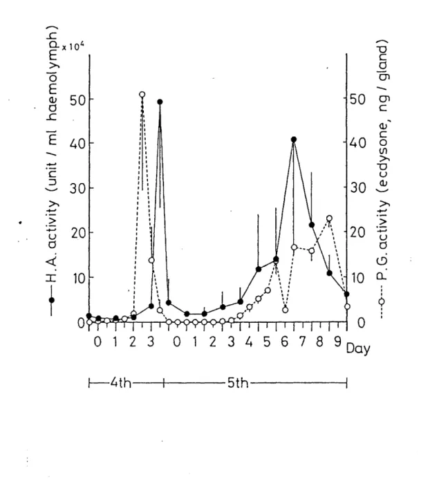
Hemagglutinating activity and ecdysteroid levels in the
hemolymph were determined from 3rd eedysis through pupation. As seen in Fig. 1, hemagglutinating activity in the hemolymph of 4th instar larvae increased from Day 1 and reached a maximum level on Day 3 just following the increase in the hemolymph ecdysteroid titer. The activity deereased rapidly after the 4th eedysis and remained at low levels through the following 2 days.
Hemagglutinating activity increased again exponentiaily from Day 2 of 5th instar and reached a maximum ievel at the time of gut purge. Ecdysteroid titer in the hemolymph of 5th instar larvae remained at iow levels and increased one day before the time of gut purge. These results indicate that hemolymph ecdysteroids may be involved in the increase in the hemagglutinating activity.
Characterizationg;fhema lutinatin ustetivit
In order to eharacterize the Bombyx leetin, we first examined the effects of bivalent cations on hemagglutinating activity. As shown in Table 1, hemagglutinating activity demands bivalent cations, especialiy Mg2+. Inhibition studies with
monosaecharides demonstrated that hemagglutinating activity was not affected at all by galactose and fruetose and slightly
inhibited by glucose and mannose, whereas it was strongly
inhibited by glucuronic acid and galaeturonic acid (Table 2).
Purification of lectin
Hemolymph eollected from larvae immediately after gut purge was used for purification of leetin. Ammonium sulfate
fractionation resulted in almost 100% yield of the activity at a
eoncentration of 30% saturation. Hemolymph was fractionated two times by 30% saturated ammonium suifate, and resulting
preeipitation was used as a sample for gel filtration of Sephaeryl S-300. As shown in Fig. 2, a single peak of hemagglutinating activity was observed at the top of elution profile, in almost the same fraction of Blue dextran (MW 2000 kDa). The recovery of hemagglutinating aetivity from gei filtration was nearly 100%. SDS-PAGE analysis of all the
fraetions revealed that 350 kDa protein band was detected only in the aetive fraetions (Fig. 3 a), indicating that 350 kDa pretein was possibly responsible for hemaggiutinating activity.
Further pur'ifieation was tried using ion-exchange
chromatography, The reeovery of the aetivity was less than 9%
and specific aetivity was not inÅëreased by this procedure.
Consequently we did not employ the ion-exchange chromatography.
Affinity ehrernatography using either glucuronie acid or
galacturonic acid as iigand was performed for further
purification. Active fraetions obtained from the gel filtration
were combined and applied to a column of giucuronie acid-
Sepharose 6B equilibrated with BIS. After washing with BIS, the
bound materiai was eluted with O.2M glucuronic acid. The
recovery of the activity was about 300/o in eluted fraetions, but
we couid not remove non-specific binding proteins under these
conditions. To remove non-specific binding proteins, the column was suceessively washed with buffer 1-3 (Table 3) and eluted with buffer 4. Under these conditions, eluted fractions with O.2M glucuronic acid contained two proteins with molecular weight of about 88 kDa and 90 kDa (Fig. 4a and Fig. 5). The similar result was obtained when galaeturonic acid was used as ligand. When Triton X-100 was removed from these eluted fractions by Bio-Beads SM-2 (BIO-RAD), slight but significant hemagglutinating activity
(about O.1% of recovery) was renatured. This result indicated that 88 and 90 kDa proteins• were components of lectin.
AAgtj,!2gÅëzntibodggalusitL1ectin
Antibody against a 350 kDa protein was prepared as deseribed in Materials and Methods. As shown in Fig. 3b, Western blotting ana!ysis of partially purified lectin confirmed the specifieity
of the antiserum raised against a 350 kDa protein.
Hemagglutinating activity was speÅëifically inhibited by the
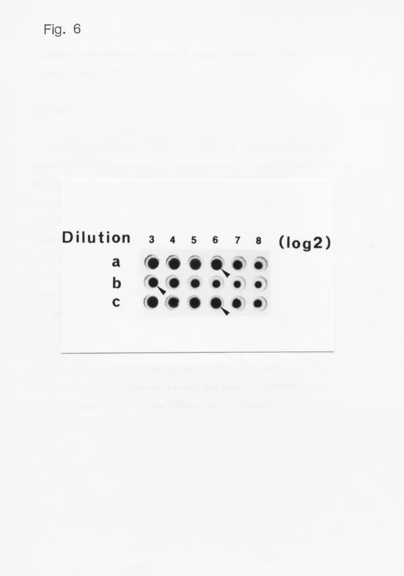
antiserum as shown in Fig. 6, These results indicated that a 350 kDa protein is responsible for hemagglutinating activity.
Western blotting analysis was carried out to confirm the same antigenicity between a 350 kDa protein and either 88 or 90 kDa protein. The antigenicity was identical between these protein$
as shown in Fig. 4b. These resuits indieate that leetin in the
hemoiymph of Bombyx mori is a 350 kDa protein eOmposed of 88 kDa
and 90 kDa subunits.
DISCUSSION
The present study clearly demonstrated that lectin in the
hemolymph of Bombyx mori is a 350 kDa protein composed of 88 and 90 kDa subunits.
Leetin in the hemolymph of Bombyx mori was previously reported by Suzuki et al. (1983) to be a 260 kDa protein but it is not
eiear whether the latter is identical to a 350 kDa protein
suggested in the present study..It is supposed that a 260 kDa protein is a proteolytic product from a 350 kDa protein because fresh hemolymph did not eontain a 260 kDa protein whereas a 260 kDa protein appeared sometimes in hemolymph after a long period of storage. It is also possible that there are several leetins
in the hemolymph of Bombyx mori as indieated in grasshopper, '
-.
Melanoplus spp. (Hapner, K. D., 1983).
Bombyx lectin reported in this paper is very different from other insect leetins in respeet of its bioehemieal
eharaeteristics. The mass molecular weight of Bombyx lectin in hemolymph was estimated to be more than 2000 kDa frorn the gel filtration on Sephacryl S-300, Such large rnolecular weight has not yet been reported exeept the !ect•in in the hemolymph of
Teleogry12as commodus (Hapner, 1981). Most lectins in inseet hemolymph (Hyalophora eecropia, Castro et al., 1987; Sarcophaga peregrina, Komano et al., 1980; MelanopZus sanguinipes, Stebbins
et al., 1985; Antheraea pernyi, Qu et al., 1987) exhibit
galaetose binding specificity and lectin in Sarcophaga peregrina and MelanopZus sanguinipes has been purified easily using
galactose-affinity chromatography (Komano et aZ., 1980; Stebbins
et aZ., 1985). Lectin in the hemolymph of Teleo.ffryllus commodus has N-acetyl-D-neuraminic acid and N-acetyl--D-glucosamine binding specifieity and was also purified easily using fetuin-affinity
chromatography (Hapner et al., 1981). In contrast, lectin in Bombyx hemolymph has glucuronic acid and galaeturonic aeid binding specificity, and detergent (Triton X-100) was need to elute leetin from affinity eoiumm using these monosaccharides as ligand. It also became clear that Bombyx hemagglutinating activity demands bivalent cations, especially magnesium ion. No other inseets hemolymph lectin has been reported to demands bivalent cations. These differenees may reflect differences in the biological role of Bombyx lectin from other inseet lectins.
Studies on Sarcophaga lectin suggested that humoral leetin of holometabolous insects play a role in immunologieal defense system and developmental processes (Komano et al., 1983).
'
Hemagglutinattng act"rifvy in the hemo!ymph of Bombyx was not increased by the inju' ry on the body wall of larvae unlike with Sarcophaga lectin (Suzuki et al, 1983). Rather, it reached a maximum level just before larvai-larval ecdysis and gut purge, suggesting that the BombJzx lectin is probably involved in the disintegration and the renewal of tissues.
The present finding that an increase in hemagglutinating
activity follows the inerease in the hemolymph ecdysteroids titer
indicates that hemagglutinating aetivity in the hemolymph qf
Bombyx larvae may be controlled developmentally by hemolymph
eedysteroids. These observations suggest Bomhyx lectin play a
role in post embryonic development rather than in immunological
defense system as suggested in Sareopha.cra.
Table 1. Effects of bivalent cations on hemagglutinating aetivity.
Dialyze buffer Relative * aetivity
Contro1
+1.5 m,M EDTA
+5.0 miYl CaC12+50 mM MgCl2
100 O.4 loe 200
Partially purified lectin fractionation was dialyzed pH6.0, at 40C for 12 h for
*
Relative hemagglutinating buffers as indicated.
by 30% ammonium against 20 mM control.
activity after
sulfate ammonlum
dialysis
acetate
against
buffer,
. various
Tab1e 2. Effects of monosaccharides on hemagglutinating aetivity.
Saccharides Concentration *
(mM)Glucuronie acid Galacturonie aeid D-Glucose
D-Ga1actose D-Mannose D-Eructose
3 6 '50 100 25
>100
Concentration of
*hemagglutinating
saccharides activity.
.
causlng 50% inhibition of the
Table 3, Composition of washing and elution buffer acid-Sepharose 6B affinity column chromatography.
for giucuronic
Buffer No. Tris O.3M NaCl 1% Triton X-1OO
O.2M acid
Glucuronic
(1) (2) (3) (4)
+ +
+ +
+ + +
+
+ +
The indicated HCI, pH 7.2, caci2).
components (+) eontaining 130mM
were NaC1
supplemented to , 5mM KCI, 50rnM
BIS (10m},vl MgC12, lmrvi
Tris-
Figure legends:
Fig. 1. Changes in hemagglutinating activity and the amount of ecdysteroids in the hemolymph of Bombyx mori during 4th and 5th stadiums. Each datum point (Åë) is an average of 5 different determinations. Open circles indicate the hemagglutinating activity averaged from 5 different detenminations, eaeh of which is indicated by closed circles. Dashed line shows the change in hemolymph eedysteroid concentration.
Fig. 2. Elution profile of hemagglutinating activity from
Sephacryl S-300 (15 x 630 mm). Partialiy purified lectin by 30%
ammonium sulfate fractionation was applied to a colu- i of
Sephacryl S-300 (1.5 x 63 cm) equilibrated with 20mM ammonium acetate buffer, pH 6.0, with 50 mM MgCl2. Fractions of 100 drops were collected,
Fig. 3. Identification of leetin. (a) SDS-polyaerylamide gel electrophoresis of eluted fraction from Sephaeryl S-300. The fractions with hemagglutinating aetivity were combined and subjected to 5% SDS-polyacrylamide gel eiectrophoresis and stained wjth Coomassie Bril!iant Blue. (b) Western blotting analysis of the aetive fraction from Sephacryl S-300 stained with anti--350 kDa protein antibody.
Fig. 4. Analysis of Bombyx leetin subunits. (a) 12.5% SDS-
polyacrylamide gel eleetrophoresis of eluted fraetions with BIS4
from glucuronic acid-Sepharose 6B affinity column (1.3 x 21.5
cm). (b) Western blotting analysis of eluted fraetions with BIS4 from glucuronic aeid-Sepharose 6B affinity column.
Fig. 5. Moleeular weight determination of the subunits of lectin by 12.50/o SDS-polyacrylamide gel electrophoresis. Molecular weight markers: (a) 30 kDa, earbonic anhydrase, (b) 43 kDa, ovalbumin, (e) 67 kDa, BSA, (d) 94 kDa, phosphorylase b.
Fig. 6. Inhibition of hemagglutinating activity by antibody
prepared against a 350 kDa protein. Partially purified lectin by 30% saturated ammonium aeetate fractionation was serially diluted as indicate in dilution amount with (A) PBS, (B) PBS with 1%
antiserum or (C) PBS with 15% non-immunized serum protein. Arrow heads denote the maximum dilution magnitude eausing
hemagglutination.
Fig. 1
A s
ax1o4
E .!r'•
o E
o 50
2
E 40
.-.-
c e 3o
:>-N-
t .i12
, U 20
q <•
z 10
i
o
: : : : : : : : :
'l l
'
: : :
, ,
:
x,,
6
.
:
iltl ll- i .Q,6 X,x,ti
}
a9 6
'
'"
9
,R
let tl
tt
ls 11 i-
x k
,
Ol23 Ol23456 789
A v c o
pt ""s
50 o c
(L}N
c 40 o m. e
30 Si
•.>--
.) 20 'v-
q q, 10 a
l e i o
Day
s
F-4th----+--- 5thny
Fig. 2
o
paco
<
2.0
1.0
o
20 40
Fraction
'
60 Number
1600
80 800
r A 9 5
'il:
Å~ >
.e. •)
< t
= <
Fig. 3
(a) (b)
350K >
Fig. 4
(a) (b)
gOKXx,
/li -=-A
eeK
lt 1l
l
I i
i' i'
(d
(c qb
(a
Fig. 5
1.0
A pp
'U
Ni
c O.5
. .9
'tii
-,,,],,,
E'
oa
b
d
c
r 88000
9OOOO
5
Motecular
10
weight (X 10-4>
Fig. 6
Di1ution
a b c
(eOOgAeNe5
/gie ee e' el
eee eN" e>
(log2)
CHAPTER 3. 0CCURRENCE OF LECTIN IN VARIOUS TISSUES OF THE SILKWORM,BOMBX\ MORI
ABSTRACT
Monoclonal antibodies against the 350 kDa lectin purified from larval hemolymph of the silkworm, Bombyx mori, were prepared.
The antibodies inhibited the hemagglutinating activity (HA
activity) and bound speeifically to the hemolymph 350 kDa lectin in Western blotting analysis. Immunohistologicai observations revealed the occurrence of lectin in the euticular intima of the anterior silk gland of fifth instar larva of Bombyx mori.
Extracts from the anterior silk glands showed HA activity and
exhibited the same biochemieal eharaeteristics as the hemolymph
350 kDa lectin. Western blotting analysis revealed that there
are several lectins with different molecular weight in epidermal
eells in addition to the anterior silk gland, but not deteeted in
fat body, and in both middle and posterior siik glands, Lectin-
like molecules in epithelial tissues may play an important role
in the hystolysis during'the molting and metamotiphosis.
INTRODUCTION
Hemagglutinins or leetins have been found in various
invertebrates as well as vertebrates (Barondes, 1984) while their funetions reported are different in investigations and speeies
used. Renwranz and Stahmer (Renwranz et al., 1983) showed that hemagglutinin isolated from the hemolymph of Mytilus edulis is linked to the hemocyte plasma membrane and involved in
reeognition of foreign bodies (Renwranz et al., 1983). The study on Sareophaga peregrina leetin also indieated that lectin has function in defense mechanisms (Komano et al., 1980, 1983, Takahashi et al., 1986). Seott (1971) found, however, that hemagglutinin did not enhanee phagpcytosis against injected vertebrate erythroeytes in Periplaneta amerieana.
Leetins are also found in the course of cell differentiation.
'
Slime mold, Dictyostelium discoideum, releases two lectins, diseoidin I and II, when starved to initiate differentiation from a vegetative form to a cohesive one (Fraizer et al., 1-975), and was suggested as a eell-adhesion moleeule in speeial
developmental stages (Berger et aZ,, 1982). The Sareophaga
lectin oecurs in the early embryonic stage and the eariy pupal
stage (Komano et al., 1980), suggesting involvement of leetin in
the post--embryonie development of this species. In vertebrates,
a elass of P-D-galactoside-binding lectins affect intereellular
interactions and have the characteristics similar to a number of
morphoregulatory proteins (Nowak et al., 1976). These studies
show that lectins in animais have a multifunction eoneerning with
the cell-cell interaction.
Oceurrence of lectin in Bombyx mori has been known (Suzuki et al., 1983). Bombyx hemolymph lectin have been aiready purified from Bombyx larval hemolymph (Amanai et al., 1990a) and
demonstrated that hemagglutinating activity (HA aetivity) in
hemolymph changed developmentally in the 4th and 5th instar. An increase in the HA aetivity was observed at the time of larval
molting aecompanied by renewal of larval tissues as well as pupal ecdysis followed by larval cell death, In addition, both these
developmental events and HA activity in hemolymph (Amanai et al., 1990a) have been demonstrated to be under the control of
ecdysteroids. Therefore, lectin could be involved in the
developmental events, espeeially at the eellular level, induced by an increase in the hemolymph eedysteroids.
To study the oceurrenee and distribution of Bombvx lectin,
monoelonal antibodies against the hemolymph 350 kDa leetin
purified frorn larval hemolty,mph were prepared. Then various
tissues of last instar larvae were inmunohistologically examined
using these monoclonal antibodies and it was found that there
were several proteins, whieh cross-react wi`Lh anti-hemolymph 350
kDa leetin monoclonal antibodies. This paper deseribed the facts
that the substances cress--reacted te the hemolyrnph 350 kDa leetin
antibodies were abundantly present in the cutieular intima of
anterior part of the silk gland as well as epidermis and wing
di$c but not in fat body. Based on these Åíindings, a possible
funetion of insect lectins will be discussed.
MATERIALS AND METHODS
Animals
Larvae of the silkworm, Bombyx mori were reared on an artificiai diet (Nihon Nosan Co.) at 25 Å} O.50C under a 12 h
light and 12 h dark photoperiodic regime. The newly ecdysed 5th instar larvae were segregated and fed from the beginning of the photophase. Under these conditions, male larvae purged their guts during the scotophase of day- 7 and pupated 3 days thereafter (Sakurai, 1984). Only male larvae were used for
immunohistologicai experiments.
SDS- ol acr !amide ggLl electro horesis
SDS-polyaerstamide gel elec'I rophoresis (SDS-PALGE') was
performed aecording' to Laemmli (1970). The sample solution mixed with one vo!ume of sample buffer (2% (w/v) SDS, 4% (w/w) 2-
mercaptoethanol, 30% (v/v) glycerol, O.04% (w/v) bromphenol blue) was denatured by heating for 5 min in boiling water. After
electrophoresis, gel was stained wtth Coomassie briliiant blue
R250 (Fairbanks et al,, 1971). Protein amount was determined by
the method of Lowry (1951) using bovine serum albumin as a
standard substance.
Monoclonaluatntibd rearation
Powder of 5% SDS-polyacrilamide gel containing hemolymph 350 kDa lectin was used as an antigen (Tjian et al., 1974). BALB/c mice (8 weeks old, female) were injected with O.25 ml mixture of gel powder (about 2 yg protein) and eomplete Freund's adjuvant.
Additional immunizations with the same amount of the mixture were carried out at 3 week intervai. Three days after the fifth
immunization, cell fusion was performed by the method of St.
Groth and Scheidegger (1980) with a minor modification. A mouse myeloma cell line (P3X63-Ag8.653) was used as a fusion partner and cultured in Dulbecco's modified Eagie's medium <DllEM, Nissui Seiyaku, Tokyo) supplemented with 10Yo fetal bovine serum (FBS, '
M.A. Bioproducts, Walkersville, MD). Exponentially growing mouse myeloma cells were mixed with 10-fold number of immunized spieen eells f•ollow•ed by centrifugation. The medium were discarded and cell pellets were loosened by fliÅëking the bottom of the tube.
Prewarmed 1.0 ml of 50%(w/w) poiyethylene glycoi 4000 (Sigma) was added over a period of 1 min and incubated for 90 sec at 37 OC.
Then le ml of serum-free DMEM was added and the mixture was further incubated for 5 min. The cell suspension was e'entrifuged and the pellet was suspended in DMEM supplemented with serum (2 x 10⁶ ceils/ml). The cell suspension (O.2 ml) was dispensed into
each well of a 96-well culture plate. After 24 hrs, medium was
changed to a growth medium eontaining 10'4 M hypoxanthine, 4 x
10-7 M amethopterine and 1.6 x 10"5 M thymidine (HAT medium,
Littlefield, 1964). HAT medium was changed every 3 days. Each
well was tested for the antibody activity as described below.
The cells in the antibody-producing wells were cloned 3 times by limiting dilution using mouse spleen eells (1-2 x 10⁶ cells/well) as feeder eells.
SSglzgggU}gcreeninctgLfuehbridom eulturemedium
The hybridoma culture media was examined for the inhibition of HA aetivity. Lectin was partially purified from day 7 of fifth
instar larvae by 30% saturated ammonium sulfate fractionation as deseribed previously (Amanai et al., 1990a). The resulting precipitate was dissolved and diluted with PBS (75rnM Na2HP04 / KH2P04, 75nhY NaCl, pH 7.2) to 1280 units/ml of HA activity and used as a sample solution for the screening. The sample solution was diluted serially 2-fold by PBS in 120 weli microtiter plate
and the same voiume of hybridoma culture mediurn 10-fold diluted with PBS was added. After mixed and stood for 10 min at 40C, 25 yl from each well w:-as transferred to an another plate. To assay the unabsorbed HA aetivity in each 25 pl mixture, 25 yl of trypsinized and giutaraldehyde fixed sheep red b!ood cell solution (1 x 10⁹ cells/ml) was added (Amanai et al., i990a).
InhibitiOn was expressed as percentage to the control.
slg class :tg2EL!ng g:fL monoelonal antibodies
Ig class typing of monoelonal antibodies was performed using
class/subelass speeific rabbit anti-mouse immunbglobulin (MnoAb-
ID EIA Kit, Zymed Co., San Fransisco, CA). 100-fold diluted
hybridoma culture medium was used as antigen in this assay,
WesterntL2tLg!tu!gtt ustl
Western blotting analysis was performed by the method of
Towbin et al. (1979). The proteins separated by 5% SDS-PAGE were electrophoretieally transferred to a nitrocellulose membrane
filter (pore size O.45 pm). The filter was first reacted with
1000-fold diluted anti-hemolymph 350 kDa lectin monoclonal anti-- '
body, and then with 200-foid diluted peroxidase conjugated anti- mouse IgM antibody (Cappel Co.). The immunoreactive peptides on a filter were visualized by ineubating the filter"in developer
(O.05M Tris-HCI, pH 7.5, O.5 mg/ml diaminobenzidin, O•3% H202)•
Immunohist, ochemical E!t2ygyd gf anterior silk g.ILIuuxd
The anterior silk glands of fifth instar larvae were dissected out and rinsed in iee-cold MES--BIS (15mM NaCl, 46TruM KCI, 12mVii
CaC12 2H20, 50nLM MgC12 6H20, 112miI GIueose, 5MM 2-(N-Morpholino) ethanesulfonic acid monohydrate, pH 6.5) for 5 min with gentie
swirling. After the rinse, the anterior siVA' glands were fixed in Bouin fixative for 2 hrs and dehydrated in ethanoi series.
Eaeh tissue was singly embedded in paraffi: and sectioned at 5
pm. Peroxidase-anti-peroxidase (PAP) cempiex staining was
performed by the method of Sternberger et aZ. (1970) with a minor
modification. The sections were incubated with 5% skimmilk in
Dulbecco's PBS for 20 min at room temperature to prevent non-
specific binding followed by an ineubation with 1000-fold diluted
anti-hemolymph 350 kDa lectin monoclonal antibody. After three
times of rinse with Dulbeceo's PBS for 5 min, sections were
incubated with 200-fold diluted goat anti-mouse immunoglobulins (IgA + IgG + IgM) antibody (Cappel Co.) for 1 hr at room
temperature. After the rinse with Dulbeeeo's PBS, sections were ineubated with 200-fold diluted mouse monoclonai PAP (Zyrned Co.) for 1 hr at room temperature and visualized by an ineubation with a developer (O.e5M Tris-HCI, pH 7.5, O•5 mglml DAB, O•3% H202) for 10 min at room temperature, As a eontrol for immunostaining, 50% (NH4)2S04 fractionated non-immunized mouse serum was used.
Pre aration gLf extraets from the anterior silk g,ILans!d
Ten pairs of anterior silk glands were disseÅëted from day 3 5th instar larvae, rinsed twice with iee-eold MES-BIS for 5 min
.'
and homogenized in 1 ml oÅí SDS-lysis buffer (10 niM Tris--HCI, pH 7,6, 1% (w/v) SDS, 2 pg/ml PMSF, 2 yg/ml antipin, 2 yg/ml
pepstatin A). The homogenate was stood for tuore than 3 hrs at room temperature and centrifuged at 10,OOO g for 15 min at 40C,
' Ciear supernatant was mixed with 1/9 volume of 1.8 M potassium phosphate buffer, pH 7.5. The mixture was stood on iee for 30 min and eentrifuged at 10,OOO g for 20 min at 40C. Detergent was mostly rernoved by this procedure. After dialysis of the
supernatant against PBS, precipitated materials were removed by
eentrifugation at 10,OOO g for 15 min. The resuXting supernatant
was used as a sample for the assay of HA aÅëtivity, SDS-PAGE and
Western blotting analysis.
Tissues extraetions
integument and fat body were dissected Åírem day 3 iarvae and rinsed twiee with MES-BIS for 5 min on iee. Epidermis of
single--eeli iayer was obtained after eompiete removai of euticle layer from the integument. Proeedure of the preparation of a sample fer SD$-PAGE was the same as deseribed above for the anterior silk glands. The resulting supernatant was centrifuged at le,eOO g for 20 min at 40C and the re$ultant preeipitates were
washed twiee with Dulbeeco's PBS and dis$olved in SDS-PAGE sample buffer. The samples for SDS-PAGE were direetily used Åíor Western bletting analysis.
'
l!IEEII!U!,1:UlSL
!Cgl4aE!gsx!Lgy2Lga!zl,gzth cte zatx sLfmQnoconalantiboie${if;aJls!pt!hs!uRsmpRkl h350
kDa leetin
b4ouse menoclenal antibodies were sueeessfuily prepared against Bembyyhemelymph 35e kDa lectin` We obtained 989 clQne$ of hybridema after twe times of oeM fusion prQcedures. When
$upernatant freff} 305 clones were tested Åíor it$ ability to inhibit HA aetivity, 51 elones significantiy inhibited the HA activity (Table 4). AÅíter two times additiQnal elonings, le
clones w,ere selected and prepared ascite from these hybridomas, Aselte was obtained from 7 ef iO clones and !g eiass typing of
the obtained monoelonal antibodies was performed using hybridoma eulture media. As shown in Figure 7, only 2 monocional
antibodies, denoted as aBH-1. and aBH-2, were ebviousiy determined as Ig M (K). Eaeh -antSbody was partiaily purified by ammonium sulÅíate fraetionation and used to examine the abiiity to inhibit HA aetivity (Fig. 8), All 7 antibedies examined signiÅíieantiy inhibited HA aetivity. Binding speeiÅíieity of aBH-1 and aBH--2 was te$ted by Western blotting analysis. As shewn in Figure 9, aBh'-i, and aBH--2• $hewed $peeific binding tQ`a 350 kDa preteSn reported as lectin in the previous paper (A• manai et aZ., i99ea).
Immunohistoio- a.e 1 gb e/rva ien gfL sula 1 ss!!Js ew
Anterior, middle and posterior silk glands were
immunohistologically obBerved us,ing aBH-1 or aBH-2. Middle and
posterior silk giands were net $tained. A$ shewn in Figure 10, cuticular intima ef the anterior siik gland in beth feeding stage
<day 3 of fifth instar) and prepupal stage (2 day$ after gut purge) was well stained, As shown in Figure 10 <b), thin layer around the euticular intima inside the anterior siik gland cells was stained strongiy than the cutieular intirna. This observation indieated. that the thin layer of the apieai eyteplasta ef the t celis as well as the eutieular intima eentains substanÅëe positive to anti-hemolymph 350 kDa ieetin moneclonai antibody. To Åíurther een.firm the eccurrenee of the positive substanee, I u$ed other 4 antibedies whieh wer'e expeeted te have different recegnitien frQm aBH-1 sinee they were prepared from totally different elones
separated at the eariy phase ef the eloning. These 4 antibodies gave the sams result (data not shewn).
.ltL{ig!ptÅíd nti"ieakion g•sf, the imaynoh`ste ica•li s2gs.l:t!z!yi ]ggptLpt!} S"t•
]tu}s!!h eu 'oular intima sLf anterior gL!:Ug gslau!;sgct
The anterior silk gland extraetg exhibited a very high HA aativity in the assay using tr•y-f,sinized and, glutaraldehyde fixed sheep ned• bloed• eelSs. Figure IZ $hew$ the typiÅëai reguit that a pair Qf the anterior silk gland of day 3 larvae eontained the HA activity of 215 units. {rhe HA activity was redueed by heat treatment and completely disappeared aÅíter triptie digestion.
CPherefore, HA acUvity of the extraets was due te pyoteSn, The HA aÅëtivity was strongly absorbed by ai}-H-1, as indieated in
F Figure 12. These resuits suggest that the anterior silk gland is
in its possession ef a protein with HA aetivity. In order to
confirm this finding, Western blotting analysis of the anterior silk gland extracts was performed using aBH-1. As shown in Figure 13(A), 12.5% SDS-PAGE and Western blotting analysis revealed mainly two bands, one was 49 kDa and the other remained near the top of the gel. In addition, several faint bands were
deteeted at the molecular weight ranging from 55 to 80 kDa. In order to analyze the molecular weight of the larger band found near the top of the gel, 5% SDS-PAGE and Western blotting analysis were performed. As $hown in Figure 13(B), the stained area was broad in a range of molecular weight from 84 to 150 kDa, and no distinguishable band was observed.
Lectin in the extracts of other tissues
To examine the occurrenee of leetin detectabie by aBH-1 in tissues other than silk g!ands, extraets from epidermis and fat body were subjeeted to Western blotting analysis using aBH-1. As shown in Figure 14, the epidermal cell extracts contained several proteins with different moieeular weight. Among them 4 bands were cross-reacted to aBH-1 elear!y, In contrast, extraets of fat body did not eontain any protein as detected by aBH-1.
t
blSCUSSION
The present $tudy elearly demonstrated that Bombyx ieÅëtin is present not cniy in hemolymph but al$e in various tissues such as anterior silk glands and epidermis.
Oecurrenee of lectin in the anterior silk gland was
immuitohistologiÅëally sbown using monoelonal antibodies. Also, the anterier silk glands eontained leetin as judged by the assay Q'f HA activity and Western blotting anaiysis using aBH-1. The s'ilk gland leÅëtin was 49 kDa whil•e the hemolymph lectin was 350 kl)a, apparentiy ghewing that these two leetins are diÅíferent though data indieated that the anterior silk giand lectin
pessesses the identieal eharaeteristies te the hemoiyniph 350 kDa leetln in respeÅët of inmunoehemieai prepertie$. Consequentiy, the anterior sitk gland leetin is homologous to the hemoiyRlph 350 kDa ieetin. Although anQther broad band larger than 49 kDa was deteeted frorn the silk glands by Western bletting analysis, it is not clear whether this banct is Zeetin sinee a di$tinguished band was net deteeted by Western blett'ing using 5% SD$-PAGE.
3that is the souree eS the 1•eetin in the cutiÅëuiar i'ntima and what is its• ftm' ction? AnterigrL sUk gland i-s a thin duet
•Åëonpesad eÅí seme 300 eells and iined with a thiek euticular intima at the surfatva faeing the lu.rnen. Eleetron migxeseopie ebservation of the anterie" silk gland eell shows that
ekiectronlueent granuieg afe 1ined mp irn the peripheral eytoplasrn and exten<l finely ramified thin tubules onto the pl-asma' :membrane faeing the lumen, forming a layer of approximately 4 pm in length
(Akai, 1984). The monoelonal antibody against the hemolyrnph 350
kDa iectin stained strQngly the peripheral thin iayer ef eytQpla$m in addition te the cuticular intima, This
immunohistelQgical ebservation suggests that the anterier siik gland lectin is accumulated at the peripheral thin iayer and the lectin in cuticular intima is seereted by the anterior siik gland eells, although whether the leetin is synthesized in the cells or results from processing of the hemelymph 350 kDa lectin is not known. The struetures found in the peripheral layer is
considered to produce fibers whieh may be the preeur$ers of the eutieular intima (Akai, 1984). AcÅëording to the eieetren
mieroscopie observations, the fibers may aggregate and form dense--body oeeurving immediately inside of the plasma membrane.
Additionally, partially purified leetin from Bombrx hemolymph tends to agglutinate and stiek to the surÅíace of a test tube (Suzuki et al., 1983). Thes.e findings show that .in eutieular intima, leetin rnay be invoived in forming the retieular strueture of cuticle by making a eomplex with other proteins and macre molecule$ $uch a$ polysaecharides.
Western blotting analyses showed that the epithelial eells eontain several pretein$ which deteeted by aBH.-1. During the molting stage, eutiele of the integument and the euticuiar intima are renewed as secreted from the epidermal eells and the anterior
$ilk gland celis (Akai, 1984), re$pectively. Zn these tissues,
lectin may be used as a component of euticle. Therefore, the
function ef both leetins may be eemmen in these two tissues
theugh the mole-eular weight is different. The similar arguments
are found in reports to denote the oeeurrence of nearly identieal
leÅëtins detected by the same antibody both in the serum and other
tissues. In rabbit, serum lectin exhibits the same
immunochemical properties and pes$esses the same $ubunit profiles as leetin from liver (Kozutsumi et aZ., 1980). Fibronectin, a
leetin known as cell adhesion moleeule, oeeurs both in
extracellular matrix and serum (Rouslahti et al., 1973, 1975,
Yamada et al., 1975, Grinnell et al., 1978). It is, therefore,
possible that Bombyx lectins in various tissues are eriginated
from the hemolymph 350 kDa ieetin. Alternatively, untii the
leetins in the various tissues are purified and characterized,
the possibility remains that the hemolymph 350 kDa lectin is
different from lectins in tissues in respect of physioiogieai
funetion and the site of'synthesis,
Table 4, Profiles of cloning and screening of hybridoma.
---nyi-t--- ---"--p--- ---.--t---pt- ---t-"---"}---e--m--i----tm---.
No. oÅí hybridoma No. of No. of positive obtained $creening clones (%>
Celi fusionA 331 120 19 (15.8) Cell fusionB 658 185 32 (17.3)
"---4---t---"---h---P---td---t--"----d---
Totai 989 305 51 (16.7)
---i.ep---hp---p---.-..---"-4b--"---.---d---e--t---k----"b----t----)---"----.--".--
CelZ fusion A and B were carried by the same proeedure exeept the
use of spleen ceils obtained from different -mice.
Figure legends:
Figure 7. Ig class typing of 6 anti-heruoiymph 35e kDa lectin monoclonal antibodies (aBH-1, 2, 4, 5, 7, 8). Hybridema eulture media wa$ diiuted with Dulbecco's PBS by 100-Åíold and used as antigen in this assay, Class oS heavy and iight ehain of
irnmuneglobulins detected by the seeond antibQdy (elass!subelass specifie rabbit anti-mouse immunoglobulin) is indicated at the tep;Yl, IgGl (gamma 1 chain);'Y2a, !gG2a (gamma 2a ehain);'Y2b, rgG2b (gan}ma 2b ehain);'Y3, rgG3 (gamrna 3 ehain);ct, IgA (aZpha Åëhain);f", igM (mu chain); re, kappa light chain; X, lautida light
ehain. NRS, normai rabbit serum as a control.
Figure 8, Inhibition of the HA aetivity by anti-hemoiymph 35e , kDa ledtin monoclonal antibQdies. Haneiymph leetin'pavtially
purified by 30% $aturated ammgnium $uXfate ÅíraÅëtienatioR was u$ed. The lectin $ample used had 12ge units of HA aÅëtivity
(Amanai et aZ,, 1990a) and this activity was set te 10e%, Eaeh menoelonal antibody was serially diiuted with PBS and 25pl was added te eaeh assay. Number with arrovg indicates the name of monoelonal antibedy.
Figure 9. Binding speeifieity oÅí anti--heinolymph 35e kDa lectin monoelonal antibedies (aBH--1 and 2). Partialiy puriÅíied hemolymph 350 kDa leeti'n by 30% saturated amonium sulfate fractionation wa$ subjeeted to 5% SDS--PAGE and, after
eleetrophoresis, the gel was divided inte 3 portions. Eaeh was
used for staining with Ceemassie brilliant blue (a), Westepn
blotting analysis with aBH-1 (b) and aBH-2 <c).
Figure 10. Immunohistochemical staining of the anterior silk gland of the fifth instar larvae using aBH-1. Transversal section. (a), x 200; (b), x 600; (e), control. SGC, anterior silk gland cell; L, lumen; CI, cuticular intima; N, nucleus; C,
cytoplasm. -
Figure 11. HA activity in the extracts from the anterior silk glands (a). SDS-lysis buffer after removed of the detergent by the same procedure (b). PBS (c). Trypsinized and glutaraldehyde fixed sheep red blood eells were used for the assay (Amanai et al., '90). Arrow head denotes the maximum dilution magnitude to cause the hemagglutination.
Figure 12. Inhibition of HA activity in the extracts of the anterior silk gland by the monoclonal antibody, aBH-1. HA activity of the extracts of the anterior silk gland was justified to 1280 units/ml and used for the assay. Other details are the same as in Figure g.
Figure 13. Western blotting analysis of proteins from the anterior silk gland. Twenty ug of proteins were subjeeted to
12.5% SDS-PAGE. Western blotting analysis were performed using
1000-fold diluted aBH-1.
Figure 14. SDS-PAGE (a) and Western blotting analysis (b) of proteins from epidermal cells and fat body cells in the fifth instar larvae of Bimbyx mori. A, epidermal eells (12.5% SDS- PAGE); B, fat body (5% SDS-PAGE). Arrow heads indicate the protein bands which detected clearly by Western blotting analysis of which molecular weight are estimated as 180, 92, 54 and 43
kDa. •
,
t
Fig. 7
aBH-5 (
NRS 7i72a 72b 73 a p K X
v'X
C N.'S ,'- OA ,A 4, ,rx
NN k''X ("Xi "N ('> ,"" rx r's tttf
aBH-4 (
<- ts (-) )1aBH-1 ( o ee 'ls
(--ii .h ee i/...)
aBH.2 t •'" O "N (Aiee •s
N . <'- c Ns ( i fr ,f "h r' xi ee 'y
aBH-7 ( (' (' C'X'( (>( •i x> ,i A .t ( (" ( C.) (), ) "j .I)
aBH-8 ( fv (" CJOOOOOO
CCCOOOOQQ
-
(l5'
s
eo HA activity(o/,)
o
> g.
.a g
=A o'
(h-`'
v ;
o - o
o
Eilll
r'--'--M-"-T--'-""-'"'----i
pt clJ
=
lw N
i
Q
:o
6
- o o
oo •Vi....
g•
$ E
E3
b
;
•
l
Q
:Fig. 9
e
, ' J q
Jl,i
i
i,l
"' i
ths
-pt
! i
[1 e "(350K
Fig. 10
'
7
7"'
t
'
5.J
;t
-:
ts'
?.
,L'i"+.:lb
tf"`';'
kliiiil,-.
t'' ''
,t.`l`t;stl•';;'i/`iKl"/L'"
--- -if;.5i),iiii;". .g'c' ,
:Si:.L' :{E :
. ----r-'M'M(h")'
if,.f,i,r:s:/s'snyg' {ll[r'1i/IRS e7 :{ :e' •r, t,, .. /
" - t . . ";-g
s"
;,1
J--
Xi/ti}/li,,,.
,
ti
L
't'-
sg
A-,
Li•
. It:yr"""
Y L
r f,.t
, ;".bt. ht 'lt' }N
,. S]F..
u,]N• 1'egsr.:., •
t LJi
ti v.
,-•-•:'ii,Lic's'`
'i.( ;';i,
-"r'J .'')", t;-i.,.:
•K
,r{l•il,St"Cl
L -
"i<iil•Litt,{:t/z,IiSili,,Sf,a,.,g.,,,f\f,X5s'}N V;1' N•
N' ,
• •::.'U:.
'N•'- ' •••,".. c
N';-
<"
. N,i:.
-i ,vs N<t,
' X '`r• -
k, (P.
(b)
.+- -- " f - . -t 4- - --. i--+ ---
,
l
i
s 1
t+ --.--
.
(c)1
.
4
, •r.
•:
'Fig. 11
Dilution
b
c
'e e'
'e (" e te eeeeee e'e e'
'e e' e t'
e e. eeeeee e'e 'v Cho
ce (e 4e ..
Fig. 12
A R
•f2i...,•
>
•.--
>
'.H'
v U
< =
100
50
o
e-o----o---Q----o--- Ptain serum
aBH •-1
10
Antibody (pg / ml)
100
Fig. 13
a b
r"
""pa-
it't' 't'
<il 49K
Fig. 14
a ii
-iiei
lgE.:l;=
,`,"ii"""i i,D,,,,i,,
1l
e•e
e• e
A
b
- -- <1111
-q . -v. (
-'.-
-" (
e
- •t •
i
B
b
CHAPTER 3. THE SITE OF HEMOLYMPH LECTIN PRODUCTION AND ITS ACTIVATION IIV VITRO BY 20--HYDROXYECDYSONE
ABSTRACT
To identify the tissues whieh produee hemolymph leetin in larvae of Bombyx mori, ovary, testis, fat body and hemocytes from 5th instar larvae were cultured in vitro and the culture medium partially purified and assayed for hemagglutinating aÅëtivity.
Among the tissues tested, hemocytes appeared to be a rnajor source of the hemolymph lectins, Ovary produeed leetins to about one tenth of the amount observed for the hemocytes, whereas testis and fat body were not produetive, To study the hormonal eontrol of hemolymph lectin production by hemoeytes, hemocytes from 4th instar larvae were eultured in vitro. Hemagglutinating aetivity in the hemolymph of 4th instar larvae was immunostainable with the monoc!onal antibody raised against 350 kDa lectin found in the 5th instar hemolymph, but their molecular sizes were larger
• than the 5th instar hemolymph leetins. When 20-hydroxyecdysone was added into the medium, produetion of the lectin by the
hernocytes was remarkably enhanced, depending upoR the hormone
concentra" tion,INTRODUCTIOLN
Lectin-like substances with hemagglutinating (HA) activity have been found in the hemolymph of a variety of insect species.
In Sareophaga, lectin production is induced through the activation of the lectin gene by a humorai faetor induced by injuries on body wall (Komano et al., 1980, Shiraishi et al., 1988, 1989), and this lectin may participate in defense
meehanisms (Komano et al., 1980). In contrast, HA aetivity in Bombyx hemolymph does not increase in response to injuries on the body wall (Suzuki et al., 1983). Rather, the HA aetivity in the
larval hemolymph of Bombyx fluctuates during the 4th and 5th instars in concert with the changes in ecdysteroid secretory activity of the prothoraeie glands (Amanai et al., 1990a).
Lectins are detected Smmunologieaily in tissues such as anterior silk gland and epidermis, and the coneen'vration of these leetins undergo drastic changes during metamorphosis (Amanai et aZ,, 1990b), This suggests that the lectin may have important functions in the renewal of cuticle, the degeneration of larval tissues and the development of adult tissues.
krhile the site of lectin produetion in Bombyx has not
previousl.v been eonfirmed, in Sarcophaga peregrina (Komano et al., 1983) and Melanoplus differentialis (Stiles et al., 1988)
leetins are produced by the fat body. In hemimetaboious inseets such as Leucophaea maderae (Ammirante, 1976), the hemocytes appear to be a major source bf lectin. Therefore, lectins may have distinet physiological toles on the different insect
t
spec1es.
To clarify the physiologieal role of Bombyx lectin in larval molting and larval-pupal metamorphosis in Bombyx, I have attempted first to identify the site of produetion of the ieetin.
To this end, 1 used mature 5th instar larvae since the hemolymph 350 kDa lectin is very abundant at this stage (Amanai et al., 1990a), and beeause these larvae are large enough to ea$ily collect both larval tissue and hemolymph.
I have feund previously that the leetin concentration of the hemolymph increases in a developmental pattern similar to that ef the secretory activity of prothoraeie glands (Amanai et al.,
i990a), However, the reiationship between the lectin titer and the ecdysteroid titer of the hernolymph is net clear during the 5th instar. The titer of the lectin rises on day 3 of the
instar, when t. he prothoracie glands'become active, but when the hemolymph eedysteroid titer has not yet risen. The leetin
reaches a maximum level Qn day 6, 3 days ear!ier than the glands exhibit the highest aÅëtivity. In contrast, during the 4th
instar, the changes in the lectip. content of the hemolymph shows a very elose correiatien with the seeretory aetivity of the
prethoracic glands. Theteefore, I have studied the aetivation of
leetin synthesis by 20•-hydroxyecdysone using hemoeytes from 4th
instar larvae.
MATERIALS AND METHODS
Anima1s
Larvae of the silkworm, Bomby.\ mori (J 124 x C 124 and Gunpo x $hugyoku) were reared on an artifieial diet (Nihen Nosan Co.) at 25 -+ O.5 OC under a 12 h iight and 12 h dark photoperiodic regSme. Larvae were staged at the 3rd and 4th larvai molting and the day of ecdysis was designated as day O of the instar
'
(Sakurai, 1983). Fifth instar larvae were selected again during the phQtophase of Day 6 aecording to spinneret pigmentatSon (Sakurai, 1984) since the larvae with pigmented spinneret were predicted to purge their gut during the following scetophase, These larvae were used for in vitro eulture of tissues and immunofluoreseense staining of the hemocytes.
tH!gipgg!mps!1h


![Fig. 5 1.0 A pp 'U Ni c O.5 . .9 'tii -,,,],,, E' oa bd c r 88000 9OOOO 5 Motecular 10 weight (X 10-4>](https://thumb-ap.123doks.com/thumbv2/123deta/5643401.2003829/31.892.96.806.48.1100/fig-pp-u-ni-tii-oooo-motecular-weight.webp)