O RIGINAL A RTICLE
Circulating Endothelial Cells in Non-small Cell Lung Cancer Patients Treated with Carboplatin and Paclitaxel
Makoto Kawaishi, MD,* Yutaka Fujiwara, MD,† Tomoya Fukui, MD,* Terufumi Kato, MD,*
Kazuhiko Yamada, MD,† Yuichiro Ohe, MD, PhD,† Hideo Kunitoh, MD, PhD,†
Ikuo Sekine, MD, PhD,† Noboru Yamamoto, MD, PhD,† Hiroshi Nokihara, MD, PhD,†
Takeshi Watabe, PhD,‡ Yuji Shimoda, PhD,‡ Tokuzo Arao, MD, PhD,§ Kazuto Nishio, MD, PhD,§
Tomohide Tamura, MD† and Fumiaki Koizumi, MD, PhD*
Introduction:Circulating endothelial cells (CECs) increase in can- cer patients and play an important role in tumor neovascularization.
Methods:This study was designed to investigate the role of CEC as a marker for predicting the effectiveness of a carboplatin plus paclitaxel based first line chemotherapy in advanced non-small cell lung cancer (NSCLC).
Results:The CEC count in 4 ml of peripheral blood before starting chemotherapy (baseline value) was significantly higher in NSCLC patients, ranging from 32 to 4501/4 ml (n
⫽
31, mean⫾
SD⫽
595⫾
832), than in healthy volunteers (n⫽
53, 46.2⫾
86.3). We did not detect a significant correlation between the CEC count and estimated tumor volume. CECs were significantly decreased by chemotherapy as compared with pretreatment values (175.6⫾
24 and 173.0⫾
24, day⫹
8,⫹
22, respectively). We investigated the correlation between baseline CEC and the clinical effectiveness of chemotherapy. CEC values are significantly higher in patients with clinical benefit (partial response and stable disease, 516⫾
458, 870.8⫾
1215, respectively) than in progressive disease patients (211⫾
150). Furthermore, a statistically significant decrease in CECs, on day 22, was observed only in patients with partial response. Patients who had a baseline CEC count greater than 400/4 ml showed a longer progression-free survival (⬎
400, 271 days关
range: 181–361兴
versus⬍
400, 34关
range: 81–186兴
,p⫽
0.019).Conclusion:CEC is suggested to be a promising predictive marker of the clinical efficacy of the CBDCA plus paclitaxel regimen in patients with NSCLC.
Key Words:Circulating endothelial cell, NSCLC, Chemotherapy.
(J Thorac Oncol.2009;4: 208 –213)
A ngiogenesis plays a critical role in the growth and me- tastasis of solid tumors.1 The clinical importance of angiogenesis in human tumors has been demonstrated by several reports indicating a positive relationship between the blood vessel density in the tumor mass and poor prognosis, i.e., survival, in patients with various types of cancers includ- ing non-small cell lung cancer (NSCLC).
2– 6 Furthermore, Natsume et al.
7reported the antitumor activities of anticancer agents to be less active against vascular endothelial growth factor-secreting cells (SBC-3/VEGF), in vivo as compared with its mock transfectant (SBC-3/Neo). In recent years, antiangiogenic agents have also been demonstrated to be active against a variety of malignancies, including lung, colorectal, and renal cancer.
8 –10 Thus, angiogenesis is a promising target for cancer treatment and is related to the prognosis and efficacy of these drugs, though the tumor vessel biomarkers which predict the effectiveness of antian- giogenic agents and other anticancer agents are not always useful and have not become well-established.
Circulating endothelial cells (CECs) have been recog- nized as a useful biomarker for vascular damage. CECs are increased in cardiovascular disease, vasculitis, infectious dis- ease, and various cancers.
11–14Recently, CECs were found to be more numerous and viable in cancer patients than in healthy subjects.
14,15Furthermore, elevated CECs in cancer patients were found to be nearly normalized when the tumor was removed surgically or with chemotherapy.
15Therefore, most CECs are considered to be disseminated tissue endo- thelial cells in the tumors and the CEC number may reflect the extent of tumor angiogenesis. Indeed, the CEC level has been demonstrated to correlate with the plasma level of VEGF, one of the pivotal factors promoting tumor angiogen- esis.
15Mancuso et al. reported that CEC kinetics and viability are promising predictors of the response to chemotherapy with antiangiogenic activity in patients with advanced breast cancer.
16Thus, CEC is likely to be a useful marker for predicting the effectiveness of chemotherapy as a noninva- sive angiogenesis marker.
NSCLC is the leading cause of cancer-related death worldwide. NSCLC accounts for approximately 50% of pa- tients presenting with unresectable advanced stage,
17and platinum-based chemotherapy offers only a small improve-
*Shien-Lab; †Medical Oncology, National Cancer Center Hospital, Chuo-ku, Tokyo, Japan; ‡Center for Molecular Biology and Cytogenetics, SRL Inc., Shinmachi, Hino-shi, Tokyo; and §Department of Genome Biology, Kinki University School of Medicine, Osaka-Sayama-shi, Osaka, Japan.
Disclosure: The authors declare no conflicts of interest.
Address for correspondence: Fumiaki Koizumi, MD, PhD, Shien-Lab, Na- tional Cancer Center Hospital, 5-1-1 Tsukiji, Chuo-ku, Tokyo, Japan.
E-mail: [email protected]
Copyright © 2009 by the International Association for the Study of Lung Cancer
ISSN: 1556-0864/09/0402-0208
ment in survival with advanced NSCLC.
18,19Over the past decade, several new agents against NSCLC have become available, including the taxanes, gemcitabine, vinorelbine, and irinotecan. The combination of platinum and these new agents has resulted in a high response rate and prolonged survival compared with older chemotherapy regimens (e.g., vindesine, mitomycin, ifosfamide, with cisplatin). Therefore, these regimens are considered standard chemotherapy for advanced NSCLC.
20 –26Although new agents have different mechanisms of action, these combination regimens have not been administered based on the biologic characteristics of each tumor.
Paclitaxel inhibits several endothelial cell functions in vitro such as proliferation, migration, morphogenesis, and metalloprotease production.
27–29These activities result in antiangiogenic activity in in vivo xenograft models.
27,30In- terestingly, human endothelial cells are more sensitive to paclitaxel than other cellular types.
29We hypothesized that the CEC value is associated with tumor neovascularization, which is one of the targets of paclitaxel. In the present study, we investigated whether the CEC count at baseline is asso- ciated with the effectiveness of the CDDP plus paclitaxel regimen in patients with advanced-stage NSCLC.
MATERIALS AND METHODS Patients
Patients with histologically or cytologically docu- mented advanced NSCLC were eligible for this study. Each patient was required to meet the following criteria: (1) no prior treatment including chemotherapy, surgery, irradiation, or any fluid drainage; (2) no prior general anesthesia for diagnostic procedures including mediastinoscopy or thora- coscopy; (3) no concomitant diseases including ischemic heart diseases, systemic vasculitis, pulmonary hypertension, or serious complications including infectious disease or dia- betes; (4) written informed consent. The trial document was approved by the institutional review board. The clinical characteristics of the patients are shown in Table 1.
Treatment Schedule and Response Evaluation All patients were treated according to the following chemotherapeutic regimen: paclitaxel at 200 mg/m
2over a 3-hour period followed by carboplatin at a dose with an area under the curve of 6 on day 1, repeated every 3 weeks. The treatment was repeated for three or more cycles unless the patients met the criteria for progressive disease (PD) or experienced unacceptable toxicity.
The major axis (a) and minor axis (b) of the tumor mass in each patient were measured with computed tomography.
Estimated tumor volume (ETV) was calculated using the following formula; ETV ⫽ 4/3 ⫻ (a/2 ⫻ b/2) ⫻ (a/2 ⫹ b/2)/2. Computed tomography examinations were performed before treatment and with every one or two cycles of chemo- therapy. Response was evaluated according to the RECIST, and tumor markers were excluded from the criteria.
31Assay for CEC
Blood samples from NSCLC patients and healthy vol- unteers were drawn into a 10-ml Cellsave Preservative Tube
(Immunicon Corp. Huntingdon Valley, PA) for CEC enumer- ation. The CEC protocol used was approved by the Institu- tional Review Board and written informed consent was ob- tained from each subject. Samples from NSCLC were obtained before (baseline) and 8 and 22 days after starting chemotherapy. Samples were kept at room temperature and processed within 42 hours after collection. All evaluations were performed without knowledge of the clinical status of the patients. The CellTracks system (Immunicon Corp) which consists of CellTracks AutoPrep system and the CellSpotter Analyzer system was used for endothelial cell enumera- tion.
32,33In this system, CD146 ⫹ /DAPI ⫹ /CD105-PE ⫹ / CD45APC- cells are defined as CECs. Briefly, cells which express CD146 were immunomagnetically captured using ferrofluids coated with CD146 antibodies. The enriched cells were then labeled with the nuclear dye 4V,6-diamidino-2- phenylindole (DAPI), CD105 antibodies conjugated to phy- coerythrin (CD105-PE), and the pan-leukocyte antibody CD45 conjugated to allophycocyanin (CD45-APC). In this system, the CD146-enriched, fluorescently labeled cells were identified as CECs when the cells exhibited the DAPI ⫹ / CD105 ⫹ /CD45- phenotype. We performed CEC enumera- tion twice, using the same sample, and calculated the mean value.
Statistical Analyses
This study was carried out as exploratory research for detecting CECs from NSCLC patients. The number of en- rolled patients was therefore not precalculated. Spearman’s correlation analysis was performed to investigate the corre- lation between CEC count and ETV. Between-group com- parisons were made using the t test. The association between CEC count and progression free survival (PFS) was estimated using the Kaplan-Meier method. The log-rank test was used to assess the survival difference between strata. Differences were considered statistically significant at p ⬍ 0.05.
TABLE 1. Baseline Characteristics of the Patients Characteristic
Nⴝ31 No. (%) Gender
Male 17 (55)
Female 14 (45)
Median age (yr) 60
Range 43–71
ECOG performance status
0 18 (58)
1 13 (42)
Stage
IIIA 2 (6)
IIIB 7 (23)
IV 22 (71)
Histology
Adenocarcinoma 23 (74)
Squamous cell carcinoma 4 (13)
Others 4 (13)
RESULTS Patient Characteristics
A total of 32 patients were enrolled in the study between August 2005 and March 2006 (Table 1). One patient withdrew consent to participate. Table 1 summarizes the characteristics of the study population. The median age of the patients was 60 years (range, 43–71). The histologic and/or cytologic diagnosis was adenocarcinoma in 23 patients (74.2%), squamous cell carcinoma in 4 (12.9%), and unclas- sified NSCLC in 4 (12.9%). There were 17 males (54.8%).
The clinical stage was IIIA in 2 patients (6.5%), IIIB in 7 (22.6%), and IV in 22 (71.0%).
Ninety-two CEC samples from 31 patients (three sam- ples per patient) were obtained and analyzed. One sample, obtained 22 days after treatment, was not examined because of inadequate collection.
Quantification of CEC
In 31 advanced NSCLC patients, CECs ranged from 32 to 4501 cells/4.0 ml of blood, mean ⫾ SD ⫽ 595 ⫾ 832 at baseline. CEC counts were elevated in a large portion of patients with NSCLC as compared with healthy volunteers (n ⫽ 53, mean ⫾ SD ⫽ 46.2 ⫾ 86.3/4 ml). Case 21 had an exceptionally high CEC count (4501 at baseline). We did not detect a significant correlation between the CEC count and ETV in the 28 assessable patients (p ⫽ 0.84, Figure 1). The analysis of CECs during the first course of treatment showed CEC levels to be reduced by CBDCA plus paclitaxel chemo- therapy as compared with pretreatment values (176 ⫾ 141 at 8 days and 173 ⫾ 189 at 22 days after treatment) (Figure 2).
These reductions were significant (p ⫽ 0.011 on day 8 and p ⫽ 0.04 on day 22), but there was no significant difference between CEC amounts on day 8 versus day 22 (p ⫽ 0.476).
There was no difference in the amount of CEC at baseline when patients were subgrouped according to characteristics, such as sex, smoking history, histologic type, and clinical
stage. Furthermore, there was no correlation of CEC amounts with the blood examination data (e.g., number of white blood cells, neutrophils, lymphocytes, hemoglobin, platelets, albu- min, LDH, CRP, CEA, CYFRA).
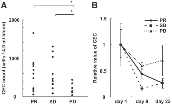
CEC Amounts and Objective Tumor Response to Chemotherapy
Thirteen (41.9%) of the 31 patients who received carbo- platin and paclitaxel therapy showed a partial response (PR) and 12 (38.7%) showed stable disease (SD). The other 6 patients (19.4%) showed PD. The amounts of CEC at baseline in the patients who showed PR and SD were 516 ⫾ 458/4 ml and 871 ⫾ 1215/4 ml, respectively, and these values were signifi- cantly higher than in PD patients (211 ⫾ 150/4 ml, p ⫽ 0.023 and p ⫽ 0.044, respectively) (Figure 3A). Although CEC dec- rements during chemotherapy were observed in all three sub- groups, the extent of the decrements tended to be greater in
0 500 1000 1500
0 50 100 150 200 250
Estimated tumor volume (cm3)
CEC (baseline) / 4 ml
FIGURE 1. Scatter plot analysis to determine the correla- tion between the number of circulating endothelial cell (CEC) and estimated tumor volume (ETV). ETV is calculated with computed tomography (CT) examination. Case 21 is not included.
0 1000 2000
baseline day 8 day 22
CEC count (cells / 4.0 ml blood)
173r189 * 176r141 *
595r832 meanrSD
day 22 day 8
baseline
FIGURE 2. Circulating endothelial cell (CEC) levels during the first course of CDDP plus paclitaxel chemotherapy. *p
⬍
0.05 versus values at baseline.0 1000 2000
PR SD PD
*
*
A B
0 0.5 1 1.5
day 1 day 8 day 22 PR SD PD
CEC count (cells / 4.0 ml blood) Relative value of CEC
FIGURE 3. A, Comparison of circulating endothelial cell (CEC) amount at baseline in non-small cell lung cancer (NSCLC) patients with different clinical responses to CBDCA plus paclitaxel chemotherapy. *p
⬍
0.05 versus values of patients with progressive disease (PD). Case 21 is not in- cluded.B, Relative change in CEC amount in patients with partial response (PR), stable disease (SD), and PD.patients with PR and SD than in those with PD (Figure 3B). In the subgroup analysis, a significant decrease in CECs was observed on day 22 only in PR patients (p ⫽ 0.018).
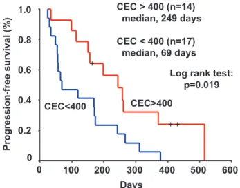
CEC Amounts and PFS
For all 31 patients, the median PFS was 154 days (range, 81–361 days). Univariate analysis indicated that pa- tients who had a CEC count of more than 400/4 ml at baseline showed a significantly improved PFS (n ⫽ 14, median; 244 days) (Log-rank test, p ⫽ 0.019, Figure 4). A CEC count below 400 at baseline was associated with a poorer PFS (n ⫽ 17, median; 69 days). The CEC count did not exceed the value of 400/4 ml in any of the healthy volunteers. When we compared the patients whose CEC counts exceeded 200 with those whose counts were less than 200, a consistent differ- ence in PFS was observed between the two groups ( ⬎ 200;
n ⫽ 22, median 227, ⬍ 200; n ⫽ 9, median 116, p ⬍ 0.039).
DISCUSSION
In the present study, we investigated the number of CEC during the first course of CBDCA plus paclitaxel chemotherapy.
To our knowledge, this is the first report of CEC in NSCLC patients before treatment. Our findings demonstrated CEC counts in advanced NSCLC at baseline level to be much higher than those in healthy subjects (595 ⫾ 832/4.0 ml versus 32.6 ⫾ 29.5/4.0 ml). Because the NSCLC patients had not yet received anticancer therapy, these increased CECs are likely to be mostly derived from the tumor site. In a previous study, it was found that the amounts of CECs correlate strongly with tumor volume in vivo in an animal model
34. Nevertheless, we did not find a significant correlation between CECs and ETV. Because the number of CECs could be influenced by many factors related to tumor vasculature, neovascularization, and localization of the tumor, our failure to identify a strong correlation in this study is not surprising. We were also unable to detect a significant direct
correlation between CEC amounts and various blood examina- tion data including tumor markers such as CEA and CYFRA. It is unclear at present what biologic characteristics of the tumor or clinical features the CEC number most closely reflects as a biomarker. Mancuso et al. reported that CECs are strongly associated with plasma levels of VCAM-1 and VEGF in breast cancer and lymphoma patients.
15,34Because VCAM-1 and VEGF are crucial factors for tumor angiogenesis, the variability in CEC values among NSCLC patients might indicate a differ- ence in the neovascularization of each tumor.
We were further able to demonstrate that elevated CECs decreased dramatically after CBDCA plus paclitaxel treatment, but did not reach the level of healthy subjects.
Decreased CEC values did not rise again during the first cycle of chemotherapy. Although myelosupression was observed on day 8 and recovered on day 22 in many patients (data not shown), CEC kinetics do not parallel those of WBC, indicat- ing that CEC kinetics might not be influenced by myelopoi- esis. Several clinical studies in the field measuring CEC found chemotherapy to be associated with either an increase or a decrease in CECs.
35–39The different tumor types, stages, prior therapy or not, the anticancer drugs used, measuring points and quantification methods of CEC might have influ- enced the CEC results after treatment. In the present study, the pretreatment CEC value was much higher than that in lung cancer with metastasis (mean ⫾ SD ⫽ 146 ⫾ 270/4 ml), as reported elsewhere.
33Although the details of the prior therapy in patients with metastatic carcinoma were not provided,
33che- motherapy can eventually decrease the CEC count.
Schiller et al. compared four standard chemotherapy regimens, cisplatin plus paclitaxel, cisplatin plus gemcitab- ine, cisplatin plus docetaxel, and carboplatin plus paclitaxel and found no significant difference in survival.
25Despite the different modes of action of each nonplatinum agent against tumors and different biologic characteristics of each tumor, we could not select the regimen based on these characteris- tics. In our small study, the patients with PR/SD and longer PFS had higher baseline CEC values. Therefore, it seems that the baseline CEC count is a promising predictor of clinical response to the CBDCA plus paclitaxel regimen and survival in advanced NSCLC. If CEC is a marker for angiogenesis and reflects tumor neovascularization, it is likely that a high CEC is associated with a poor prognosis and lower effectiveness of antiangiogenic therapy. Paclitaxel and docetaxel are catego- rized as mitotic spindle agents with potent antiangiogenic properties.
27–30This is why a paclitaxel based regimen might be more effective against tumors with high CEC values.
Nevertheless, CEC counts have also been reported to be increased in several clinical syndromes, such as cardiovascu- lar diseases, infectious diseases, and vasculitides.
11–13The CEC counts in patients with vasculitides have been reported to be dozens of fold higher than those in healthy subjects,
12therefore, we have to consider the patient condition carefully while interpreting the CEC counts in individual patients, although there were no patients with vasculitis in the present study. Further clinical investigation, with a similar approach, including other nonplatinum anticancer agents, such as
CEC>400CEC<400
Log rank test:
p=0.019 CEC > 400 (n=14)
median, 249 days CEC < 400 (n=17)
median, 69 days
Progression-free survival(%)
Days 1.0
0.8
0.6
0.4
0.2 0
0 100 200 300 400 500 600
FIGURE 4. Progression-free-survival according to circulating endothelial cell (CEC) count at baseline. The median dura- tion of progression-free survival was greater in patients whose CEC count exceeded 400 (median, 244 days) than in patients whose CEC count was less than 400 (69 days).
CDDP plus gemcitabine, is essential for the clinical applica- tion of CEC for made-to-order chemotherapy in NSCLC.
Antiangiogenic therapy targeting the VEGF pathway such as bevacizumab and VEGFR inhibitors have shown promise in the treatment of solid tumors.
8,39These agents inhibit endothelial cells through inhibition of the VEGF pathway. It was recently demonstrated that the addition of bevacizumab to CBDCA plus paclitaxel in advanced NSCLC patients produces a significant survival benefit as compared with chemotherapy alone.
40Considering the outstanding clin- ical trial and our present study, it would be of great interest to investigate the role of CEC in this regimen.
In conclusion, CECs were measured in NSCLC patients before treatment. Our small clinical study indicates that the CEC count at baseline is a potential biomarker for predicting the response to chemotherapy and PFS, but further clinical evaluation is needed. In the near future, we will start a clinical investigation, using a similar approach, to examine other chemotherapeutic regimens.
ACKNOWLEDGEMENTS
This study was supported in part by a Grant-in-Aid for the 3rd Term Comprehensive 10-year Strategy for Cancer Control from the Ministry of Health, Welfare and Labour, Japan.
REFERENCES
1. Folkman J. Anti-angiogenesis: new concept for therapy of solid tumors.
Ann Surg1972;175:409 – 416.
2. Gasparini G, Harris AL. Clinical importance of the determination of tumor angiogenesis in breast carcinoma: much more than a new prog- nostic tool.J Clin Oncol1995;13:765–782.
3. Dickinson AJ, Fox SB, Persad RA, Hollyer J, Sibley GN, Harris AL.
Quantification of angiogenesis as an independent predictor of prognosis in invasive bladder carcinomas.Br J Urol1994;74:762–766.
4. Takahashi Y, Kitadai Y, Bucana CD, Cleary KR, Ellis LM. Expression of vascular endothelial growth factor and its receptor, KDR, correlates with vascularity, metastasis, and proliferation of human colon cancer.
Cancer Res1995;55:3964 –3968.
5. Williams JK, Carlson GW, Cohen C, Derose PB, Hunter S, Jurkiewicz MJ. Tumor angiogenesis as a prognostic factor in oral cavity tumors.
Am J Surg1994;168:373–380.
6. Koukourakis MI, Giatromanolaki A, Thorpe PE, et al. Vascular endo- thelial growth factor/KDR activated microvessel density versus CD31 standard microvessel density in non-small cell lung cancer.Cancer Res 2000;60:3088 –3095.
7. Natsume T, Watanabe J, Koh Y, et al. Antitumor activity of TZT-1027 (Soblidotin) against vascular endothelial growth factor-secreting human lung cancer in vivo.Cancer Sci2003;94:826 – 833.
8. Hurwitz H, Fehrenbacher L, Novotny W, et al. Bevacizumab plus irinotecan, fluorouracil, and leucovorin for metastatic colorectal cancer.
N Engl J Med2004;350:2335–2342.
9. Yang JC, Haworth L, Sherry RM, et al. A randomized trial of bevaci- zumab, an anti-vascular endothelial growth factor antibody, for meta- static renal cancer.N Engl J Med2003;349:427– 434.
10. Johnson DH, Fehrenbacher L, Novotny WF, et al. Randomized phase II trial comparing bevacizumab plus carboplatin and paclitaxel with car- boplatin and paclitaxel alone in previously untreated locally advanced or metastatic non-small-cell lung cancer.J Clin Oncol2004;22:2184 –191.
11. Mutin M, Canavy I, Blann A, Bory M, Sampol J, Dignat-George F.
Direct evidence of endothelial injury in acute myocardial infarction and unstable angina by demonstration of circulating endothelial cells.Blood 1999;93:2951–2958.
12. Woywodt A, Streiber F, De Groot K, Regelsberger H, Haller H, Haubitz M. Circulating endothelial cells as markers for ANCA associated small- vessel vasculitis.Lancet2003;361:206 –210.
13. Mutunga M, Fulton B, Bullock R, et al. Circulating endothelial cells in patients with septic shock.Am J Respir Crit Care Med2001;163:195–200.
14. Beerepoot LV, Mehra N, Vermaat JS, Zonnenberg BA, Gebbink MF, Voest EE. Increased levels of viable circulating endothelial cells are an indicator of progressive disease in cancer patients.Ann Oncol2004;15:
139 –145.
15. Mancuso P, Burlini A, Pruneri G, Goldhirsch A, Martinelli G, Bertolini F. Resting and activated endothelial cells are increased in the peripheral blood of cancer patients.Blood2001;97:3658 –3661.
16. Mancuso P, Colleoni M, Calleri A, et al. Circulating endothelial-cell kinetics and viability predict survival in breast cancer patients receiving metronomic chemotherapy.Blood2006;108:452– 459.
17. Bu¨lzebruck H, Bopp R, Drings P, et al. New aspects in the staging of lung cancer. Prospective validation of the International Union Against Cancer TNM classification.Cancer1992;70:1102–1110.
18. Grilli R, Oxman AD, Julian JA. Chemotherapy for advanced non-small- cell lung cancer: how much benefit is enough?J Clin Oncol1993;11:
1866 –1872.
19. Non-small Cell Lung Cancer Collaborative Group. Chemotherapy in non-small cell lung cancer: a meta-analysis using updated data on individual patients from 52 randomised clinical trials.BMJ1995;311:
899 –909.
20. Kubota K, Watanabe K, Kunitoh H, et al. Phase III randomized trial of docetaxel plus cisplatin versus vindesine plus cisplatin in patients with stage IV non-small cell lung cancer: the Japanese Taxotere Lung Cancer Study Group.J Clin Oncol2004;22:254 –261.
21. Le Chevalier T, Brisgand D, Douillard JY, et al. Randomized study of vinorelbine and cisplatin versus vindesine and cisplatin versus vinorel- bine alone in advanced non-small cell lung cancer: results of a European multicenter trial including 612 patients.J Clin Oncol1994;12:360 –367.
22. Belani CP, Lee JS, Socinski MA, et al. Randomized phase III trial comparing cisplatin-etoposide to carboplatin-paclitaxel in advanced or metastatic non-small cell lung cancer.Ann Oncol2005;16:1069 –1075.
23. Yana T, Takada M, Origasa H, et al. New chemotherapy agent plus platinum for advanced non-small cell lung cancer: a meta-analysis.Proc Am Soc Clin Oncol2002;21:328a.
24. Baggstrom MQ, Stinchcombe TE, Fried DB, Poole C, Hensing TA, Socinski MA. Third-generation chemotherapy agents in the treatment of advanced non-small cell lung cancer: a meta-analysis.J Thorac Oncol 2007;2:845– 853.
25. Schiller JH, Harrington D, Belani CP, et al; Eastern Cooperative On- cology Group. Comparison of four chemotherapy regimens for advanced non-small-cell lung cancer.N Engl J Med2002;346:92–98.
26. Ohe Y, Ohashi Y, Kubota K, et al. Randomized phase III study of cisplatin plus irinotecan versus carboplatin plus paclitaxel, cisplatin plus gemcitabine, and cisplatin plus vinorelbine for advanced non-small-cell lung cancer: Four-Arm Cooperative Study in Japan.Ann Oncol2007;
18:317–323.
27. Belotti D, Vergani V, Drudis T, et al. The microtubule-affecting drug paclitaxel has antiangiogenic activity.Clin Cancer Res1996;2:1843–1849.
28. Hayot C, Farinelle S, De Decker R, et al. In vitro pharmacological charac- terizations of the anti-angiogenic and anti-tumor cell migration properties mediated by microtubule-affecting drugs, with special emphasis on the organization of the actin cytoskeleton.Int J Oncol2002;21:417– 425.
29. Wang J, Lou P, Lesniewski R, Henkin J. Paclitaxel at ultra low concentrations inhibits angiogenesis without affecting cellular microtu- bule assembly.Anticancer Drugs2003;14:13–19.
30. Vacca A, Ribatti D, Iurlaro M, et al. Docetaxel versus paclitaxel for antiangiogenesis.J Hematother Stem Cell Res2002;11:103–118.
31. Therasse P, Arbuck SG, Eisenhauer EA, et al. New guidelines to evaluate the response to treatment in solid tumors. European Organiza- tion for Research and Treatment of Cancer, National Cancer Institute of the United States, National Cancer Institute of Canada.J Natl Cancer Inst2000;92:205–216.
32. Smirnov DA, Foulk BW, Doyle GV, Connelly MC, Terstappen LW, O’Hara SM. Global gene expression profiling of circulating endothelial cells in patients with metastatic carcinomas.Cancer Res2006;66:2918 –2922.
33. Rowand JL, Martin G, Doyle GV, et al. Endothelial cells in peripheral blood of healthy subjects and patients with metastatic carcinomas.
Cytometry A2007;71A:105–114.
34. Mancuso P, Calleri A, Cassi C, et al. Circulating endothelial cells as a novel marker of angiogenesis.Adv Exp Med Biol2003;522:83–97.
35. Beaudry P, Force J, Naumov GN, et al. Differential effects of vascular endothelial growth factor receptor-2 inhibitor ZD6474 on circulating endothelial progenitors and mature circulating endothelial cells: impli- cations for use as a surrogate marker of antiangiogenic activity. Clin Cancer Res2005;11:3514 –3522.
36. Fu¨rstenberger G, von Moos R, Lucas R, et al. Circulating endothelial cells and angiogenic serum factors during neoadjuvant chemotherapy of primary breast cancer.Br J Cancer2006;94:524 –531.
37. Rademaker-Lakhai JM, Beerepoot LV, Mehra N, et al. Phase I pharmacokinetic and pharmacodynamic study of the oral protein kinase C beta-inhibitor enzastaurin in combination with gemcitabine
and cisplatin in patients with advanced cancer. Clin Cancer Res 2007;13:4474 – 4481.
38. McAuliffe JC, Trent JC. Biomarkers in gastrointestinal stromal tumor:
should we equate blood-based pharmacodynamics with tumor biology and clinical outcomes?Clin Cancer Res2007;13:2643–2650.
39. Hanrahan EO, Heymach JV. Vascular endothelial growth factor receptor tyrosine kinase inhibitors vandetanib (ZD6474) and AZD2171 in lung cancer.Clin Cancer Res2007;13:S4617–S4622.
40. Sandler A, Gray R, Perry MC, et al. Paclitaxel-carboplatin alone or with bevacizumab for non-small-cell lung cancer.N Engl J Med2006;355:
2542–2550; 2007;356:318.