*岩手医科大学第二内科/循環器医療センター 受付:15 年 10 月 3 日
最終稿受付:16 年 1 月 8 日
別刷請求先:岩手県盛岡市内丸 19–1 (0 020–8505) 岩手医科大学第二内科
西 山 理 I. 緒 言
不安定狭心症 (Unstable Angina Pectoris; UAP) は 急性心筋梗塞症に移行しやすく,来院時診断の確 かさが予後に関わる.しかし,UAP は重篤な病 態ながら,非虚血性胸痛症例との鑑別が困難であ り,心電図や心エコー図,および心筋逸脱酵素測 定などの既存検査でも確診が得られないことがあ る.
心筋の血流イメージに 99mTc-標識製剤が用いら れるようになり,救急医療現場での検査が可能に なって以来,同製剤が救急外来での胸痛患者の鑑
別診断に有用とした報告が多い1〜5).さらに,心 電図同期心筋 Single Photon Emission Computed Tomography (SPECT) 法が急性心筋梗塞症の診断 に有用であるとした報告もなされている6〜8).し かし UAP 診断に有用であるか否かは未知であり,
心電図同期心筋 SPECT 法で心筋虚血を検出する 試みは,新たな心筋虚血評価法として期待され る.
そこで本研究では,UAP の疑診症例に心電図 同期心筋 SPECT 法のひとつである Quantitative Gated SPECT (QGS) を行い,従来の血流イメージ の評価に機能イメージ評価を加味して心筋虚血の 有無の診断を試みた.
II. 対象と方法
1. 対 象
1997 年 6 月から 2000 年 5 月までに, UAP の 疑いで岩手医科大学附属循環器医療センター
《原 著》
99m
Tc-標識製剤を用いた安静時心電図同期シンチグラムによる
不安定狭心症の診断精度と予後の推測に関する研究
西山 理* 上嶋 健治*
要旨 〔目的〕 不安定狭心症 (UAP) の疑診例に心電図同期心筋 SPECT 検査 (QGS) を行い,診断精度 と入院期間中の予後を評価した.〔方法〕 対象は UAP の疑診例で,CCU 入室直後に 99mTc-tetrofosmin で QGS を行い,かつ入院中に冠動脈造影 (CAG) を施行した連続 57 症例で,SPECT 血流イメージか ら Defect score (DS) を用いて虚血の程度を 4 段階に,また機能イメージから局所壁運動異常の有無を 視覚的に評価した.CAG の結果をもとに,1) 血流イメージ解析 (P-解析), 2) 機能イメージ解析 (F-解 析),3) P-解析と F-解析を加味する方法 (P+F 正常;両解析がともに正常のみを虚血陰性,P+F 異常;
両解析がともに異常のみを虚血陽性,P+F 複合;P-解析で DS が 2 未満でかつ F-解析で正常のみを虚 血陰性) の,それぞれの精度を評価した.また入院中の転帰について検討した.〔結果〕 35 例に有意冠 動脈病変を認めた.P+F 複合が感度 83%, 特異度 82%, と高精度であった.また,本法での陽性例 は陰性例に比し早期に血行再建術を要した (45% vs. 8%).〔結論〕 QGS は UAP の診断と入院期間中の 心事故の発生予測に有用である.
(核医学 41: 101–107, 2004)
102 核 医 学 41巻2号(2004)
CCU に収容された連続 57 症例 (男性 37 例,女 性 20 例,平均年齢 62±7 歳) を対象とした.心 筋梗塞症と狭心症の既往症例および冠攣縮性狭心 症と確診した症例は除外した.なお,UAP の診 断基準には Branuwald の分類9) を用いたところ,
クラス IA 群 26 例,クラス IIA 群 1 例,クラス IIIA 群 30 例であった.
2. 方 法
CCU 入室直後に平均 590 MBq の 99mTc-標識製 剤 (99mTc-tetrofosmin) を静脈注射し,30 分後に GE 横河社製 Optima NX-2 検出器に低エネルギー 汎用コリメータを用いて QGS の撮像を開始した.
撮像条件は 16×2 step, 180 度収集,30 秒/step,
64×64 matrix とし,データ前処理には Butterworth filter, 再構成には Ramp filter を用いた.心電図 同期は,R-R 間隔を 8 分割し,QGS 法により心 筋血流イメージと機能イメージを再構成した.
血流イメージには,SPECT 断層像のグレース ケールの短軸と水平および垂直長軸像を用いて,
心筋灌流低下部位を左前下行枝 (LAD), 左回旋枝
(LCX), 右冠動脈 (RCA) の各領域に分類した.
さらに color polar map を用いて灌流低下の程度を 視覚的に Defect score として,4 段階 (0. 正常,1.
軽度から中等度灌流低下,2. 高度灌流低下,3. 完 全欠損) にスコア化した.機能イメージには,3D シネモード表示から,視覚的に局所壁運動の低下 の有無と部位 (LAD 領域,LCX 領域,RCA 領域) を評価した.いずれの解析も,読影経験を積んだ 3 名の医師のうち 2 名以上の合意で評価を定めた.
なお,入院中に冠動脈造影検査 (CAG) を行 い,心筋虚血の責任病変部位を L A D, L C X,
RCA に分類し,75% 以上の狭窄を有意冠動脈病 変とした.多枝病変例で血流イメージと機能イ メージの異常部位が複数部位一致した場合は,
CAG で最も高度な狭窄病変部位を責任病変部位 とし,血流イメージと機能イメージの異常部位が 不一致の場合は虚血陰性とした.
CAG の結果をもとに,SPECT イメージを以下 の 5 通りの方法で解析した.すなわち,1) 従来通 り血流イメージのみの解析 (P-解析), 2) 機能イ Table 1 Evaluation of P+F (comb) analysis
Defect score F-analysis stenosis (+) stenosis (−) total
≧2 asynergy 11 1 12
no asynergy 6 1 7
→
Ischemia (+)asynergy 12 2 14
<2 no asynergy 6 18 24
→
Ischemia (−)total 35 22 57
The negative case assumed to the inside of a thick line
→
→
Table 2 Accuracy of P-analysis and F-analysis
P-analysis F-analysis
CAG CAG
QGS stenosis stenosis
total QGS stenosis stenosis
total
(+) (−) (+) (−)
positive 29 12 41 positive 21 3 24
negative 6 10 16 negative 14 19 33
total 35 22 57 total 35 22 57
P-analysis: Perfusion analysis only; sensitivity 83%, specificity 45%, accuracy 69%
F-analysis: Functional analysis only; sensitivity 60%, specificity 86%, accuracy 70%
103
メージのみの解析 (F-解析), 3) 両解析ともに正 常例のみを虚血陰性例とする解析 (P+F 正常―解
析), 4) 両解析ともに異常例のみを虚血陽性例と
する解析 (P+F 異常―解析), 5) P-解析で Defect
score が 2 未満かつ F-解析で正常例のみを虚血陰 性例とする解析 (P+F 複合―解析; Table 1) の 5 方法とし,それらの精度を比較検討した.次に,
最も高精度であった評価法を用いて,入院中の心 事故発生を予測できるか否かを評価した.なお冠 動脈血行再建術 (coronary artery bypass grafting;
CABG と percutaneous coronary intervention; PCI) に至る病態を心事故と定義した.
また,重症冠動脈病変や,薬物治療に抵抗した ため冠動脈血行再建術を施行した症例のうち,
CCU 入室中に施行した例を緊急例とし,一般病 棟へ転出後の諸検査の結果をみて CABG または PCI を施行した症例を待機例とした.さらに最終 的に薬物治療とした症例を加えて転帰を評価し た.
III. 結 果
57 例中 35 例の 50 枝に有意冠動脈病変 (LDA 23 例,LCX 4 例,RCA 8 例,一枝病変 24 例,二 枝病変 7 例,三枝病変 4 例) を認めた.責任血管 の狭窄度は 100% 5 例,99% 8 例,90% 18 例,
75% 4 例であった.また,最終発作から検査まで の平均時間は有意冠動脈病変のある例で 8.5±7.1 時間,有意冠動脈病変のない例で 8.3±6.7 時間で あった.
Table 3 Accuracy of P+F analysis P+F analysis is classified the following 3 analyses.
1) In P+F (nl-nl) analysis: negative findings of both P- and F-analysis were considered as the negative cases, and the others were positive cases.
sensitivity 89%, specificity 45%, accuracy 72%
CAG
QGS stenosis stenosis
total
(+) (−)
positive 31 12 43
negative 4 10 14
total 35 22 57
2) In P+F (ab-ab) analysis: positive findings of both P- and F-analysis were considered as the positive cases, and the others were negative cases.
sensitivity 54%, specificity 86%, accuracy 67%
CAG
QGS stenosis stenosis
total
(+) (−)
positive 19 3 22
negative 16 19 35
total 35 22 57
3) In P+F (comb) analysis: less than 2 of defect score in P-analysis with negative F-analysis (no asynergy) were considered as the negative cases, and the others were positive cases.
sensitivity 83%, specificity 83%, accuracy 82%
CAG
QGS stenosis stenosis
total
(+) (−)
positive 29 4 33
negative 6 18 24
total 35 22 57
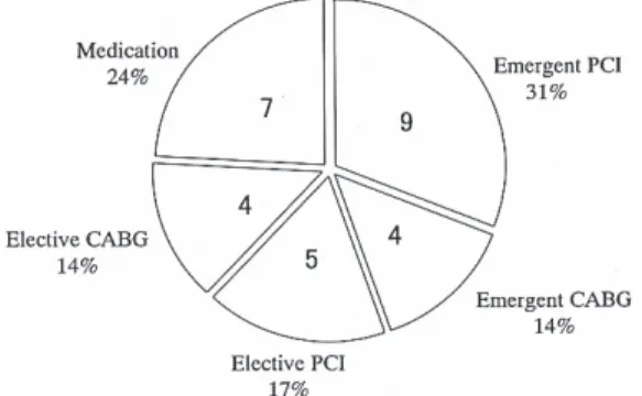
Fig. 1 Prognosis of 29 cases with positive P+F (comb) analysis and significant coronary stenosis (true positive). Almost half of the patients with positive P+F (comb) analysis needed emergent coronary revascularization.
104 核 医 学 41巻2号(2004)
P-解析では,感度 83%,特異度 45%,正確度
69% と特異度が低く,F-解析では,感度 60%,
特異度 8 6 %,正確度 7 0 % と感度が低かった (Table 2).P+F 正常―解析は P-解析と比較して 感度は 89% と向上したが,特異度は変わらず,
正確度は 72% であった.P+F 異常―解析は F-解 析と比較して特異度は 86% と不変であったが,
感度が 45% と低く,正確度は最も低く 67% で あった.いずれの解析方法も感度と特異度に隔た りがみられた.P+F 複合―解析は,感度 83%,
特異度 82%, 正確度 82% であり,他の解析に比 べて最も高い精度であった (Table 3).
そこで,最も精度の高かった P+F 複合―解析 で,虚血が陽性または陰性と判断された症例の入
院中の転帰を検討した. P+F 複合―解析で異常 を認めた 29 症例のうち13 例 (45%) が緊急の冠動 脈血行再建を必要とし,薬物療法のみで治療可能 であった症例は 7 例 (24%) のみであった (Fig. 1).
また 4 例の心筋虚血偽陽性例のうちの 2 例は,後 に問診から冠攣縮性狭心症疑いと診断された (Table 4).心筋虚血偽陰性 6 例のうち 5 例は LCX 領域に有意狭窄がある症例で,2 例に緊急血行再 建術を必要とした (Table 5).なお有意冠動脈病変 がなかった 22 症例の最終診断は,問診やアセチ ルコリン負荷検査の結果から冠攣縮性狭心症疑い 16 例,胸痛症候群 3 例,一過性心房細動 1 例,
気管支喘息 1 例,急性胃炎 1 例であり,いずれも 内科的治療により症状の軽快をみた.
IV. 考 察
従来の 201Tl-標識製剤による心臓核医学検査で
は,緊急への対応が困難とされていた.しかし,
標識が簡便で各施設での調製が可能である99mTc- 標識製剤が登場し,心臓核医学検査の緊急時対応 を可能にした.しかも,同製剤は短半減期 6 時間 で比較的大量投与が可能で,かつ 140 keV という 高いエネルギーピークを有しているため,鮮明な 血流画像を描出可能である.また心筋集積後には 臨床的に有意な再分布がなく,投与時の心筋血流 の状態を保持 (freezing image) するとされている.
さらに,従来の心筋 SPECT から得られる情報は 血流イメージのみであったが,心電図同期心筋 SPECT 法は,壁運動評価や左室駆出率などの心 筋機能イメージが動画像として得られることか Table 4 Four cases with positive P+F (comb) analysis and no significant stenosis (false positive)
cases P-analysis F-analysis
final diagnosis
(Defect score) (Asynergy)
65 y.o. male Inf, 2 None hypertensive heart
52 y.o. female Ant, 2 Ant-sep s/o vasospastic angina
65 y.o. male Inf, 1 Inf s/o vasospastic angina
57 y.o. female Inf, 1 Inf chest pain syndrome
Three of 4 cases with positive P+F (comb) analysis had the abnormalities in F-analysis accordance with P-analysis.
Table 5 Six cases with negative P+F (comb) analysis and significant stenosis (false negative)
cases CAG prognosis
RCA 99%
50 y.o. male LCX (#11) 99%* emergent PCI LCX (#14) 90%*
76 y.o. female LCX 90% emergent PCI 75 y.o. male LAD 90%
elective PCI LCX 90%*
63 y.o. female RCA 90% elective PCI
56 y.o. male LCX 99% medication
74 y.o. female LCX 90% medication
*ischemia related artery
Five of 6 patients with false negative P+F (comb) analysis were LCX lesion, and two of these patients needed emergent coronary revascularization.
105 ら,心筋血流と機能イメージを同時に非侵襲的に
評価することが可能となった.しかし,心電図同 期心筋 SPECT 法はデータ収集や処理に時間がか かり,かつてはルーチンでの施行が困難であっ た.1995 年になって,Germano らによって開発 された心電図同期心筋 SPECT 解析ソフトプログ ラムである QGS が,簡便かつ短時間のデータ処 理による心機能解析を可能にした10).
従来より,心臓核医学検査は冠動脈疾患の診断 や治療に際して過剰な冠動脈造影や PCI を防ぐ ゲートキーパとして期待されている11〜13).中で も急性冠症候群 (acute coronary syndrome; ACS) に
99mTc-標識製剤を用いて心筋 SPECT 法で心筋虚血
を解析する試みは,迅速かつ簡便な心筋虚血評価 法であると評価されている1〜4).とくに UAP で は,運動をはじめ種々の負荷検査が禁忌であり,
安静時のみのイメージから診断を下す必要があ る.Parodi らは14),安静時狭心症などの重篤な心 筋虚血を有する症例では 90% の例に血流イメー ジでの灌流低下を認め,症状出現時の陽性率はさ らに高いと報告している.本研究でも P-解析の感 度は 83% と良好であったが,特異度が 45% と低 く満足な精度は得られなかった.
血流イメージの読影で最も注意すべき点は虚血 性変化とアーチファクトとの鑑別であり,この アーチファクトは冠動脈疾患診断の偽陽性の原因 となる.アーチファクトによる固定性欠損の原因 は,1995 年に DePuey らが15),前壁は女性の乳 房,下壁は主に男性の横隔膜による γ 線の減弱に よるものが多いと報告している.とくに,横隔膜
による γ 線の減弱や肝臓左葉および胆囊などの心
臓隣接臓器へのトレーサの高集積によるアーチ ファクトは,右冠動脈支配領域と一致することが あり,判定に窮する症例が少なくない.
Taillefer ら16) や Smanio らは17), QGS による 冠動脈疾患の診断能は機能イメージの追加で向上 すると報告している.また Choi らは18)機能イ メージの追加で読影者間の虚血判定の相違は少な くなると報告している.これらの QGS の代表的 報告では視覚的半定量法を用いている.その精度
は既知と考え,本研究も読影経験を積んだ複数の 検者による視覚的半定量的評価とコンセンサス法 を用いて心筋虚血の評価とした.しかし今回の検 討では,従来の血流イメージに機能イメージを単 純に加味するだけの方法では,診断精度の向上は 得られなかった.とくに,下壁で血流イメージ読 影の際の Defect score が 2 未満の症例に偽陽性例 が多かった.したがって Defect score が 2 未満を 血流イメージでは正常とし,それに左室収縮能を 評価した機能イメージを加味した P+F 複合―解 析が診断精度を高めるのではないかと考えた.そ の P+F 複合―解析を行った結果,P-解析と比較 して感度は不変で,偽陽性例を 12 例から 4 例に まで減少させえた.とくに P-解析のみでは偽陽性 と判定され,P+F 複合―解析では正常と判定さ れた 8 例中の 6 例 (男/女=5/1) が,最もアーチ ファクトとの鑑別が困難とされる下壁領域に血流 低下を認める症例であった.その結果,特異度を 45% から 82% に,正確度を 62% から 82% にま で向上させえた.なお,偽陰性例は LCX に有意 狭窄のある症例に多かった.これは L 型検出器に よる 180 度収集法では側壁が検出器に最も接近す るために同部へのトレーサの集積を過大評価する こと,また機能イメージ上も,心室の中隔壁より も自由壁の壁運動を過大評価することが原因と考
えられた19〜21).また,偽陰性例で緊急血行再建
術が必要であった症例は LCX 領域の 2 例のみ で,偽陰性例の予後はおおむね良好であった.以 上より P+F 複合―解析は,血流イメージ評価法 を工夫し,さらに機能イメージを追加すること で,虚血性変化とアーチファクトとの鑑別による 偽陽性例を低下させ,特異度を向上させることで 診断精度を改善させたと考えられる.
さらに P+F 複合―解析で異常と判定した例の 約半数が緊急の冠動脈血行再建を必要とし,薬物 療法のみで対応できた症例は 4 分の 1 にすぎな かった.したがって,本法で異常と判定された症 例は早期に血行再建術を要する可能性が高く,入 院期間中の転帰の推測にも有用と考えられた.一 方偽陽性 4 例のうち 3 例には P-解析での異常部
106 核 医 学 41巻2号(2004) 位に一致して F-解析による壁運動異常があり,冠
攣縮性狭心症の関与を完全には否定できなかっ た.以上のことから心電図同期心筋 SPECT 法 は,UAP 例を診断する上で信頼性の高い検査法 と考えた.
V. 総 括
99mTc-標識製剤を用いた心電図同期心筋 SPECT
法は UAP の診断と入院期間中の心事故の発生予 測に有用であった.
稿を終えるに当たり,終始ご指導とご校閲を賜り ました岩手医科大学内科学第二講座平盛勝彦教授に 深く感謝いたします.さらに本研究にご協力いただ きました現西神戸医療センター循環器内科の谷口泰 代先生に厚く御礼申し上げます.
文 献
1) Itchhaporia D: Using radionuclide techniques for evaluating patients with chest pain or unstable angina.
Eur J Nucl Med 1995; 22: 185–188.
2) Varetto T, Canadialupi D, Altieri A, Orrlandi C:
Emergency room technetium-99m Sestamibi imaging to rule out acute myocardial ischemic events in patients with non diagnostic electrocardiograms. J Am Coll Cardiol 1993; 22: 1804–1808.
3) Bilodeau L, Theroux P, Gagnon D, Arsenault A:
Technetium-99m sestamibi tomography in patients with spontaneous chest pain: Correlations with clinical, electrocardiographic and angiographic findings. J Am Coll Cardiol 1991; 18: 1684–1691.
4) Kotos MC, Josse RL, Schmidt KL, Ornato JP, Tatum JL: Value of Acute Rest Sestamibi Perfusion Imaging for Evaluation of Patients Admitted to the Emergency Department with Chest Pain. J Am Coll Cardiol 1997;
30: 976–982.
5) Ritchie J, Bateman TM, Bonow RO, Crawford MH, Gibbons RJ, Hall RJ: AHA/ACC task force report:
Guidelines for clinical use of cardiac radionuclide imaging. Circulation 1995; 91: 1278–1303.
6) Toba M, Ishida Y, Jukuchi K, Noguchi T, Itoh A, Nonogi H: Use of ECG-gated SPECT to assess the evolution of perfusion after acute myocardial infarc- tion. Eur J Nucl Med 2000; 27: 517–523.
7) Tnaka R, Nakamura T: Times Course Evaluation of Myocardial Perfusion After Reperfusion Therapy by 99m-Tetrofosmin SPECT in Patients with Acute Myocardial Infarction. J Nucl Med 2001; 42: 1351–
1358.
8) Neumann FJ, Kosa I, Dickfeld T, Blasini R, Gawaz M, Hausleiter J: Recovery of myocardial perfusion in acute myocardial infarction after successful balloon angioplasty and stent placement in the infarct-related coronary artery. J Am Coll Cardiol 1997; 30: 1270–
1276.
9) Braunwald E: Unstable angina—A classification.
Circulation 1989; 80: 410–414.
10) Germano G, Kiat H, Kavanagh PB, Moriel M, Mazzanti M, Su HT: Automatic quantification of ejection fraction from gated myocardial perfusion SPECT. J Nucl Med 1995; 36: 2138–2147.
11) Brown KA: Prognostic value of thallium-201 myocar- dial perfusion imaging: A diagnostic tool comes of age. Circulation 1991; 83: 363–377.
12) Iskander S, Iskandrian AE: Risk assessment using single photon emission computed tomographic technetium-99m sestamibi imaging. J Am Coll Cardiol 1998; 32: 57–62.
13) Fletschmann KE, Hunink MG, Kuntz KM, Douglas PS: Exercise echocardiography or exercise SPECT imaging: A meta-analysis of diagnostic test perform- ance. JAMA 1998; 280: 913.
14) Parodi O, Uthurralt N, Severi S, Bencivelli W, Michelassi C, Labbate A: Transient reduction of regional myocardial perfusion during angina at rest with ST-segment depression and normalization- waves. Circulation 1981; 63: 1238–1247.
15) DePuey EG, Rozanski A: Using gated technetium- 99m-sestamibi SPECT to characterize fixed myo- cardial defect as infarct or artifact. J Nucl Med 1995;
36: 952–955.
16) Taillefer R, DePuey EG, Udelson JE, Beller GA, Latour Y, Reeves F: Comparative diagnostic accuracy of Tl-201 and Tc-99m sestamibi SPECT (perfusion and ECG gated SPECT) in detecting coronary artery disease in woman. J Am Coll Cardiol 1997; 29: 69–
77.
17) Smanio PE, Watson DD, Segalla DL, Vinson EL, Smith WH, Beller GA: Value of gating of technetium- 99m-sestamibi single-photon-emission computed tomographic imaging. J Am Coll Cardiol 1997; 30:
1687–1692.
18) Choi JY, Lee KH, Kim SE, Kim BT, Lee SH: Gating provides improved accuracy for differentiating artifacts from true lesions in equivocal fixed defects on technetium 99m tetrofosmin perfu-sion SPECT. J Nucl Cardial 1998; 5: 395–401.
19) 汲田伸一郎,趙 恵一,水村 直,木島鉄仁,石 原眞木子,鳥羽正浩: 心電図同期併用の 201TlCl/
99mTc-tetrofosmin dual SPECT による運動負荷心筋 シンチグラフィー 1 回撮影法――罹患冠動脈枝自
107 動診断を含めて――.Jpn J Clin Radiol 1997; 42:
181–186.
20) Yong AA, Imai H, Chang CN, Axel L: Two-dimen- sional left ventricular deformation during systole using magnetic resonance imaging with spatial modulation of magnetization [published erratum
Summary
Diagnostic and Prognostic Evaluation of Unstable Angina Pectoris Using ECG-Gated Single Photon Emission Computed Tomography (SPECT) with
99mTc-Tetrofosmin
Osamu N
ISHIYAMAand Kenji U
ESHIMASecond Department of Internal Medicine, Iwate Medical University Memorial Heart Center
appears in Circulation 1994 Sep; 90 (3): 1584].
Circulation 1994; 89: 740–752.
21) 樋口隆弘,中嶋憲一,滝 淳一,辻 志郎,利波 紀久: 心電図同期 SPECT 解析による局所収縮機能 指標の検討.核医学 1999; 36: 445–451.
[Purpose] The purpose of this study is to the diag- nostic accuracy and the value as prognostic predictors based on the perfusion and/or functional analysis us- ing the QGS program on 99mTc-tetrofosmin gated SPECT (Tf・QGS-SPECT) in patients with unstable angina pectoris (UAP) who admitted to the CCU.
[Methods] Fifty seven consecutive patients (37 males and 20 females, mean age: 62 ± 7 years) with sus- pected UAP, who underwent Tf・QGS-SPECT and coronary angiography, were entered into this study.
We compared the accuracy for diagnosing UAP in the following 3 analyses: 1. Perfusion analysis alone, 2.
Functional analysis alone, 3. Perfusion analysis with functional analysis. In addition, we evaluated the prognostic values of the perfusion and/or functional analysis with Tf・QGS-SPECT. [Results] Thirty-five of 57 patients with suspected UAP had significant
coronary stenosis. The highest accuracy was obtained by adequate combination of perfusion and functional analysis (sensitivity 83%, specificity 82%, accuracy 82%). Thirteen of 29 patients with positive findings in this analysis needed emergent or urgent coronary revascularization. Five of 6 patients with false neg- ative findings in this analysis were left circumflex artery lesions, and two of these patients needed emer- gent coronary revascularization. [Conclusion] Evalu- ation with perfusion and functional images using Tf・ QGS-SPECT is useful to improve the accuracy of di- agnosis and to predict the prognosis of patients with UAP who admitted to the CCU.
Key words: Quantitative gated SPECT, Perfusion images, Function images, Accuracy, Unstable angina pectoris.