Theoretical Studies of the Dependence of
Chemical Reaction on Tautomeric Form of His64 in the Active Site of Human Carbonic Anhydrase
?
著者 ムハマド コイマツ
著者別表示 Muhamad Koyimatu journal or
publication title
博士論文本文Full 学位授与番号 13301甲第4143号
学位名 博士(理学)
学位授与年月日 2014‑09‑26
URL http://hdl.handle.net/2297/40524
Theoretical Studies of the
Dependence of Chemical Reaction on Tautomeric Form of His64 in the
Active Site of Human Carbonic Anhydrase II
Muhamad Koyimatu
July 4
thDissertation
Theoretical Studies of the
Dependence of Chemical Reaction on Tautomeric Form of His64 in the
Active Site of Human Carbonic Anhydrase II
ヒト由来炭酸脱水酵素
II
の化学反応性の活性部位His64
互変異性依存性に関する理論的研究Graduate School of Natural Science and Technology Kanazawa University
Major Subject: Mathematical and Physical Sciences Course: Computational Science
1123102011
Name: Muhamad Koyimatu
Chief Advisor: Hidemi Nagao
Acknowledgement
This doctoral thesis is a summary of my research from October 2011 to June 2014 at Graduate School of Natural Science and Technology, Kanazawa University.
I would like to convey my gratefulness to Professor Hidemi Nagao for the guidance, discussion, and encouragement throughout the course of this thesis. I also want to express a deep sense of gratitude to Professor Hideto Shimahara at Japan Advanced Institute of Science and Technology for various discussion and advices. I would like to thank Dr. Hiroaki Saito, Dr. Kazutomo Kawaguchi, and Dr. Kimikazu Sugimori.
The financial support of the Asahi Glass Scholarship Foundation is gratefully acknowledged.
Lastly, I thank almighty, my family, friends, and colleagues for their constant
encouragement.
Contents
Acknowledgement ... ii
Contents ... iv
Part I. General Introduction ... 1
1 Introduction to the Thesis ... 3
1.1. Assessing Properties of Amino Acid Residues on Human Carbonic Anhydrase II ... 3
1.2. The energy profile of tautomeric forms and imidazolium form of His64 on Human Carbonic Anhydrase ... 4
1.3. The interaction energy profile of tautomeric forms and imidazolium form of His64 on Human Carbonic Anhydrase ... 4
2 Introduction to Studies on Carbonic Anhydrase and ab initio calculation ... 5
2.1 Carbonic Anhydrase ... 5
2.2 Human Carbonic Anhydrase II ... 7
2.3 Hydrogen bonding Interaction ... 8
3 Ab initio calculation ... 11
3.1 Density Functional Theory ... 11
3.2 Møller-Plesset Perturbation Theory ... 15
3.3 Hybrid Meta-generalized gradient-approximation (hybrid meta-GGA) exchange functional ... 17
Part II. The Properties of Carbonic Anhydrase ... 21
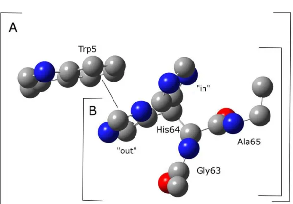
4 The -stacking interaction between Trp5 and His64 on Human Carbonic
Anhydrase II ... 23
4.1 Introduction ... 23
4.2 Computational Details ... 24
4.3 Result and Discussion ... 26
Method and basis set comparison ... 26
Substitution of Imidazole with Benzene ... 28
4.4 Conclusions ... 30
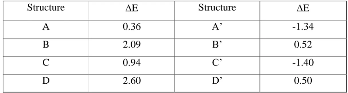
5 Tautomeric forms of His64 and The Difference Energies ... 31
5.1 Introduction ... 31
5.2 Computational Details ... 32
5.3 Results and Discussion ... 34
The Energy Profile of B3LYP Method ... 34
The Energy Profile using MP2 method ... 37
5.4 Conclusion ... 40
6 Interaction energy of Trp5 to Tautomeric forms and imidazolium ion of His64 41 6.1 Computational Details ... 41
6.2 Results and Discussion ... 43
M06-2X calculation ... 43
MP2 calculation ... 46
6.3 Conclusion ... 49
Part III. General Conclusions ... 51
7 General Conclusion ... 53
7.1 The π-stacking interaction to His64 ... 53
7.2 Potential Energy Profile ... 53
7.3 The Interaction Energy Profile ... 53
7.4 Future works ... 54
Appendix ... 55
Appendix A. Approaching Proton Relay in Human Carbonic Anhydrase II ... 57
A.1 Introduction ... 57
A.2 Computational Details ... 58
A.3 Results and Discussion ... 60
A.3.1 Geometry Optimization ... 60
A.3.2 Potential energy profiles ... 62
A.4 Conclusion ... 64
List of Publications ... 65
References ... 67
Part I. General Introduction
1 Introduction to the Thesis
This doctoral thesis is a summary of my research interest in carbonic anhydrase. The big determination was used to the structural, energetic, and interaction of residues in carbonic anhydrase.
Three years of my research at Graduate School of Natural Science and Technology Kanazawa University between October 2011 and June 2014 can be summarized in the three steps, based on my three publications as the first author.
1.1. Assessing Properties of Amino Acid Residues on Human Carbonic Anhydrase II
We have investigated the presence of the -stacking interaction that could stabilize the local structure of protein by using the density functional theory (DFT) and second order of Møller-Plesset Perturbation theory (MP2) calculation to understand the structural geometry of human carbonic anhydrase II.
M. Koyimatu, H. Shimahara, M. Iwayama, K. Sugimori, K. Kawaguchi, H. Saito,
and H. Nagao, Theoretical Model For Assessing Properties of Local Structures in
Metalloprotein, American Institute of Physics AIP Conference Proceeding, Volume
1518, pp. 626-629 (2013).
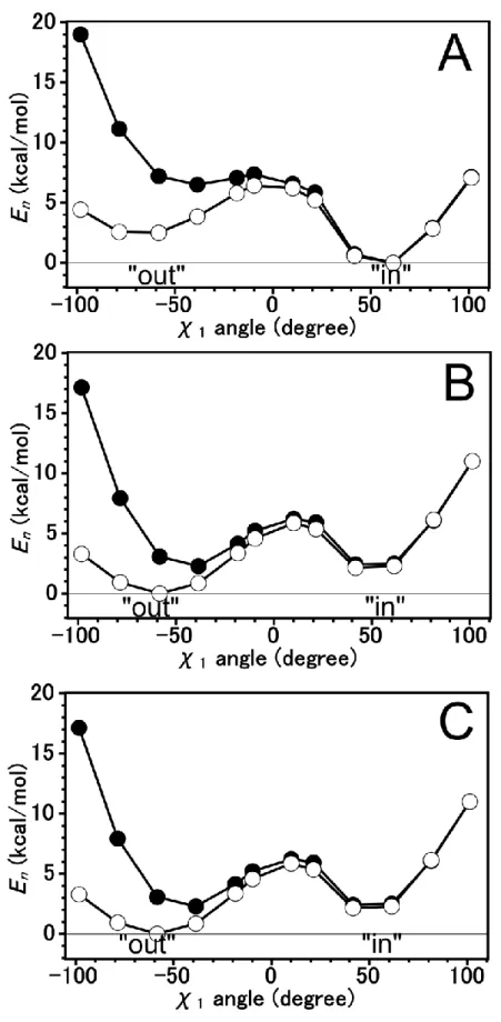
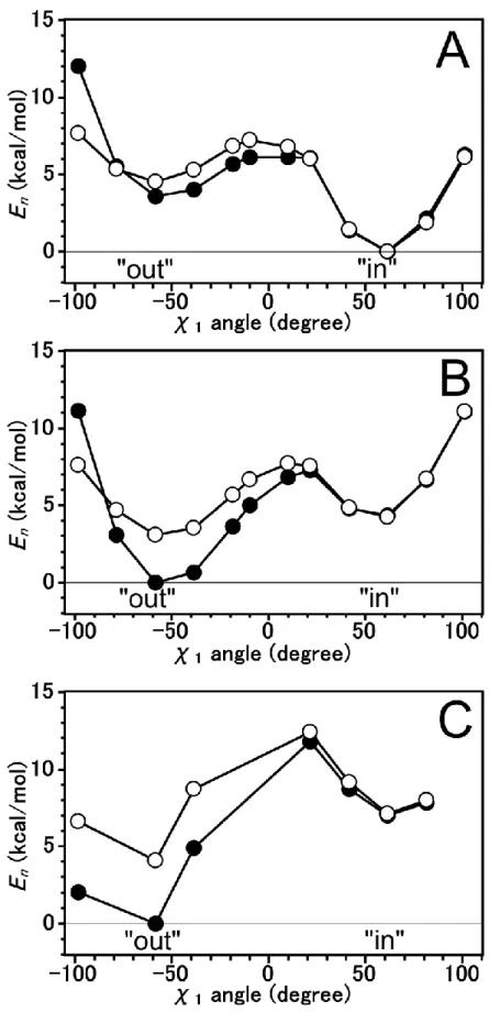
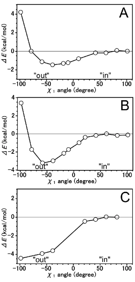
1.2. The energy profile of tautomeric forms and imidazolium form of His64 on Human Carbonic Anhydrase
We have investigated the difference energy and energy profile for N2-H tautomeric form of His64 on Human carbonic anhydrase II by using MP2 method. The energy profile at the “out” region is stabilized by -stacking interaction.
Muhamad Koyimatu, Hideto Shimahara, Kazutomo Kawaguchi, Hirosaki Saito,
Kimikazu Sugimori, and Hidemi Nagao, Theoretical Study of a -stacking interaction in carbonic anhydrase, Recent Development in Computational Science, Volume 4, pp. 87-93 (2013).
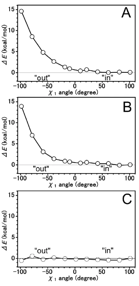
1.3. The interaction energy profile of tautomeric forms and imidazolium form of His64 on Human Carbonic Anhydrase
We have investigated the interaction energy in of the addition of Trp5 to His64 by applying binding energy equation and counterpoise method. We applied the M06-2X, a better DFT theory than B3LYP theory for geometry optimization.
Muhamad Koyimatu, Hideto Shimahara, Kimikazu Sugimori, Kazutomo
Kawaguchi, Hirosaki Saito, and Hidemi Nagao, -stacking Interaction between
Heterocyclic Rings in a Reaction Field of Biological System, The Physical Society of
Japan JPS Conference Proceeding, Volume 1 (2014).
2 Introduction to Studies on Carbonic Anhydrase and ab initio calculation
2.1 Carbonic Anhydrase
Carbonic anhydrase (CA) are enzymes that helps regulate the acid-base balance and pH in blood and other animal tissues. It is known for its role in facilitating the transport of carbon dioxide and protons in the intracellular space, across biological membranes and in the layers of the extracellular space. This enzyme is present in most organisms, from bacteria to plant.
CA was first discovered in vertebrate erythrocytes [1]. It was found that a catalyst was present in blood that could dehydrate bicarbonate and allow it to escape as CO
2[2]. The first CA was purified from erythrocytes in 1933 followed by characterization of several mammalian isozymes [3]. The existence of CA in plant has been discovered in 1947 [4], however cannot overcome the domination of CA research in mammalian until recently.
Carbonic anhydrase (CA) catalyze the reversible reaction between carbon dioxide hydration and bicarbonate dehydration, by the following reaction [5].
H
2O + CO
2⇌ H
++ HCO
3-(2.1)
CA was found to contain bound zinc, associated with catalytic activity. This
discovery made carbonic anhydrase the first known zinc-containing enzymes [6]. The
zinc-hydroxide mechanism is widely accepted to explain the role of zinc ion in by
two-step mechanisms [7, 8]. In the first step, the zinc-bound hydroxide bind to the
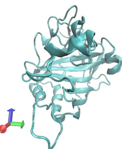
Figure 1. Visualization of HCAII (PDB file, code: 2CBA)
intermediate is replaced by a water molecule. In the second step, the active site is regenerated by the ionization of the zinc-bound water molecules and transferring a proton to an exogenous proton acceptor such as buffer (B) solution, equation (2.3).
EZnOH
-+ CO
2+ H
2O ⇌ HCO
3-+ EZn-H
2O (2.2)
EZnH
2O + B ⇌ EZnOH
-+ BH
+(2.3)
2.2 Human Carbonic Anhydrase II
Human Carbonic Anhydrase II (HCAII) has the fastest catalytic rate among CA isozymes [9, 10]. For HCAII, Histidine at position 64 (His64) has been considered to mediate the transfer of proton from zinc-bound water to a buffer molecule (Equation 2.3). Replacing it with another residue could drastically decrease the maximal turnover rate [11, 12]. X-ray crystallography data shows that the active site of HCAII has two orientations of His64 and water-bridge that connected the zinc-bound water and His64 [13]. The two orientation of His64 (“in” and “out” conformation) has been assumed to be related to the swinging or rotational motion of His64 as a final step of the proton transfer.
Due to the weakness of diffraction signal, the hydrogen atoms position and
occupancies cannot be easily obtained by X-ray crystallography. Increasing the X-ray
dose cannot enhanced the signal of hydrogen atom easily [14]. The position of
hydrogen atom is important is necessary to determine the form of His64 (N1-H
tautomer, N2-H tautomer, or protonated form). The position of hydrogen atom can
be determined by using NMR information data of HCAII. The NMR study also
suggest that the interconversion of two tautomeric form and one protonated form can
be associated with the proton transfer [15]. It believed that two alternate conformation has a relation to the rotational motion as a final step of proton transfer.
2.3 Hydrogen bonding Interaction
Hydrogen bond is a noncovalent, attractive interaction between a proton donor and a proton acceptor. Hydrogen bond occurs when H atom is bonded to electronegative atoms such as N, O, and F atoms. However, in the stabilization of weak hydrogen bonding interaction, the C-H can be involved in hydrogen bond and the p electron can act as a proton acceptors [16, 17]. The three types of hydrogen bonding interactions, which are most often discussed, are weak, moderate, and strong. The strength of weak, moderate, and strong hydrogen bonds are vary from 1-4 kcal/mol, 4-15 kcal/mol, 15-40 kcal/mol, respectively [16, 17, 18, 19]. The strength of hydrogen bond is depending on its length and angle and directionality.
Most biological molecules have so many hydrogen bonding group that hydrogen bonding is important in determining their three-dimensional structures and their intermolecular association. The internal hydrogen bonding group of protein are arranged in nearly all possible hydrogen bond are possibility formed. Hydrogen bonds has a major influence on the structures of proteins. In protein, the hydrogen bonding interactions always between C-O and H-N group, that give a shape to alpha helices and beta pleated sheet.
Other than protein, water molecules also associate through hydrogen bonds. The
hydrogen bond length between two water molecules is 1.8 Å. This interaction is
crucial to both the properties of water and to its role as a biochemical solvent. Water
dissolves more types of substances and in greater amount than any other solvent. The
polarity of water makes it an excellent solvent for polar and ionic materials.
The dissolve process can be related to Coulomb’s law:
𝑈 = 𝑘𝑞
1𝑞
2𝑟
(2.4)
Where U is the energy association, q
1and q
2are two electronic charges, r is the distance, and k is a proportionality constant. The dielectric constant of a solvent is measured of its ability to keep the opposite charges apart. The dielectric constant of water is among the highest of any pure liquid, whereas those of nonpolar substances, such as hydrocarbons, are relatively small [20].
When electrical current passes an ionic solution, the ion migrate toward the opposite electrodes at a rate, depending on to electrical field and frictional drag of ion and solution. The mobility of H
3O
+and OH
-are faster compared to other ions [21]. The high mobility rate of H
3O
+(or H
+, proton) resulting from the ability of protons jump rapidly jumps from water molecule to another water molecules. This proton movement ability also responsible in acid-base reactions in aqueous solution and probably have an important role in biological proton-transfer reactions.
In HCAII, the exceed proton from CO
hydration in the active site is transferred to a
bulk buffer molecules via His64. The distance between His64 and zinc atom is
approximately 7.5 Å, and the proton cannot directly jump at such a long distance. X-
ray crystallography shows several water molecules are visible between them. The
water molecules form a hydrogen bond network that help the mobilization of proton
from the active site to the His64.
3 Ab initio calculation
The development of quantum theory has begun since 20
thcentury. It solved many problems in science with a good precision and accuracy. However the Schrödinger equation, a fundamental equation in quantum mechanics, cannot be solved by analytically, and cannot be applied to large-scale system. To solve a difficult equation, a numerical method is required.
3.1 Density Functional Theory
Density functional theory (DFT) tried to simplify the work to find the energy of a system [22]. In the Hartree-Fock (HF) theory, the position variable of each electron was needed to build a wave function of atom or molecule to determine the energy.
The DFT theory tried to approach that system by using one variable, a density function for atom in a space. Thus the energy of a system can be solved easily by computation.
The DFT method was constructed from Hohenberg-Kohn theory, by proposing two theorems [23, 24]. The first theorem states that the ground state of electron density of many electron systems in the presence of an external potential uniquely determines the external potential. The second theorem states that the functional that delivers the ground state energy of the system, delivers the lowest energy if and only the density is true ground state density.
Using both theorem, Kohn and Sham assemble an equation of electron density and energy.
𝐸[𝜌(𝑟)] = 𝑇
𝑛𝑖[𝜌(𝑟)] + 𝑉
𝑛𝑒[𝜌(𝑟)] + 𝑉
𝑒𝑒[𝜌(𝑟)] + 𝐸
𝑥𝑐[𝜌(𝑟)] (3.1)
That equation express that the energy of the system is the total energy of kinetic of electron with the assumption that there is no interaction, potential between nucleus and electron, potential repulsive between electrons, and the correction of the total energy caused by non-classical interaction between electrons. The real system can be approach using this unreal system, because both systems has the same density when the number of atoms and position is same.
With the assumption that no electrons interaction in the unreal system, the kinetic energy for the system can be represented by the total of kinetic energy for each electrons. Energy as a function of density that including electron orbital can be described in equation
𝐸[𝜌(𝑟)] = ∑ (〈𝑥
𝑖|− 1
2 ∇
𝑖2| 𝑥
𝑖〉 − 〈𝑥
𝑖| ∑ 𝑍𝑘
|𝑟
𝑖− 𝑟
𝑘|
𝑛𝑢𝑐𝑙𝑒𝑖
𝑘
|〉)
𝑁
𝑖
+ ∑ 〈𝑥
𝑖| 1
2 ∫ 𝜌(𝑟
′)
|𝑟
𝑖− 𝑟′| 𝑑𝑟′| 𝑥
𝑖〉
𝑁
𝑖
+ 𝐸
𝑥𝑐[𝜌(𝑟)] (3.2)
Density and electron orbital can be associated with
𝜌 = ∑〈𝑥
𝑖|𝑥
𝑖〉
𝑁
𝑖=1
(3.3)
E
xcis a difference of total energy between real system and unreal system.
That energy equation can be solved by finding the x which minimalize the energy value. If x is constructed by single orbital electron x
i, we can obtain the equation
ℎ
𝑖𝐾𝑆𝑥
𝑖= 𝜀
𝑖𝑥
𝑖(3.4)
With the Kohn-Sham operator that can defined as
ℎ
𝑖𝐾𝑆= − 1
2 ∇
𝑖2− ∑ 𝑍
𝑘|𝑟
𝑖− 𝑟
𝑘| + ∫ 𝜌(𝑟
′)
|𝑟
𝑖− 𝑟′| 𝑑𝑟
′+ 𝑉
𝑥𝑐𝑛𝑢𝑐𝑙𝑒𝑖
𝑘
(3.5)
and
𝑉
𝑥𝑐= 𝛿𝐸
𝑥𝑐𝛿𝜌
(3.6)
That equation can be solved if the function of E
xcis known. E
xcan be approach by the following equation:
𝜀
𝑥[𝜌(𝑟)] = − 9𝛼 8 ( 3
𝜋 )
1
3
𝜌
13(𝑟)
(3.7) This approach known as Local Density Approximation (LDA) because equation (3.7) explicitly refer to the
cvalue at a position can be calculated from the density of that position. The value of can be vary, a=2/3 for LDA method, =1 for Slater method, and =3/4 for X method.
All method above can be expanded by adding the polarization spin effect, became:
𝜀
𝑥[𝜌(𝑟), 𝜉] = 𝜀
𝑥0[𝜌(𝑟)] + {𝜀
𝑥1[𝜌(𝑟)]
− 𝜀
𝑥0[𝜌(𝑟)]} [ (1 + 𝜉)
4/3+ (1 − 𝜉)
4/3− 2 2(2
13− 1)
] (3.8)
that know as Local Spin Density Approximation (LSDA).
Vosko, Wilk, and Nusair (VWN) propose an approach for
cthat similar with the
LSDA [25]. The value for
ccan be described in equation (3.9).
𝜀
𝑐𝑖(𝑟
𝑠) = 𝐴
2 {𝑙𝑛 𝑟
𝑠𝑟
𝑠+ 𝑏√𝑟
𝑠+ 𝑐
+ 2𝑏
√4𝑐 − 𝑏
2tan
−1( √4𝑐 − 𝑏
22√𝑟
𝑠+ 𝑏 )
− 𝑏𝑥
0𝑥
02+ 𝑏𝑥
0+ 𝑐 {𝑙𝑛 [ (√𝑟
𝑠− 𝑥
0)
2𝑟
𝑠+ 𝑏√𝑟
𝑠+ 𝑐 ]
+ 2(𝑏 − 2𝑥
0)
√4𝑐 − 𝑏
2tan
−1( √4𝑐 − 𝑏
22√𝑟
𝑠+ 𝑏 )}} (3.9)
The most common variation for the
cis VNW and VNW5. The combination of
cand Slater
xis known as SVWN.
In a molecular system, the electron density is not homogenous. A better variable of E
xcthan LDA and LSDA is needed. A way to obtain a better LSDA value is adding density gradient component to the
xcvalue, known as Generalized Gradient Approximation (GGA).
𝜀
𝑥/𝑐𝐺𝐺𝐴[𝜌(𝑟)] = 𝜀
𝑋𝐶
𝐿𝑆𝐷𝐴
[𝜌(𝑟)] + Δ𝜀
𝑥/𝑐[ ∇𝜌(𝑟) 𝜌
43(𝑟)
] (3.10)
Some applications of
xGGA function that popular are B, CAM, FT97, etc. The most popular of
cGGA function is LYP (Introduced by Lee, Yang, and Parr) [26]. The more advanced function improvement is by adding the component of second derivative density to the
xcvalue, known as meta-GGA.
Another approach is by combine the E
xfrom HF with E
xcfrom DFT. This
combination is very famous and well known as B3LYP method, which can be
defined as equation (3.11) [27, 28].
𝐸
𝑥𝑐𝐵3𝐿𝑌𝑃= (1 − 𝑎)𝐸
𝑥𝐿𝑆𝐷𝐴+ 𝑎𝐸
𝑥𝐻𝐹+ 𝑏∆𝐸
𝑥𝐵+ (1 − 𝑐)𝐸
𝑐𝐿𝑌𝑃+ 𝑐𝐸
𝑐𝐿𝑌𝑃(3.11)
By using E
xcfunction, the Kohn-Sham (KS) equation can be solved by self-consistent method. At the first step, the electron density is defined. Then that electron density is used to solve KS equation for every electron in the system. The result is a new electron density value, and it used to solve KS equation. The iterative process is continue until the new electron density value is same with the previous value.
3.2 Møller-Plesset Perturbation Theory
Christian Møller and Milton S. Plesset described the Møller-Plesset Perturbation (MP) Theory in 1934 [29]. MP takes the zeroth-order Hamiltonian for an atom or molecule as the sum of the one particle Fock operator.
𝐻 ̂
0= ∑ 𝐹̂(𝑖) = ∑ ℎ(𝑖) + 𝑉
𝐻𝐹(𝑖)
𝑁
𝑖=1 𝑁
𝑖=1
(3.12)
We write the perturbation as the difference between the perturbed and unperturbed Hamiltonian
𝑉̂ = 𝐻 ̂ − 𝐻
0(3.13)
The Hamiltonian is expressed as
𝐻 ̂ = ∑ 𝑓(𝑖) + ∑ 𝑔(𝑖, 𝑗)
𝑁
𝑖<𝑗 𝑁
𝑖=1
(3.14)
The Fock operator
𝐹̂(𝑖) = 𝑓̂(𝑖) + 𝑉
𝐻𝐹(𝑖) (3.15)
The one electron operator, f in H and H are identical and can cancel each other in
taking the difference in the perturbation
𝑉̂ = ∑(𝑟
𝑖𝑗−1− 𝑉
𝐻𝐹)
𝑁
𝑖<𝑗
(3.16)
The first-order correction the energy average of the perturbation over the unperturbed wavefunction can be described as:
𝐸
0(1)= 〈𝜓
0|𝑉̂|𝜓
0〉 = 〈𝜓
0|( 1
𝑟
12− 𝑉̂
𝐻𝐹)|𝜓
0〉 (3.17)
making the total first energy
𝐸
𝑛= 𝐸
𝑛0+ 𝐸
𝑛(1)= ∑ 𝜀
𝑎− 1 2
𝑁
𝑎
∑〈𝑎𝑏||𝑎𝑏〉
𝑁
𝑎𝑏
(3.18)
The energy throught first-order is simply Hartree-Fock energy.
The second-order MP, or MP2, is the most widely used method in quantum chemistry. The second-order correction to the ground state energy depends on the first order correction to the wavefunction. The first order wavefunction may be expanded as
𝜓
𝑛0(1)= ∑ 𝐶
𝑛,𝑎𝑏𝑟𝑠(1)𝑁
𝑎>𝑏;𝑟>𝑠
𝜓
𝑎𝑏𝑟𝑠(3.19)
Where 𝜓
𝑎𝑏𝑟𝑠respresent a wavefunction which has electron excited from spin orbital a and b (occupied) into spin orbital r and s (unoccupied), respectively. The coefficient C
abrsare determined by
𝐶
𝑛,𝑎𝑏𝑟𝑠(1)= 〈𝜓
𝑎𝑏𝑟𝑠|𝜓
𝑛(1)〉 = ∑ 〈𝜓
𝑎𝑏𝑟𝑠|𝜓
𝑛(0)〉 𝜖
𝑎+ 𝜖
𝑏− 𝜖
𝑟− 𝜖
𝑠𝑁
𝑎>𝑏;𝑟>𝑠
(3.20)
This wave function can be placed in the second order energy equation.
𝐸
0(2)= ∑
|〈𝜓
0(0)| 1
𝑟
12|𝜓
𝑎𝑏𝑟𝑠〉|
2
𝜖
𝑎+ 𝜖
𝑏− 𝜖
𝑟− 𝜖
𝑠𝑁
𝑎>𝑏;𝑟>𝑠
(3.21)
3.3 Hybrid Meta-generalized gradient-approximation (hybrid meta-GGA) exchange functional
The DFT fail for some kinds of systems, such as the polarizability of conjugated systems, and the ground-state energy of protonated polyenes. This failure contribute to the incorrect long-range behavior of the effective potentials generated by the density functional function. In order to correct the long-range error in DFT is by mixing the full Hartree-Fock correlation and the DFT correlation, the hybrid exchange-correlation energy, which can be written as equation (3.22).
𝐸
𝑥𝑐ℎ𝑦𝑏= 𝑋
100 𝐸
𝑥𝐻𝐹+ (1 − 𝑋
100 ) 𝐸
𝑥𝐷𝐹𝑇+ 𝐸
𝑐𝐷𝐹𝑇(3.22)
Where 𝐸
𝑥𝐻𝐹is the nonlocal Hartree-Fock exchange energy, X is the percentage of the HF exchange, 𝐸
𝑥𝐷𝐹𝑇is the local DFT exchange energy, and 𝐸
𝑐𝐷𝐹𝑇is the local DFT correlation energy.
The M06 functionals may be classified as hybrid meta-generalized gradient- approximations (hybrid meta-GGAs), which developed by Zhao and Truhlar [30, 31].
These functionals composed of four functional that have similar functional forms for the DFT part, but each parameter optimized to be used with a different percentage of HF exchange. The M06-2X has 54% HF exchange with a high nonlocality functional with double of the amount of nonlocal exchange (2X). This functional is good for all area in chemistry including thermochemistry and reaction kinetic, but excluding multireference system such as many systems containing transition metals.
The M06 and M06-2X functionals depend on three variables; spin density ( 𝜌
𝜎),
reduced spin density gradient (𝑥
𝜎), and spin kinetic energy density (𝜏
𝜎).
𝑥
𝜎=
|∇𝜌𝜎|𝜌𝜎4/3
𝜎 = 𝛼, 𝛽 (3.23)
𝜏
𝜎=
12
∑
𝑜𝑐𝑐𝑢𝑝𝑖|∇Ψ
𝑖𝜎|
2(3.24) The M06 functional form is a linear combination of the functional form of the M05, the first meta-GGA functional, and van Voorhis and Scuseria exchange correlation functional (VSXC) exchange functional is given by equation (3.25).
𝐸
𝑥𝑀06= ∑ ∫ 𝑑𝑟 [𝐹
𝑥𝜎𝑃𝐵𝐸(𝜌
𝜎, ∇𝜌
𝜎)𝑓(𝑤
𝜎) + 𝜀
𝑥𝜎𝐿𝑆𝐷𝐴ℎ
𝑋(𝑥
𝜎, 𝑧
𝜎)]
𝜎
(3.25)
where 𝐹
𝑥𝜎𝑃𝐵𝐸(𝜌
𝜎, ∇𝜌
𝜎) is the exchange energy density of the Perdew-Burke-Ernzerhof (PBE) exchange model [32], 𝑓(𝑤
𝜎) is the spin kinetic energy density enhancement factor
𝑓(𝑤
𝜎) = ∑ 𝑎
𝑖𝑤
𝜎𝑖𝑛
𝑖=0
(3.26)
𝑤
𝜎is a function of 𝜏
𝜎, and 𝜏
𝜎is a function of the spin kinetic energy density 𝜏
𝜎and spin density 𝜌
𝜎.
𝜔
𝜎= (𝑡
𝜎− 1) (𝑡
𝜎+ 1)
(3.27)
𝑡
𝜎= 𝑡
𝜎𝐿𝑆𝐷𝐴𝑡
𝜎(3.28)
𝑡
𝜎𝐿𝑆𝐷𝐴≡ 3
10 (6𝜋
2)
2/3𝜌
𝜎5/3(3.29)
𝜀
𝑥𝜎𝐿𝑆𝐷𝐴is the local spin kinetic energy density enhancement factor.
𝜀
𝑥𝜎𝐿𝑆𝐷𝐴= 3 2 ( 3
4𝜋 )
1/3