Acta med. nagasaki. 8 : 81-93
Chromosomal Projections from the Resting Nuclei of Cells of Watanabe II Tumor
Fumitomo WATANABE*
Laboratory of Animal Tumor, Nagasaki University,
School of Medicine, Nagasaki, Japan
Received for publication March 1, 1964
Ascites tumor cells of Watanabe II tumor of rats usually have a chromosomal projection or projections protruded from the nuclear surface in about 20 per cent of tumor cell population in every transplant genera- tion. They are covered with nuclear membrane and contain a number of fine chromatin granules similar to those of the nuclei from which they have projected. Although they are parts of the nuclei at resting stage, they begin to show chromosome constitution at prophase and become longer chromosome thereafter.
It is highly probable that a extremely long and other larger chromosomes characteristic to Watanabe II tumor occur as chromosomal projections in resting nuclei because of their larger size, judging from the observation of the mitotic course of the tumor cells having such marker chromosomes.
Recently fine structure of the resting nuclei of somatic and malig- nant cells have been intensively studied. For instance, observations
of the sex chromatin have been made by Barr and his associates", Klinger", Ishizaka and Kosin3) and James4), and so-called drumsticks of leucocytes were studied by Davidson et al2) in detail. The present paper reports the results of the observation of chromosome projections from the resting nuclei of ascites tumor cells of Watanabe II. This strain of ascites tumor was established from the spontaneous tumorous ascites derived from the hemoangioendothelioma originating from the cysticercus cyst in liver of a female rat of Ohmura strain receiving repeated injections of hot water.
MATERIALS AND METHODS
Hemorrhagic tumorous ascites as pure cultures of free tumor cells were used as materials. They were taken in the 18th to 20th transplant
*渡 辺 文 友
82 CHROMOSOMAL PROJECTION Vol. 8.
generation from the abdominal cavities of 6 rats of Ohmura strain 4 or 5 days after intraperitoneal inoculation of 0.8 cc of ascites tumor. A drop of fresh hemorrhagic ascites tumor was put on the slide. 0.3 cc of 35 per cent acetic dahlia was added to the tumorous fluid on the glass. Both fluids were mingled with the cover glass edge. After approximately 10 minutes, it was pressed strongly with the finger tip.
Then, it was mounted with balsam-paraffin and observed under micro- scope. Some materials of tumorous fluid were pretreated with water.
Chromosomal projection from the resting nuclei of tumor cells in some other materials were observed by the help of the phase-contrast micro- scope.
RESULTS
A) Numbers of the Tumor Cells Having Chromosomal Projections from Their Resting Nuclei:
Table 1 indicates numbers of the tumor cells showing chromosomal projections from their resting nuclei among 2025 cells observed. Numb- ers of chromosomal projections ranged from one to three projections per cell. Cells containing one projection were 342 (16.88%), largest in number. Cells having two were 70 (3.45%) in number and cells having three were 7 (0.34%). The more the number of projections from one nucleus increased, the less the incidence rate becomes. Total number of cells containing chromosomal projections was 419 (20.68%). Large and middle-sized cells presenting the projections showed the largest numbers of incidence (142 and 139 respectively). A small number of binucleated cells, giant mononucleated cells and small-sized cells showed chromosomal projections of their resting nuclei, as indicated in Table 1. These projections observed ranged in shape from a hemisphere-like process to rod-shaped projection protruded outside the surface of resting nuclei treated by acetic-dahlia technique. When the tumor cells are pretre- ated with water or hypotonic solution, the chromosomal projections become clear and the incidence rate of hemisphere-like small projections increase, because of the probable pushing out the projection from the nuclear surface by the pressure of swollen nuclear content through permeation of water added. Incidence rates of chromosomal process of
resting nuclei of tumor cells pretreated with water or hypotonic solution will be reported elsewhere.
B) Cytological Behavior of Chromosomal Projection of Resting Nuclei of Tumor Cells of Watanabe II :
One chromosomal projection can usually protrude from one resting nucleus, but rarely one nucleus has 2 or 4 projections (Table 1). The
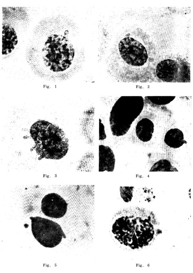
surface of the chromosomal projection of resting nuclei is always cover- ed with nuclear membrane (Fig. 1). Contour of projections protruding
1963 V. WATANABE $3
Table 1.
Incidence Rate of Chromosomal Projections of 2025 Resting Tumor Cells of Watanabe II
One
(3.45%)
1
Number of chrom.
projection 2 3
Total of tumor celled observed
Both nuclei havingnucleus having a project. 2 a chro m. project.
Bin ucleated cells 1
One nucleus having a project. •••••••••••• 1
Mononucleated 2 ' 2 1
giant cells
2025 Large cells27 3
Middle-sized cells27 1
Small cells13 2
70 7Total
(16.88%)(0.34%)
142
139
56
342
Total of cells having chromosomal projection ... 419 (20.68%)
from the upper surface of nuclei can be observed infrequently in the nuclei (Fig. 2). Size, shape and distribution of chromatin granules in the projection are usually similar to those in the resting nuclei from
which projection protruded (Fig. 3). But in some cases the projection have less chromatin distribution than that within nucleus (Fig. 1).
However, in other cases there were more chromatin granules in the tip area and scanty in basic part of chromosomal projections. The author could not find in chromosomal projection such top enlargement as that of drumstii-en found in neutrophile leucocytes. From the above described findings the chromosomal projection is suggested to be a nuclear part originating from the chromosome portion which could not be drawn completely into telophasic nucleus through some mitotic disturbances.
C) Chromosomal Projection Found in Small-sized Cells in Prepar- ation of Tumorous Ascites :
Beside the tumor cells, chromosomal projections of small-sized resting cells resembling lymphocytes or monocytes were rarely observed in acetic-dahlia squash preparation made of same materials. The projection found in small-sized cells also ranged in shape from hemispheric to rod-shaped process (Fig. 4). Some of the projections protruded slant- ingly from the surface of the nuclei of small-sized cells (Fig. 5).
Nature of these small-sized cells can not be immediately determined,
84 CHROMOSOMAL PROJECTION Vol. 8.
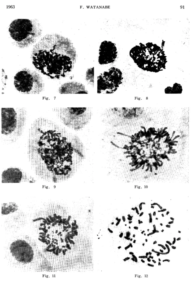
because the strain of Watanabe II tumor is of angioendotheliomal origin and show characteristic morphology. In the late prophase, the continued part of projection existing in nucleus is clearly outlined and becomes with the projected part a whole chromosome thead (Figs, 6, 7, 8).
When the nuclear membrane disappeared, the projection as a part of chromosome exhibited spiral structure specific to chromosome (Fig. 9).
In prophase some chromosomal projections demonstrated a constriction indicating a centromere near the end of chromosome thread. These chromosomal projections seem to be parts of the subtelocentric chromo- somes (Fig. 7). In some of the cells at early metaphase, length split becomes clearer in the chromosomal projection which extruded from the plate (Fig. 8).
D) Irregularly Arranged Metaphase Plates and Karyotype in Con- nection with Chromosomal Projection :
One or two long arms of J-shaped chromosomes which projected from the metaphase plates were rarely observed in some of the tumor cells of this strain (Fig. 10). A chromosome thread shifted out of their regular position in metaphase plates also can be seen infrequently in tumor cells squashed (Fig. 11). It must be under consideration that some parts of 4 or 6 long chromosomes threads including extremely long one specific to the stem cells of this tumor showed protrusion from the outline of metaphase plates (Fig. 12, 13).
E) Chromosome Bridges at Anaphase and Telophase in Relation to Formation of Chromosomal Projections in Resting Nuclei of Tumor Cells :
It is well known fact that because of the increased adhesiviness of chromosome of tumor cells ana- and telophasic chromosome-bridges frequently occur. One, two or even three bridges of chromosomes could be seen in some of the ana- and telophasic cells (Fig. 14, 15). In spite
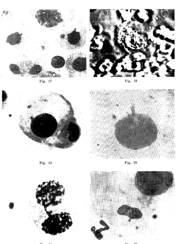
of the artificial separation owing to the acetic-dahlia squash technique, a long clear chromosome bridge can be formed between two daughter cells (Fig. 16). In such cases, after breakage of a chromosome bridge the retarded chromosomes should protrude from the surface of the new daughter nuclei (Fig. 17).
F) Chromosomal Projections Observed in the Telophase Nuclei of Two Separated Cells :
When the cells at telophase are squashed through acetic-dahlia squash technique, two separated daughter cells being formed as a result of artefact should have no nuclear membrane. In such separated daug- hter cells, both nuclei have not yet entered the resting stage, but showed an aggregated condition of chromosome in both seemingly daughter cells. Sometimes in incomplete nuclei of both cells there appear clear
1963 F. WATANABE 85 chromosomal projections protruded from the aggregated masses of chromosomes (Fig. 17).
G) Phase-Contrast Microscopic Observation of Chromosomal Proje- ctions of Resting Nuclei of Tumor Cells :
Chromosomal projections of resting nuclei of tumor cells in the materials obtained from the abdominal cavity of a rat 4 days after the intraperitoneal transplantation at the 19th generation. A clear-cut figure being presumed as chromosomal projection were observed (Fig. 18).
In another nucleus of the resting tumor cell, a clear chromosomal proje- ction existing near the middle of nuclear surface. It protruded outside the contour of the nucleus, preserving chromosome structure. In other two cases there could be seen somewhat obscure projection which was indistinguishable from outflow of the nuclear material.
DISCUSSION
1) It is true that chromosome configuration can not be recognized in the resting nuclei of somatic and malignant cells. Effects of some chromosomal and mitotic irregularities upon the formation of resting nuclei are expressed as chromosomal projections in nuclei of resting cells. Morphological observation of chromosomal projection and the elucidation of the mechanism of the occurrence of the projection found in the resting nuclei of the Watanabe II ascites arising in the female rat seem to be of importance in the studies of chromosome and the mitotic cycle. Davidson and Smith" described the drumstick which is believed to be the projection of X-chromosome protruded from the lobe of polymorphonuclear neutrophile leucocytes in female animals. Although the "drumstick" shows usually tip enlargement, the chromosomal projection found in tumor cells of the Watanabe II has no such morpholo- gical peculiality. From the genesis of the drumstick, the possibility of the X-chromosomal origin of the chromosomal projection of resting nuclei of tumor cells of Watanabe II can not be excluded, because this strain of the ascites tumor originated in the female rat. However, multiple occurrence, morphological variability and the mitotic behaviors of chromosomal projections seem to contradict the X-chromosomal origin. The prophasic projections seem to show no heteropycnosis expressed by X-chromosome at prophase in some of the tumor cells, which was suggested by Ohno, Kovacs and Kinoshita7', ", Ohno and Hauschka".
2) Pomerat'°>> and Nakanishi observed the difference in incidence rate of nuclear bud or outpushing between control Hela S. clone and r-irradiated one. The nuclear bud or outpushing named by Pomerat found in Hela S:, clone resembles in shape the chromosomal projection suggested by the author. From the name of nuclear bud or out pushing,
86 CHROMOSOMAL PROJECTION Vol. 8.
Pomerat seem to have the idea that it is a result of the outflow of the nuclear material perhaps from the data obtained by r-irradiation.
Recently Ruddle"' reported nuclear blebs protruded from interphase nuclei in an established line of pig kidney cells. The chromosomal proj- ection of resting nucleus is clearly different from the drumstick of polymorphonuclear neutrophil leucocyte and nuclear bud or outpushing observed in Hela S3 cells in that the chromosomal projection is a part of a chromosome and behaves as a chromosome in mitotic cycle as demontrated in the present materials.
3) Although as later described, chromosomal projection can infre- quentiy be observed in embryonal cells of a rat or somatic cells of an adult woman, high incidence (20.68%) of the chromosomal projection in tumor cells of Watanabe II seems to be based on the intensive stic- kiness of the chromosome of the tumor cells. Makino and Yoshida"
suggested that the increased stickness of chromosome and the spindle abnormality being recognized as the common feature of the tumor induces the fusion on the terminal contact point of two separated chromosomes.
This telosynapsis at anaphase seems to form a chromosome bridge resulting in retarded chromosome which becomes extruded element in telophasic nucleus. On the other hand, non-disjunction is apparently one of the important causes of the occurrence of the projection. Accord-
ingly, it is easily considered that chromosomal projections of resting nuclei of both daughter cells result from the extruded chromosome element of telophasic nuclei. It is unknown that X-chromosomes being capable of special condensation express the increased stickiness leading to form protruded elements in tumor cells. The suggestion about the formation of the chromosomal projection has realized by author's inve- stigation" I""4" on the stepwise transformation of chromosome bridge from anaphase to telophase forming extruded elements of telophasic nuclei and resulting chromosomal projections of both daughter cells, as shown by several photographs.
4) Another important aspect concerning the appearance of chromo- somal projection is the karyotype characteristic to the stem cells of Watanabe II tumor. Although chromosomal projections were observed rarely in the resting nuclei of Watanabe I tumor, in tumor cells of Watanabe II the projections were observed in the high incidence of 20.68 per cent (Table 1). This fact cannot exclude the effects of the characteristic karyotype containing extremely long chromosomes resulting from the translocation. Therefore, occurrence of 1 to 3 chro- mosomal projections protruded from the resting nuclei of tumor cells of Watanabe II suggests the participation of long chromosomes in the karyotype.
5) Hemispheric projections protruding from the outline of the resting nuclei seemed to be more frequent and obscure in the materials pretreated with water than in non-pretreated ones only prepared by
1963 F. WATANABE 87 acetic-dahlia squash technique. This fact indicates that the projections can be pushed out from the surface of the nuclei through the swelling of nuclear content by absorbed water. If this is true, projected chro- mosome should have a peculiar condensation. Chromatin distribution in the chromosomal projection indicates that the projection has changed its structure to a part of nuclei originating from the chromosome element. It is interesting that nuclear membrane can be seen through the projections protruding out of the upper surface of the nuclei and laying across the nuclear contour (Fig. 2). In some of the materials pretreated with water, chromosome contour and even longitudinal fibre-like structure could be observed (Fig. 19). This findings suggest that contour of chromosome is maintained in the resting nuclei in special cases of tumor cells (Fig. 20). Results of the observations by means of phase-contrast microscope, demonstrated. the presence of chromosomal projection which was similar to that made by the squash technique (Fig. 18). This finding means that the projection can be seen in living cells and is different from the nucleard bud or outpushing described by Pomerat10)' .
6) The behaviors of chromosomal projection in mitosis seem to determine its nature and origin. One of the significant features is that invisible chromosomal part in the nucleus together with chromosomal process makes a whole chromosome thread expressing spiral figure at the prophase. On the other hand chromosomal projections connecting two nuclei of binucleated cells also show the spiral structure of chrom- osome at prophase (Fig. 21). From the behavior of chromosomal proje- ction above described, it is strongly suggested that in the resting nuclei there are chromosomal unit areas corresponding to the chromo- somes, although their contours are lost in the nuclei.
7) A long chromosomal projection could be observed in the resting round nuclei of a monocyte-like small cell in the buccal smears obtained from the female adult woman, which were treated by acetic-dahlia squash technique. One or two chromosomal projection of resting nuclei of a monocyte also could be seen in tumorous ascites of a rat. Some of the projections showed the clear-cut structure of a chromosome (Fig.
4). A chromosomal projection protruding from the middle of the side surface of long ovoid nuclei of a cell could be found in ernbryonal brain of a rat of Ohmura strain (Fig. 22). The infrequent occurrence of the condition of resting nuclei of normal 'cells can be interpreted by the fact that extruded element which can change to chromosomal projection in resting nuclei is observed more frequently in tumor cells containing skicky chromosomes. From the interpretation about the formation of chromosomal projection, there should be parallel incident e between chromosomal projection of resting nuclei and chromosome bridges at ana- and telophase as suggested by personal communication of Dr.
88 CHROMOSOMAL PROJECTION Vol. S.
Tonomura, Nakanishi and Kinoshita. Present investigation seems to have solved partly the question whether the chromosomal projection have hereditary significance, in other words, whether the projection as a marker chromosome can be transmitted from a cell to its daughter cells in succession of stem cells of Watanabe II ascites tumor.
SUMMARY
1) Chromosomal projections of resting nuclei of ascites tumor cells was observed in the materials treated with acetic-dahlia squash techni- que during from 18th to 20th generation of Watanabe II tumor origina- ting in female rat. The incidence rate of the projection was in 20.68 per cent.
2) Projections were discussed in comparison with the drumstick in the lobe of polymorphonuclear neutrophile leucocytes and the nuclear bud or outpushing suggested by Pomerat in the observation of cells of Hela S3 clone.
3) They appear as parts of chromosomes protruded into cytoplasm of resting nuclei and have the same chromatin distribution as those in resting nuclei. These conditions Lare also demonstrated by using phase contrast microscope.
4) Formation of the chromosomal projection was discussed based on the fate of chromesome bridge and retarded chromosome owing to increased stickiness or spindle abnormalities and of the extremely long chromosome being characteristic to the Watanabe II tumor cells.
5) Behavior of the chromosomal projection during the mitotic stages suggested the chromosome area in resting nucleus.
6) Chromosomal projections could be observed in round nuclei of monocytes of female human adult and in some cells in a brain of a rat embryo of Ohmura strain.
7) The present investigation seems to demonstrate that chromoso- mal projections of resting nuclei of Watanabe II tumor cells can be transmissible as the marker condition from the parent cells to their daughter cells in successive passage of the tumor.
The author wishes to express his thanks to Mrs. Hashiguchi for her technical assistance.
REFERENCES
1) BARR, M. L. and BERTRAM, E. G. : A morphological distinction between nucleus of the male and female, and the behavior of the nucleolar satellite
during accelerate nucleoprotein synthesis. Nature. 163 : 676, (1949).
2) DAVIDSON, W. M, and SMITH, D. R. : A morphological sex difference in the polymorphonuclear neutrophile leucocytes.British Med. Jour. 6 : (1954).
3) IsHIZAKT, H. and KOSIN, I. L. : Sex chromatin in early chick embryos. Exp.
Cell Res. 21 : 197-200, (1962).
1963 F. WATANABE 89 4) JAMES, J. : The "sex chromatin" and the nucleic acids. Exp. Cell Res . 21
205-208, (1960).
5) KLINGER, H. P. : The fine structure of the sex chromatin body. Exp. Cell Res. 14 : 207, (1958).
6) MAKINO, S. and YOSHIDA, T. H. : Cytological studies on cancer, I. Morpholo- gical and statistical observations on the abnormal mitosis in tumor cells of
the Yoshida sarcoma through a transplant generation. Jour. Fac. Sci. Hokkaido
Univ. Ser. VI. Zool. 10 : 209-224, (1951).
7) OHNO, S. KOVACS, E. T. and KINOSHITA, R. : On the X-chromosomes of mouse mammary carcinoma cells. Exp. Cell. Res. 16 : 462, (1959).
8) O:NO, S. and HAUSCHKA, T.S. : Allocycly of the X-chromosome in tumor and normal tissues. Cancer Res. 20 : 541-545, (1960).
9) OHNO,, S . , KAPLAN, W. D. and KINOSHITA, R, : X-chromosome behavior in germ and samatic cells of rattus norvegicus Exp, Cell Res. 22: 535, (1961).
10) POMERAT, C. M. : Cellular changes induced by radiation. Annals of the New
York Academy of Sci. 71 : 1143-1155, (1958).
11) RtJDDLE, F. H. : NUCLEAR BLEB : A stable interphase marker in established line of cells in vitro. r. Natl. Cancer Inst. 28 : 1247-1252, (1962).
12) WATANAB'S, F. : Cancer as a monocellular organism, Report 1. An intermediate form of an ascites tumor of a rat (Watanabe II tumor). Acta Path. Jap. 7
No. .4, 342-343, (1957).
13) WATANABE, F. and AZUMA, M. : A new rat ascites tumor showing fragments with histoid structure in the ascites fluid. Gann 48 : 547-549, (1957).
14) WATANABE, F., AZUMA, M. and KANEKO, M. : Chromoscmal projections of cell nuclei of Watanabe 11 ascites tumor (ascites endothelioma of a rat). The
Japanese Journal of Genetics. 34 : 9, 315, (1959).
Explanation of plates
Fig. 1. Surface of the chromosomal projection of resting nucleus of a tumor cell is covered nucleus membrane.
Fig. 2. Contour of the projection protruded from the upper surface of nuclei can be seen.
Fig. 3. Chromatin distribution of the projection is similar to that in nucleus.
Fig. 4. Projection of a nucleus of a small-sized cell resembling lymphocyte in tumorous fluid.
Fig. 5. Projection slantingly projected from the nuclear contour.
Fig. 6-9. Projection at prometaphase.
Fig. 10. Two long projected chromosome at metaphase plate.
Fig. 11. J-shaped chromosome protruded from a contour of metaphase plate.
Fig. 12. Extremely long chromosome found in a metaphase plate of the W-II tumor cell.
Fig. 13. Karyotype analyse of chromosome of W-II tumor cells showing extremely 1 ong chromosomes.
Figs. 14-17. Chromosomebridge at ana-and telophase probably causing the projection of resting nuclei.
Fig. 18. Chromosomal projection of a tumor cell observed by phase contrast micros- cope.
Fig. 19. Fibre-like structure of a projection of the water treated nucleus.
Fig. 20. Chromosomal contour of projecting preserved in water treated cells.
Fig. 21. Spiral structure of the connecting projection of a binucleated tumor cell.
Fig. 22. Projection of the long, ovoid nucleus of a cell found in the embryonal brain of a rat.
90 CHROMOSOMAL PROJECTION Vol. 8.
Fig. 1 Fig. 2
Fig. 3 Fig. 4
Fig. 5 Fig. 6
1963 F. WATANABE 91
Fig. 7 Fig. 8
Fig. 9 Fig'. 1C
Fig. 11 Fig. 12
92 CHROMOSOMAL PROJECTION V ol. 8.
Fig. 13
Fig. 14 Fig
, 15 Fig. 16
1963 F. WATANABE 93
Fig. 17 Fig. 18
Fig. 19 Fig.. 20
Fig. 21 Fig. 22