Acta Med. Nagasaki. 22 : 50-62
Ultrastructural Study of the Dorsal Root Ganglia in Fetal Organic Mercury Intoxicated Rats
Masao KISHIKAWA*
Department of Pathology, Atomic Disease Institute, Nagasaki University School of Medicine
Nagasaki, Japan
Received for publication, January 5, 1977
A part of this study was presented at the 18th Kyushu district meeting of Japanese Society of Electron Microscopy, December 4, 1976.
Methylmercuric chloride (MeHg) was given in pregnant rats during the last week of gestation. Newborn and infant rats treated with MeHg during intrauterine life were sacrificed at birth, and 5th, 10th and 30th day after birth.
Dorsal root ganglia (DRG) of the baby rats were examined light and electron mi- croscopically. The neurons were more various in shape and had eccentric nuclei more often than these in control baby rats. Interface between the neurons and their satellite cells showed less interdigitations even at 30 days of age. The rough endoplasmic reticulum (RER) was shortened with dilated cisternae and irregular branching. At 30-day-old rat, the dilated endoplasmic reticulum was decreased in number. The alterations of RER are an evidence of morphological aspects for the protein synthesis decreasing. By 10th day, the neurofibrils were not so prominent in DRG neurons of MeHg treated rat at 5th day, while they were increased in number to form fibrillar bundle and separated the RER into small stacks at 30 days of age. This may reflect early, ongoing regenerative changes in injured DRG neurons.
The DRG from the MeHg-baby rats disclosed extensive injuries of neurons, associated with developmental retardation of the ganglia.
*岸 川 正 大
INTRODUCTION
Despite the fact that the toxicity of mercurial compounds causes community health problems, these compounds continue in present use because of their extreme usefulness in medicine, agriculture, and industry.
Recently, the investigations on mercury poisoning in intrauterine life are getting increased. Alkyl-mercury exhibits the fetotoxic effects both to the victims of fetal Minamata disease")' 30), 31) and to the laboratory animals")' 19), 24), 32), 33) . The neuropathological observations on the fetal Minamata disease have been throughly performed by TAKEUCHI et al.29)-31)
On the other hand, the most patients of the adult cases of Minamata disease examined by TOKUOMI34) revealed a essential clinical manifestation of peripheral neuropathy.
There have been many morphological discriptions both on human victims13)' 29) and on experimental animals2)' 3). 5)-s), 11), 12), 14), 20)-23). However, there have been no published pathologic investigations of dorsal root ganglia (DRG) in animals poisoned congenitally by mercury. The reports of MURAKAMI et al .25),21) on mercurial contraceptives point to the need for an understanding of the effects of alkyl mercurials on the fetus and newborn.
The purpose of present study was to represent the morphological changes on the DRG poisoned by Methylmercuric chloride (MeHg).
MATERIALS AND METHODS
Wistar rats were used for this study. Estrous female rats were put into a cage with a male and the female was examined for copulation plug. The day on which the plug was found was considered day 1 of pregnancy. The five pregnant rats were given 10 mg MeHg (WAKO Pure Chemical Industries, LTD, Osaka, Japan) per body kilogram by a single, very slow injection into the tail vein on the 14th or 15th of pregnancy. The MeHg was dissolved in ethyl alcohol and 0.9% saline solution, (1:1, v/v).
The baby rats were sacrificed without anesthetic after birth at the following times : five animals each at birth, and 5th, 10th and 30th day postnatally. Eight normal baby rats were sacrificed as the same time schedule.
Lumbar laminectomy was performed quickly and the tissues were immersed in 1.5%
glutaraldehyde in 0. 1 M. phosphate buffer (pH 7.4). While immersed in the fixative,
the DRG (T12-L3) were dissected free and kept in fresh fixative. The tissues were rinsed
in 0.1 M. phosphate buffer (pH 7.4), postfixed in 1% Os04 in 0. 1 M. phosphate buffer
(pH 7.4), dehydrated in ascending concentrations of ethyl alcohol and embedded in Epon
812 resin. Thick sections were cut lp in thickness and stained with toluidine blue for
light microscopy. After trimming, ultrathin sections were made with Porter-Blum MT-I
ultra-microtome. These sections were stained with uranyl acetate and lead citrate, and
examined with JEM 7A and JEM 100B electron microscopes.
RESULT
LIGHT MICROSCOPIC OBSERVATIONS: Two types of neuron were not differ- entiated in the newborn MeHg poisoned rats, although these could be identified with some difficulty in the control DRG. These two types could be observed in the poisoned DRG of ten-day-old rats. There was a distinct variation in the size of DRG neuron in all the animals, treated and control alike, at birth, but at older age the neurons in control became a uniform size and shape with almost centrally located nucleus (Fig. la) However, the affected neurons remained moderate abnormalities, which were in shape and in location of the nucleus (Fig. 1b). Especially, the diameter of large clear neuron was slightly shorter than that in control (Fig. 1b). The eccentric nuclei were observed frequently in the DRG of MeHg treated animals (Fig. 1b).
ELECTRON MICROSCOPIC OBSERVATIONS : It is difficult to distinguish large clear neurons from small neurons at birth by electron microscopy, both in the control and the influenced rats. It was sometime possible in control DRG when both cell types being in the same field, but not in the MeHg treated rats at all. The cytoplasm of the satellite cell which enveloped the neuron was very thin in both control rats and MeHg poisoned group at birth. While there could be observed several layers of the cytoplasm of satellite cell (Fig. 2a) in control newborn rat, there were only few layers among two neurons (Fig. 2b) in MeHg poisoned newborn rat. The interdigitations between the cytoplasmic membrane of neuron and the cytoplasm of satellite cell in control rat became complicated day by day with its growth (Fig. 3a). This intercellular specialization in MeHg poisoned rat revealed less prominent than the control (Fig. 3b, 7b).
Among the cytoplasmic organelles, the Nissl substances showed the most prominent changes. The RER, which corresponds to the Nissl substance by light microscopic observation, was shortened, had markedly dilated cisternae and irregular branching (Fig.
2b, 3b). The stacks of RER were sometimes disorganized while the RER of control animals was arranged often parallel to the cell membrane or to the nuclear membrane without any abnormal dilation and branching (Fig. 2a, 3a). The markedly dilated cisternae of RER lost attached ribosomes in many neurons and the free ribosomes were increased in these neurons (Fig. 2b, 3b, 4). These abnormal changes of RER disappeared by 30 days of age.
The amount and distribution of neurofibrils in the cytoplasm were prominently different in the clear large neuron of control rat (Fig. 5) and in that of 30-day-old poisoned rat (Fig. 6). In the treated 30-day-old rats, the fibrils were markedly proliferated and separated the stacks of RER into scattered tiny Nissl areas (Fig. 6). The measurements of the individual fibrils in size and in length appeared to be the same as that of the control neurons. There were no differences on the microtubules, both in quantity and in size, between control neurons and these in treated animals. Although the mitochondria of neuron appeared generally normal in structure and in number, it appeared very occasionally
increased in number (Fig. 3b).
The Golgi apparatuses were well developed without abnormal arrangement and with usual frequences. Electron dense oval or round vesicles, which are so called "lysosome" , were also appeared without any differences from control neurons.
The nuclear envelopes and chromatin distributions are almost same as in the control.
The nuclear pores were not increased generally.
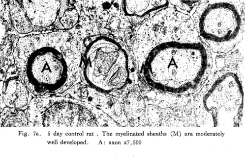
The myelinated sheaths had fewer laminae in the treated rats (Fig. 7b) than in the controls (Fig. 7a).
DISCUSSION
Since HUNTER et al.") reported four clinical cases of mercury compound poisoning and four different groups of animal experiment, there have been many investigations both on human cases and on the laboratory animals. Many of them were done on the adult cases and adult animals. Some of them were carried out in fetal human cases30), 31) and the fetal animal models"),1°>,z4)-zs>,32),33>
While there are some descriptions on the DRG of mercury poisoned adult casesl3) and animal2), 3). 5), 7), 8), 11), 12), 14), 22), 23) , the DRG of fetal poisoned materials has not been completely examined yet. Therefore, the purpose of this communication was accurate investigation of the DRG of the baby rats of which the mother rats were poisoned by MeHg during late gestation days.
HATAI°)•10) described two types of neurons in the DRG. The larger, less compact neuron is stained lightly with aniline dyes ; the smaller, very compact neuron is stained darkly. Although the light microscopic studies showed it is difficult to distinguish the two types of neurons in MeHg treated rats at birth, the differentiation of two cell types was possible in treated 5-day-old animals.
The eccentricity of the nucleus, and the variety of the size and shape were more distinct than the controls. HUNTER et al. 12) (their exp. I, II), HERMAN et al. 11) and JACOBS et al. 14) described the eccentricity of nucleus of neuron in their adult rats, and CARMICHAEL et al.') found the same nuclei in their 13 day experimental rabbits. On the other hand, CAVANAGH and CHEN3) reported the nuclei often remained centrally placed, and CHARLTON7) described there were a few slightly swollen neurons with central or eccentric and mor- phologically normal nuclei. However, neither eccentricity of neuronal nuclei nor amorphous, eosinophilic appearance of the cytoplasm are specific for mercury poisoning.
There are some repo,.•ts of satellite cell changes which consisted of "residual nodules"
of activated satellite cells in the higher dosed rats'), of proliferation"),12), and of enlargement with forming clusters12). These alterations were not observed in this experiment. There were neither satellitosis nor neuronophagia in this experimental series.
The structural change of interdigitation between plasmalemmae of the neurons in
DRG and these of satellite cells is said to be one of the index of maturation35). The
attachment among these two cells in the MeHg poisoned rats remained smooth and complex
than in the control. One of this result indicates that the intrauterine administration of
MeHg induces to inhibit the complexity of these membrane folds. Conversely, CHANG and HARTMANN5 detected the extensive proliferation of the capsule cell processes which plicated arround the nearly neurons. It is not certain at this point whether the difference in the two observations is related to age or to the amount of toxin.
CHANG and HA' _:TMANN6) described that many Schwann cells became hypertrophied and had degenerative materials in their cytoplasms in peripheral nerve sheath. Those materials were not observed yet in this experiment, while in our previous report15)''s) the degenerative materials were detected in cytoplasm of DRG of rats which had been poisoned by MeHg at early intrauterine life.
Main ultrastructural changes were observed in so called "N issl substances". The structure and arrangement of RER were disorganized in the cytoplasm of McHg poisoned neurons. These markedly dilated cisternae of RER lost attached ribosomes in many neurons and the free ribosomes were increased in number in DRG neurons. These findings, however, did not mean ribosomes or polysomes decreased in total number in the cytoplasm of treated neuron. It was also noticed that the initial changes of Nissl substances almost disappeared by 30 days of age. HERMAN et al."' have also described the obvious changes of Nissl substances which consisted of the irregular branching cisternae, the short stacks of RER with focal dilation. They also described the free ribosomes or polysomes were at first preserved, but with time those were disappeared. Other authors also observed the changes of Nissl substances')-') -1),7),12),14). JACOBS et al. 14) noticed a numerous free lying single ribosomes at their later stage. In the aspect of biochemical field, YO ,HIND et al.36 and BRUBAKER et al. 1) have all reported decreased protein synthesis in the central nervous system of adult animals after doses of alkyl mercurials ; KARK et al.") have described the same observation in brains of baby rat which were given MeHg at their uterine days.
CAVANAGH and CHEN4) have demonstrated that methylmercury can reduce the rate of conversion of 14C-glycine to protein in adult rat DRG before histological alterations were seen in the neurons. The alterations of RER and ribosomes, as observed in this report, are an evidence of morphological aspects for the protein synthesis decreasing.
The mitochondria of neuron in MeHg poisoned DRG were scattered in normal distribution but they were sometimes increased in number by 10 days of age, while CHANG and HARTMANN5) described the increase of mitochondria with vacuolarization for mercury intoxication. The Golgi complexes showed no alterations in structure and in number as those of control DRG neurons, while HERMAN et al.") noticed the progressive decrease initially arround the nuclei and finally throughout the whole cytoplasm at later stage of intoxication. JACOBS et al. 14) observed no obvious changes of Golgi complexes except often hyperactive appearance of it in small dark neurons.
By 30 days of age, as mentioned before, the changes of RER were rarely seen,
while the neurofibrils were markedly increased in the poisoned DRG neurons. Similar
changes have been described in the DRG neurons in the rat after resection of brachial
plexus37), and after crushing of the dorsal root fiber of cauda equina27). They suggested
that it was a feature of early recovery in chromatolytic processes. Present study also suport
their suggestion because of the following reasons : 1) other intracytoplasmic organelles, other than RER and neurofibrils, remained still in almost usual appearances throughout the whole, 2)the neurofibrils were less in newborn rats and more in 30-day-old rats than in the controls, and 3)even if the neurons were smaller than the control neurons, the size of these neurons was increased gradually with increase of neurofibrils.
The electron microscopic features of the large clear neuron consists of the sparsely scattered stacks of RER and numerous neurofibrils, whereas the small dark neuron has a diffuse distribution of RER35) . The cells mainly in this study seemed to be a cell group of the clear neuron, because the small dark neuron did not decrease in total number and remained generally normal. JACOBS et al. 14) and CARMICHAEL et al.') have reported that the changes of Nissl substances and all these cellular changes such as the neurofibrillar prolif- eration were more readily seen in the large neurons. The large neuron was said to be somatic afferent neurons and the small one was said to be splanchnic afferent neurons28>.
Therefore as the large clear neuron itself is mainly affected, the suggestion on the function of two cell types is sustained partially and our result is reasonable for the initial sensory disturbance of mercury poisoning.
It is still unknown item whether the degeneration is a primary event in the cell body of neuron") or in the axons) . In present study, there was no particular abnormality in axon of newborn MeHg poisoned rat, but marked changes of organella were detected in the cell body of poisoned DRG neurons at birth. At later days, the myelin sheath itself grew up with the regenerative changes of poisoned neuron. Our study may be one of the aspects on primary cell body injury at least in the ultrastructural level.
ACKNOWLEDGEMENT
The author gratefully acknowledge Professor ISSEI NISHIMORI and Associate Professor NOBUO TSUDA for their valuable advices and encouragements. The author would like to express his gratitude to Professor W. JANN BROWN, Division of Neuropathology, University of California at Los Angeles, for his encouragement.
REFERENCES
1) BRUBAKER, P.E., KLEIN, R., HERMAN, S.P., LUCIER, G.W., ALEXANDER, L.T., LONG, M. D.: Exp. Mol. Pathol. 18:263-280, 1973
2) CARMICHAE'sL, N., CAVANAGH, J. B., RODDA, R. A. : Acta Neuropathol. (Berl.) 32: 115-125, 1975.
3) CAVANAGH, J. B., CHEN, F. C. K. : Acta Neuropathol. (Berl.) 19 : 208-215, 1971.
4) CAVANAGH, J. B., CHEN, F. C. K. : Acta Neuropathol. (Berl.) 19: 216-224, 1971.
5) CHANG, L. W., HARTMANN, H. A. : Acta Neuropathol. (Berl.) 20: 122-138, 1972.
6) CHANG, L. W., HARTMANN, H. A. : Acta Neuropathol. (Berl.) 20: 316-334, 1972.
7) CHARLTON, K. M., : Can. J. Comp. Med. 38 : 75-81, 1974.
8) GARMAN, R. H. , WEISS, B. , EVANS, H. O.: Acta Neuropathol. (Berl.) 32: 61-74,1975.
9) HATAI, S. : J. Comp. Neurol. 11: 1-24, 1901.
10) HATAI, S. : J. Comp. Neurol. 12:107-124, 1902.
11) HERMAN, S. P., KLEIN, R., TALLEY, F. A., KRIGMAN, M. R.: Lab. Invest. 28:
104-118, 1973.
12) HUNTER, D., BOMFORD, R. R., RUSSELL, D. S. : Q. J. Med. 9:193-213, 1940.
13) HUNTER, D., RUSSELL, D. S. : J. Neurol. Neurosurg. Psychiatry 17:235-241, 1954.
14) JACOBS, J. M., CARMICHAEL, N., CAVANAGH, J. B.: Neuropath. Appl. Neurobiol.
1: 1-19, 1975.
15) KARK, P., Mr-,NON, N . , KISHIKAWA, M. : Proc. Int. Assoc. for Scientific Study of Mental Deficiency (1976/77 in press)
16) KISHIKAWA, M., BROWN, W. J., KARK, P., MENON, N. : in press (1976) 17) MATSJMOTO, H., KOYA, G., TAKEUCHI, T. : Trans. Soc. Path. Jap. 54:187-188,
1965 (Japanese)
18) MATSUMOTO, H., KOYA, G., TAKEUCHI, T. : J. Neuropathol. Exp. Neurol. 24 563-574, 1965.
19) MATSUMOTO, H., SUZUKI, A., MORITA, C., NAKAMURA, K., SAEKI, S. : Life Sci. 6 : 2321-2326, 1967.
20) MATSUMOTO, H., ToMIO, N . , KAMEDA, T. : Adv. Neurol. Sci. 13 : 660-669, 1969 (Japanese)
21) MATSUMOTO. H., KAMEDA, T. : Saishin Igaku 25 : 939-950, 1970. (Japanese) 22) MIYAKAWA, T., D::SHIMARU, M., SUMIYOSHI, S., TCRAOKA, A., UDO, N . ,
HATTORI, E., TATETSU, S. : Acta Neuropathol. (Berl.) 15: 45-55, 1970.
23) MIYAKAWA, T., DESHIMARU, M., SUMIYOSHI, S., TERAOKA, A., TATETSU, S.
Acta Neuropathol. (Berl.) 17: 6-13, 1971.
24) MOR:KAWA, N.: Kumamoto Med. J. 14: 87-93, 1961.
25) MURAKAMI, U., KAMEYAMA, Y., KATO, T. : Ann. Rep. Environ. Med. Nagoya Univ. 4:88-99, 1956.
26) MURAKAMI, U. : Adv. Exp. Med. Biol. 27: 301-336, 1972.
27) NATHANIEL, E. J. H., NATHANIEL, D. R. : J. Ultrastruct. Res. 45: 168-182, 1973.
28) PRETO PARVIS, V. : Monit. zool. ital. 63, Suppl. 352-354, 1954.
29) TAKEUCHI, T. , MORIKAWA, N . , MATSUMOTO, H., SHIRAISHI, Y.: Acta Neuropathol.
(Berl.) 2: 40-57, 1962.
30) TAKEUCHI, T., MATSUMOTO, H., KOYA, G. : Trans. Soc. Path. Tap. 52: 144-145, 1963 (Japanese)
31) TAKEUCHI, T., MATSUMOTO, H., KOYA, G.: Adv. Neurol. Sci. 8 : 867-883, 1964.
(Japanese)
32) TATESU, S., TAKAGI, M., MIYAKAWA, T., SUMIYOSHI,, S., DESHIMARU, M.
Psychiat. Neurol. 70: 162, 1968 (Japanese)
33) TATESU, S., TAKAGI, M., MIYAKAWA, T., SUMIYOSHI,S., DESHIMARU, M.: Adv.
Neurol. Sci. 13: 130-134, 1969 (Japanese)
34) TOKUOMI, H. : in Minamata Disease. ed. KUTSUNA, M., pp. 48-76.
Study group of Minamata disease, Kumamoto Univ., Japan, 1966 (Japanese) 35) YAMADORI, T. : Acta Anat. (Nippon) 45: 191-205, 1970
36) YOSHINO, Y., MOZAI, T., NAKAO, K.: J. Neurochem. 13: 1223-1230, 1966.
37) ZELENA, J. : Z. Zellforsch. 117: 191-211,1971.
Fig. la. 30 day control rat. Two types of neuron are obviously seen. The neurons are
uniform in size and in shape with almost centrally located nucleus. x200
Fig. lb. 30 day MeHg poisoned rat. The affected neurons remained moderately abnormal-
ities, which are in shape and in location
of nucleus. The large clear neuron is
smaller than that in control. x200
Fig. 1 Thick sections (lp) of dorsal root ganglia stained with toluidine blue.
Fig. 2a. Newborn control rat. There are several layers (-+) of the satellite cell cytoplasm
(S), which envelops the neuron (C).
The RER (*) are arranged parallel to the
cell membrane or to the nuclear membrane
without any abnormal dilation and bran-
ching. N : nucleus of neuron 41,000
Fig. 2b. Newborn MeHg poisoned rat. There are only few layers (-*) among two neurons
(C). The cisternae of RER (*) are
markedly dilated with many abnormal
branching. X11,000
Fig. 3a. 5 day control rat. The interdigitations (~ ) between the cytoplasmic membrane of neuron (C) and the cytoplasm of satellite cell (S) become
complicated. N nucleus of neuron x11,000
Fig. 3b. 10 day MeHg poisoned rat. The interdigitations (T ) are less prominent. The RER (*) are shortened, have markedly dilated
cisternae and irregular branching.
A : axon, C: cytoplasm of neuron, N : nucleus of neuron x11,000
Fig. 4 10 day MeHg poisoned rat. The markedly dilated cisternae of RER (*) lose attached ribosomes and the free ribosomes are
increased in number in cytoplasm of neuron.
F : neurofibril x25,000
Fig. 5 30 day control rat. The RER, which corresponds to the Nissl substance by light microscopic observation, are well developed
in large clear neuron.
F: neurofibril, n: Nissl substance, N : Nucleus of neuron x7,500