https://doi.org/10.1007/s00383-018-4398-y ORIGINAL ARTICLE
Acquired isolated hypoganglionosis as a distinct entity: results from a nationwide survey
Satoshi Obata
1,2· Koichiro Yoshimaru
1,2· Kosuke Kirino
1,2· Tomoko Izaki
1,2· Satoshi Ieiri
1,3· Atsuyuki Yamataka
1,4· Tsugumichi Koshinaga
1,5· Jun Iwai
1,6· Hitoshi Ikeda
1,7· Hiroshi Matsufuji
1,8· Yoshinao Oda
1,9· Tomoaki Taguchi
1,2Accepted: 18 October 2018 / Published online: 19 November 2018
© Springer-Verlag GmbH Germany, part of Springer Nature 2018
Abstract
Purpose Acquired isolated hypoganglionosis (A-IH) is a late-onset intestinal pseudo-obstruction disorder and shows different pathophysiological findings from congenital isolated hypoganglionosis (C-IH). In this study, we retrospectively examined five cases of A-IH and investigated the features of A-IH.
Methods Five cases of A-IH were extracted from a nationwide retrospective cohort study in 10 years, from which totally 355 cases of Allied Disorders of Hirschsprung’s Disease (ADHD) were collected.
Results Ages of onset were between 13 and 17 years in three cases, and 4 years and 4 months in ones. Initial symptoms were abdominal distension and/or chronic constipation in 4 cases, whereas one exhibited intestinal perforation. Affected lesions varied from case to case, extending various length of intestinal tracts. All cases underwent multiple operations (average: 4.6 times), such as enterostomy, resection of dilated intestines, and/or pull-through. Pathological findings showed the decreased numbers of ganglion cells and degeneration of ganglion cells, whereas the size of the plexus was normal. Currently, all cases were alive and almost all eat regular food without requiring parenteral feeding.
Conclusion A-IH is rare, but distinct entity characterized by different clinical courses and pathological findings from those of C-IH. The outcome is considered to be favorable after a resection of affected intestine.
Keywords Isolated hypoganglionosis · Acquired · Allied disorders · Hirschsprung’s disease · Ganglion cell
* Tomoaki Taguchi
1 Japanese Study Group for Allied Disorders
of Hirschsprung’s Disease, 3-1-1 Maidashi, Higashi-ku, Fukuoka 812-8582, Japan
2 Department of Pediatric Surgery, Reproductive and Developmental Medicine, Faculty of Medical Sciences, Kyushu University, 3-1-1 Maidashi, Higashi-ku, Fukuoka 812-8582, Japan
3 Department of Pediatric Surgery, Field of Developmental Medicine, Graduate School of Medical and Dental Sciences, Kagoshima University, 8-35-1 Sakuragaoka, Kagoshima 890-8520, Japan
4 Department of Pediatric General and Urogenital Surgery, Juntendo University School of Medicine, 2-1-1 Hongo, Bunkyo-ku, Tokyo 113-8421, Japan
5 Department of Pediatric Surgery, Nihon University School of Medicine, 30-1 Ooyaguchi Kami-cho, Itabashi-ku, Tokyo 173-8610, Japan
6 Department of Pediatric Surgery, Chiba Children’s Hospital, 579-1 Heta-cho, Midori-ku, Chiba 266-0007, Japan
7 Department of Pediatric Surgery, Dokkyo Medical University Koshigaya Hospital, 2-1-50 Minami-Koshigaya, Koshigaya, Saitama 343-8555, Japan
8 Department of Pediatric Surgery, St. Luke’s International Hospital, 9-1 Akashi-cho, Chuo-ku, Tokyo 104-8560, Japan
9 Department of Anatomic Pathology, Pathological Sciences, Graduate School of Medical Sciences, Kyushu University, 3-1-1 Maidashi, Higashi-ku, Fukuoka 812-8582, Japan
Introduction
Isolated hypoganglionosis (IH) is proposed to be one of the allied disorders of Hirschsprung’s disease (ADHD), which is rare and resembles Hirschsprung’s disease (HD) and associated with decreased numbers of intestinal gan- glion cells [1]. Historically, the existence of this entity has been questioned [2, 3]. Some researchers have doubted the existence of IH because they have never encountered the disease. We previously reported that IH had two distinct entities, with different clinical characteristics and histolog- ical findings: congenital IH (C-IH) and acquired IH (A-IH) [4]. A-IH, which is characterized by its late-onset, shows the particular pathological findings that are distinct from those of C-IH. These include the degeneration of ganglion cells, a decrease in ganglion cell numbers, and gliosis in Auerbach’s plexus with the preservation of the size of the plexus. These findings strongly suggest that ganglion cells were initially present, and that they has gradually disappeared. In this study, to investigate the incidence, the clinical symptoms and the outcomes of A-IH in Japan, we extracted the cases of pathologically proven A-IH from the data of a nationwide retrospective cohort study of ADHD that was performed in Japan from 2000 to 2009.
Patients and methods
The following diagnostic criteria could be used to define A-IH: (1) the absence of a congenital predisposing factor;
(2) the onset of symptoms (e.g., prolonged constipation,
abdominal distension without vomiting) after the neonatal period; (3) the pathological findings showing the degenera- tion of ganglion cells and decrease in their number; (4) a satisfactory outcome after the resection of the involved intes- tine. In the present nationwide retrospective cohort study, which was supported by the Ministry of Health and Welfare, Japan, preliminary questionnaires were sent to the 161 major institutes of pediatric surgery or gastroenterology (represent- ing the core members of the Japanese Society of Pediatric Surgeons, the Japanese Society of Pediatric Nutrition, Gas- troenterology and Hepatology and the Japanese Study Group of Pediatric Constipation). The questionnaires asked about the number of ADHD cases that were treated from January 2000 to December 2009. Replies were obtained from 157 out of the 161 institutes (98%). A total of 355 ADHD cases were collected [5]. We found 5 cases of A-IH regarding the crite- ria described above. Then, we sent secondary questionnaires that asked about the background characteristics, the clinical and pathological findings, and treatments and outcomes of each case of A-IH. Based on the results of the secondary questionnaire, we collected the clinical and pathological information of 5 cases of A-IH, and investigated the details.
Results
The male to female ratio was 3:2. With the exception of two cases (Case 2: necrotic enterocolitis in neonate, Case 3:
influenza encephalitis in early childhood and mental retarda- tion), none of the patients had a specific past medical his- tory before the onset of symptoms. The onset of symptoms
Table 1 The characteristics and examination results of each case of acquired isolated hypoganglionosis
Case no. 1 2 3 4 5
Sex Female Male Male Male Female
Past medical history None None Influenza encephalitis
Mental retardation None None
Onset of symptoms School-age Infant Early childhood School-age School-age
Primary symptoms Chronic constipation Abdominal distension Vomiting
Intestinal perforation Chronic constipation Abdominal distension Vomiting
Associated malforma-
tions None None None None None
Chromosomal abnor-
malities None None None Not examined Not examined
Family history Prader-Willi syndrome None None None None
Abdominal X-ray
image Abnormal intestinal
dilatation Abnormal intestinal dilatation /air-fluid level
Free air Abnormal intestinal
dilatation Air-fluid level
Contrast enema Megacolon Normal Not examined Megacolon Megacolon
Rectosphincteric
reflex Negative Not examined Positive Positive Unknown
Rectal mucosal biopsy Normal Not examined Not examined Normal Unknown
occurred in infancy (n = 1, case 2), early childhood (n = 1, case 3), and childhood (in school-age children; n = 3, cases 1, 4, and 5). The initial clinical symptoms were chronic con- stipation (n = 2, cases 1 and 4), abdominal distension (n = 1, case 2), and abdominal distension and vomiting (n = 1, case 5); one case showed intestinal perforation and vomiting (case 3) (Table 1). The abdominal X-ray images showed abnormal intestinal dilatation (n = 3, cases 1, 2, and 4), air-fluid level (n = 2, cases 2 and 5), and free air (n = 1, case 3). Contrast enema showed megacolon (n = 3, cases 1, 4, and 5) and nor- mal results (n = 1, case 2); contrast enema was not conducted in case 2 because of the bad condition. Three cases under- went anorectal manometry, which indicated rectosphincteric reflex positivity (n = 2, cases 2 and 3), and negativity (n = 1, case 1). Two patients underwent rectal mucosal biopsy;
the findings were normal in both cases (Table 1). Initially, except for case 3 with immediate surgical intervention due to intestinal perforation, all four cases were managed with
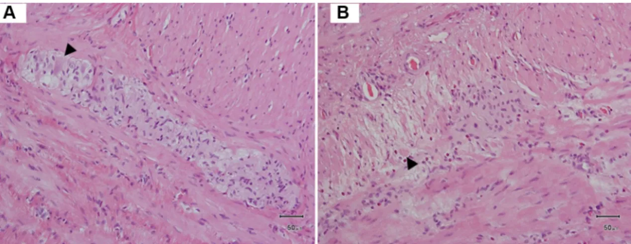
conservative medical treatment; however, their conditions did not improve. Finally, all five patients underwent mul- tiple operations (average: 4.6 operations per case) because multiple intestinal segments were affected. The procedures included creation of enterostomy, resection of the involved segments of the dilated intestine, and/or pull-through because the involved segments spread from the stomach to the rectum (partially or totally) in each case (Table 2). Hematoxilin and eosin (H&E) staining revealed that all five cases had gan- glion cell degeneration and decreased ganglion cell numbers with a normal size Auerbach’s plexus in the resected intes- tine (Fig. 1); however, it should be noted that in 2 of the 5 cases the numbers and sizes of ganglion cells were normal at the first resection. Immunohistochemical staining, which was performed using HuC/D, S-100 and SOX10 antibodies, was effective in identifying the remaining ganglion cells, the preservation of the size of Auerbach’s plexus, and increased glial cell numbers in the plexus (Fig. 2). Currently, all five
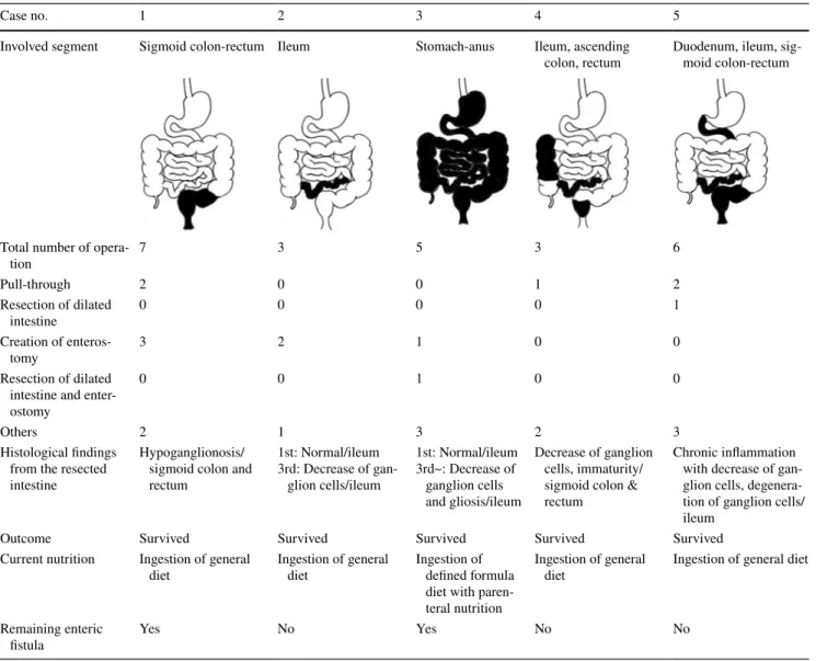
Table 2 The surgical treatments and outcomes of each case
Case no. 1 2 3 4 5
Involved segment Sigmoid colon-rectum Ileum Stomach-anus Ileum, ascending
colon, rectum Duodenum, ileum, sig- moid colon-rectum
Total number of opera-
tion 7 3 5 3 6
Pull-through 2 0 0 1 2
Resection of dilated
intestine 0 0 0 0 1
Creation of enteros-
tomy 3 2 1 0 0
Resection of dilated intestine and enter- ostomy
0 0 1 0 0
Others 2 1 3 2 3
Histological findings from the resected intestine
Hypoganglionosis/
sigmoid colon and rectum
1st: Normal/ileum 3rd: Decrease of gan-
glion cells/ileum
1st: Normal/ileum 3rd~: Decrease of ganglion cells and gliosis/ileum
Decrease of ganglion cells, immaturity/
sigmoid colon &
rectum
Chronic inflammation with decrease of gan- glion cells, degenera- tion of ganglion cells/
ileum
Outcome Survived Survived Survived Survived Survived
Current nutrition Ingestion of general
diet Ingestion of general
diet Ingestion of
defined formula diet with paren- teral nutrition
Ingestion of general
diet Ingestion of general diet
Remaining enteric
fistula Yes No Yes No No
Fig. 1 The typical pathology of acquired hypoganglionosis (Case 5).
a The colon. The plexus shows a mixture of almost normal ganglion cells and gliosis. b The ileum. The ganglion cells have disappeared
and glial cells increased. Arrow-heads indicate degenerated ganglion cells; hematoxylin & eosin staining
Fig. 2 The immunohistochemical findings in a patient with acquired hypoganglionosis (Case 5). a HuC/D immunostaining. b S-100 pro- tein immunostaining. c SOX10 immunostaining. HuC/D immu- nostaining highlights the indistinguishable ganglion cells in the
increased glial cell numbers in the plexus. S-100 protein is pan-neu- ronal marker and shows the size of enteric plexus. Glial cells were positively stained by SOX10
cases are alive, and 2 enteric fistulas remain. All but one of the patients are capable of ingesting a general diet without the need for parenteral feeding (Table 2).
Discussion
Isolated hypoganglionosis (IH) remains a diagnostic chal- lenge [2, 3] and preoperative correct diagnosis of IH is dif- ficult because the clinical symptoms of IH resemble those of Hirschsprung’s disease (HD). Both patients with HD and those with IH present delayed passage of meconium, abdominal distension, and vomiting. Hypoganglionosis as an isolated disease represent only 5% of neuronal intestinal malformations [6]. Meanwhile, we previously reported IH had two entities, with different clinical characteristics and histological findings: congenital IH (C-IH) and acquired IH (A-IH) [4]. The present study revealed that incidence of A-IH was very low (5 cases from a 10-year nationwide survey), in comparison with the other categories of ADHD [5]. The definitive diagnosis of A-IH requires pathological examination of resected bowel [7]; however, if the symp- toms are not so severe, physicians may be hesitant to per- form surgical resection. Thus, it is possible that potential patients with A-IH are present among patients with chronic constipation.
The onset of the symptoms of A-IH patients in the present study occurred at various ages in childhood after the neona- tal period (from infants to school children); the main symp- tom was constipation, which was prolonged and gradually progressive. In contrast, the onset of symptoms in patients with C-IH is in the early neonatal period, and the symptoms of C-IH include severe abdominal distension and intestinal obstruction after ileostomy in the neonatal period [5, 8].
Thus, the onset and symptoms of A-IH seem different from those of A-IH.
The intestinal segments that were involved in the cases with A-IH in the present study were wide-ranging. In line with the observations of a previous report [4], the lesions were seemed like “skip segments”, spreading from the stomach to rectum (Table 2). A previous systematic review showed that the involved intestinal segments of C-IH were localized, from total intestinal to rectosigmoidal (especially colonic) [6]. In contrast, Watanabe et al. reported the opera- tive findings of 90 cases of C-IH and noted the presence of dilated intestine with caliber changes starting at the small intestine in 63 cases [8], indicating that the involved location spreads sub-totally from the small intestine to the rectum in patients with C-IH.
In the description regarding hypogenetic-type C-IH reviewed by Friedmacher et al. [7], the number of gan- glion cells decreases and the size of plexus is also small.
In the present study, all of the patients with A-IH showed
ganglion cell degeneration and decreased ganglion cell num- bers, while the size of Auerbach’s plexus was preserved.
These findings are similar to the previous report in which the size of Auerbach’s plexus was normal, whereas ganglion cell degeneration, decreased ganglion cell numbers, and the proliferation of glial cells were prominent [4]. The report also noted that these findings were evidence that ganglion cells were initially present, and that they gradually disap- peared. In the present study, in 2 out of the 5 cases the size and the number of ganglion cells were normal at the first resection, and then the ganglion cell numbers decreased.
Therefore, it is possible that some factors may affect the ganglion cells [4]. Chagas disease, an infection caused by the protozoan parasite Trypanosoma cruzi, is known to cause the degeneration and decrease of ganglion cells. Previous reports on colonic involvement in Chagas disease patients have described that particular pathological features such as the degeneration of the intrinsic myenteric neurons and a decrease in their numbers, as well as reduced numbers of nitric oxide-containing myenteric neurons, deficiency of the interstitial cells of Cajal, T lymphocyte-induced ganglion cell damage, increased fibrosis, and increased numbers of mast cells [9]. The results of the previous reports [4] and the present study suggest that factors which are causative of A-IH, such as severe chronic constipation, chronic enterocol- itis due to chronic constipation, peritonitis due to intestinal perforation, or infection, might exist, in other words, the degeneration of ganglion cells and decrease in their number might occur secondary to prolonged constipation, systemic shock or intestinal ischemia on the involved intestines.
Immunohistochemical examinations are useful for the definitive pathological diagnosis of ADHD [10]. S-100 pro- tein is pan-neuronal marker that is not found in ganglion cell, which also shows the pattern of distribution and the size of enteric plexus, SOX10 is positive in the nuclei of the enteric glia but negative in the nuclei of ganglion cells [11]. HuC/D protein is routinely used as a neuronal marker in both the central and the peripheral nervous system. HuC/D staining offers a practical advantage, making qualification of individ- ual neurons easy [12]. In the present study, although H&E staining plays an important role in the identification of gan- glion cells and glial cells in the plexus, the use of HuC/D, S-100 protein and SOX10 were also helpful for identifying the numbers of remaining ganglion cells, the preservation of the size of Auerbach’s plexus, and increased glial cell numbers in the plexus of A-IH.
Although the A-IH patients required multiple operations
which were similar to those required by C-IH patients, their
postoperative outcomes were far better. In C-IH, dysmotility
does not improve over time, re-enterostomy was required in
most cases, and the combination of continuous parenteral
nutrition and partial enteral nutrition was necessary for the
patients to survive [4]. On the other hand, while the A-IH
cases in the present study required multiple operations, sat- isfactory outcomes were eventually obtained, with the oral intake of a general diet without the need for parenteral nutri- tion in all but one of the patients.
In conclusion, the findings of the present study prove that A-IH is a rare, but distinct entity. The pathological characteristics include a decrease in ganglion cell numbers and gliosis, and the preservation of the size of Auerbach’s plexus. The onset of symptoms occurs in patients of various ages. The clinical outcome after the resection of the involved intestine, which usually shows dilatation, is considered to be favorable. Patients with A-IH may require multiple opera- tions due to the varying extent of the associated lesions.
Acknowledgements This study was supported by a grant from The Ministry of Health, Labour and Welfare of Japan (Health and Labour Sciences Research Grants for Research on Intractable Diseases (H23- 042, H24-037, and H26-045)). The authors thank all members of The Japanese Society of Pediatric Surgeons, The Japanese Society of Pedi- atric Nutrition, Gastroenterology, and Hepatology, and The Japanese Study Group of Pediatric Constipation. The authors thank Dr. Brian Quinn for editing the manuscript.
Compliance with ethical standards
Ethical approval All procedures performed in studies involving human participants were in accordance with the ethical standards of the insti- tutional and/or national research committee and with the 1964 Helsinki declaration and its later amendments or comparable ethical standards.
And, for this type of study formal consent was not required. This ret- rospective study was also approved by the ethics committee for clinical research of Kyushu University Hospital (No. 28–155).
References
1. Tiffin ME, Chandler LR, Faber HK (1940) Localized absence of the ganglion cells of the myenteric plexus in congenital megaco- lon. Am J Dis Child 59(5):1071–1082
2. Martucciello G, Pini Prato A, Puri P, Holschneider AM, Meier- Ruge W, Jasonni V, Tovar JA, Grosfeld JL (2005) Controversies concerning diagnostic guidelines for anomalies of the enteric nervous system: a report from the fourth International Symposium
on Hirschsprung’s disease and related neurocristopathies. J Pedi- atr Surg 40 (10):1527–1531. https ://doi.org/10.1016/j.jpeds urg.2005.07.053
3. Schäppi MG, Staiano A, Milla PJ, Smith VV, Dias JA, Heuschkel R, Husby S, Mearin ML, Papadopoulou A, Ruemmele FM, Van- denplas Y, Koletzko S (2013) A practical guide for the diagnosis of primary enteric nervous system disorders. J Pediatr Gastro- enterol Nutr 57(5):677–686. https ://doi.org/10.1097/MPG.0b013 e3182 a8bb5 0
4. Taguchi T, Masumoto K, Ieiri S, Nakatsuji T, Akiyoshi J (2006) New classification of hypoganglionosis: congenital and acquired hypoganglionosis. J Pediatr Surg 41(12):2046–2051. https ://doi.
org/10.1016/j.jpeds urg.2006.08.004
5. Taguchi T, Ieiri S, Miyoshi K, Kohashi K, Oda Y, Kubota A, Watanabe Y, Matsufuji H, Fukuzawa M, Tomomasa T (2015) The incidence and outcome of allied disorders of Hirschsprung’s disease in Japan: results from a nationwide survey. Asian J Surg.
https ://doi.org/10.1016/j.asjsu r.2015.04.004
6. Dingemann J, Puri P (2010) Isolated hypoganglionosis: systematic review of a rare intestinal innervation defect. Pediatr Surg Int 26(11):1111–1115. https ://doi.org/10.1007/s0038 3-010-2693-3 7. Friedmacher F, Puri P (2013) Classification and diagnostic criteria
of variants of Hirschsprung’s disease. Pediatr Surg Int 29(9):855–
872. https ://doi.org/10.1007/s0038 3-013-3351-3
8. Watanabe Y, Kanamori Y, Uchida K, Taguchi T (2013) Isolated hypoganglionosis: results of a nationwide survey in Japan. Pedi- atr Surg Int 29(11):1127–1130. https ://doi.org/10.1007/s0038 3-013-3378-5
9. Iantorno G, Bassotti G, Kogan Z, Lumi CM, Cabanne AM, Fisogni S, Varrica LM, Bilder CR, Munoz JP, Liserre B, Morelli A, Villanacci V (2007) The enteric nervous system in chagasic and idiopathic megacolon. Am J Surg Pathol 31(3):460–468. https ://doi.org/10.1097/01.pas.00002 13371 .79300 .a8
10. Park SH, Min H, Chi JG, Park KW, Yang HR, Seo JK (2005) Immunohistochemical studies of pediatric intestinal pseudo- obstruction: bcl2, a valuable biomarker to detect immature enteric ganglion cells. Am J Surg Pathol 29(8):1017–1024
11. Walters LC, Cantrell VA, Weller KP, Mosher JT, Southard-Smith EM (2010) Genetic background impacts developmental poten- tial of enteric neural crest-derived progenitors in the Sox10Dom model of Hirschsprung disease. Hum Mol Genet 19(22):4353–
4372. https ://doi.org/10.1093/hmg/ddq35 7
12. Desmet AS, Cirillo C, Vanden Berghe P (2014) Distinct subcel- lular localization of the neuronal marker HuC/D reveals hypoxia- induced damage in enteric neurons. Neurogastroenterol Motil 26(8):1131–1143. https ://doi.org/10.1111/nmo.12371