Compositional Analysis of the Oligosaccharide Units of Xyloglucans Isolated from the Cell Walls
of Fruit Vegetables
.3::~**~WJJmmip i? 7tlJl ~ t:";f- V 0 ~·)V 7J :/ (J)
;;d- ~ =tM $ {ll (J) tJlJJX 7t fJT
Yoji KATO*, Teruko KONISHI* t, and Yasushi MITSUISHI* *
:JJoJ1i
~m*· IJ\~ !1~l=F*t. ~EY!:**
ABSTRACT
The oligosaccharide units of xyloglucans from some fruit vegetables were compara- tively analyzed by enzymatic digestion followed by anion-exchange chromatography with pulsed amperometric detection. The enzymes used were a xyloglucan-specific endo-1, 4-
P - D- glucanase (xyloglucanase) from Penicillium sp. M 451 and an isoprimeverose- producing oligoxyloglucan hydrolase from Eupenicillium sp. M9. The oligosaccharide units of the polysaccharides were XXXG, XXLG, XLXG, XXFG, XLLG, and XLFG [where each (1-4)-p-linked D-glucosyl residue in the backbone is given a one-letter code accord- ing to its substituents: G, p-D-G1c; X, a- D-Xyl-(l-6)-P-D-G1c; L, p-D-Gal-(l-2)-a- D- Xyl-(l-6)-P-D-G1c; F, a-
L-Fuc-(l-2)-p-D-Gal-(l-2)-a- D-Xyl-(l-6)-P-D-G1c] in an approximate molar ratio of 36 : 4 : 6 : 31 : 1 : 22 for cucumber, of 37 : 8 : 5 : 30 : 1 : 19 for mung bean sprouts, and of 35 : 4 : 7 : 25 : 4 : 25 for pumpkin.
*
5L-mtk~~1f~{ffi*i&~f4~~Laboratory of Food Science, Department of Home Economics, Faculty of Education, Hirosaki University, (1 Bunkyo-cho, Hirosaki 036-8560, Japan)
**I~~~~~~I~I~~~~~M
National Institute of Bioscience and Human Technology, AIST, (1-1 Higashi, Tsukuba 305- 0046, Japan)
t ffl.:tE
Jj(1'fftk~*~f4~~~MPresent address: Wood Research Institute, Kyoto University, (Gokasho, Uji, Kyoto 611-0011, Japan)
Abbreviations: UA, uronic acid; Rha, rhamnose; Fuc, fucose; Ara, arabinose; Xyl, xylose; Man,
mannose; G1c, glucose; Gal, galactose; IP
[a-0 - Xyl-(1-6)- 0 -G1c] , isoprimeverose; IPase,
isoprimeverose-producing oligoxyloglucan hydrolase; HPAEC, high-performance anion-exchange
chromatography; PAD, pulsed amperometric detection. FG, LG, LXG, LFG, LLG, XXXG, XXFG,
XLFG, XLXG, XXLG, and XLLG are the abbreviated names for xyloglucan oligosaccharides
according to the nomenclature of Fry et
at.[reLl) Each (l-4)-p-linked O-glucosyl residue in
the backbone is given a one-letter code according to its substituents: G,
j3-o-G1c; X, a-O- Xyl-(1
-6)-j3-O-G1c; L, j3-0-Gal-(1-2)-a-0- Xyl-(1-6)-j3-0-G1c; F, a-
L-Fuc-(1-2)-j3-0-Gal-(1-2)-a-
0-Xyl-(1-6)-j3-0-G1c] .
Key words: xyloglucan; xyloglucan oligosaccharide; fruit vegetables; cell walls
INTRODUCTION
Xyloglucan is the predominant hemicellulosic polysaccharide of the primary cell walls of dicotyledonous plants, typically forming 20 to 25% of dry mass of the cell wall.
2)A marked characteristic of the xyloglucan is that the polysaccharide consist of a linear backbone chain of 1, 4-linked p-D-G1c residues and side chains of single
a-D-Xyl residues, a-L-Fuc-(l
~2)-p-D-Gal-(l~2)-a-D-Xyl residues, p-D-Gal-(l
~2)-a-D-Xyl residues, and others that are attached to the 0-6-positions of the backbone chain. Treatment of xyloglucan with endo-p-1, 4-glucanase generates mainly oligosaccharide subunits (XXXG, XLXG, XXLG, XLLG, XXFG, XLFG, and so on) of the xyloglucan polymer.
3)Xyloglucan polysaccharides and some oligosaccharides playa role in the regulation of plant growth.
H )Furthermore, xyloglucan oligo- and polysaccharides were demonstrated to have some physiological effects on animals.
7-9)
From this point of view, the compositional analysis of oligosaccharide units in xyloglucan polymer is very important. In the previous work, we characterized the oligosaccharide units of xyloglucans isolated from the cell walls of leaf and root vegetables.
lO)The present work was conducted to elucidate the composition of oligosaccharide units of xyloglucans isolated from the cell walls of commercially available fruit vegetables.
MATERIALS AND METHODS
Cell wall polysaccharides - - The cell wall polysaccharides of cucumber, mung bean sprouts and pumpkin were fractionated respectively into four fractions, PS (pectic sub- stance), HC (hemicellulose)-I, HC-II, and CL (cellulose) fractions, by using successive extraction with 0.5% ammonium oxalate, 4% KOH, and 24% KOH, and subsequent dialysis of the individual extracts as described in previous papers.ll) The HC-II fractions (24% KOH extracts)ll) were used for the present work.
Resolution of HC-II fractions - - Each HC-II fraction (l8.0mg for cucumber, 21.2mg
for mung bean sprouts, and 14.1 mg for pumpkin) was dissolved in 1 ml of 20 m M N a-acetate
buffer (pH 5.0), then centrifuged to remove insoluble material. The supernatant was applied
to a column (1.8 x 8cm) of DEAE-Sephadex A-25 equilibrated with 20 m M Na-acetate
buffer (pH5.0) and eluted stepwise with 45ml of the same buffer, 63ml of 1.0M NaCl in the
same buffer, and 45ml of 0.5M NaOH. Fractions of 3.0ml each were collected and· assayed
for carbohydrate by the phenol-H
2S0
4method.
12lTubes 4-10 were combined, dialyzed
against distilled water, and freeze-dried to give HC-II-1 (xyloglucan) fraction. Yields of
HC-II-1 fractions of cucumber, mung bean sprouts, and pumpkin were 9.5mg, 12.1mg, and
5.5 mg, respectively.
Sugar composition analysis of HC - II -] fractions - - HC-II -1 fractions (about 0.1 mg) were hydrolyzed with 2 M trifluoroacetic acid (TFA) for 4 h at 100°C. In each case the hydrolyzate was evaporated to dryness. Alditol trifluoroacetates derived from sugars were analyzed by gas liquid chromatography on a column (0.4 x 200 em) packed with 1.5% QF -Ion Chromosorb W at 140°c.
13)Dronic acid was determined by the carbazole-H
2S0
4method.
14)Iodine staining of HC-II-] fractions - - An aqueous solution of HC-II-l (0.5ml, containing about 0.2mg) was combined with 0.5ml of a solution containing iodine (0.5%, w/
v) and potassium iodide (1%, w/v). A 2.5ml solution of sodium sulfate (20%, w/v) was then added under shaking. After 1 h in the dark absorption of the reaction mixture at 450-800
d . h . bl k
15)nm was rea map otometer agamst a an.
Methylation analysis of HC-II -] fractions - - Each sample (about 2mg) in dimethylsul- foxide (0.5ml) was methylated with methylsulfinyl carbanion (0.5ml) and methyliodide (0.5 ml) by the method of Hakomori.
16)After methylating, the reaction mixture was dialyzed against tap water, and the non-dialyzable materials were concentrated to dryness. The methylated HC- II -1 was hydrolyzed with 2 M TFA at 120°C for 1 h, the acid removed by evaporation. The methylated sugars were converted into their corresponding alditol acetates
17)and then analyzed by gas liquid chromatography in a glass capillary column (15 m x 0.32 mm) of DB-225. The column oven temperature was raised linearly from 140 to 200°C at a rate of 2°C/min.
Enzymatic hydrolysis of HC- II -] fractions - - A xyloglucan-specific endo-1,4-P- D-glucanase (xyloglucanase) from Penicillium sp. M451
18)and an isoprimeverose-produc- ing oligoxyloglucan hydrolase (IPase) from Eupenicillium sp. M9
19) ,purified respectively to the electrophoretically pure state, were used.
Each HC-II-l fraction (1mg/mI20mM Na-acetate buffer, pH5.5) was hydrolyzed at 40°C for 24 h with Penicillium sp. M451 xyloglucanase. A portion of xyloglucanase-treated xyloglucan (0.5mg/0.5ml 20mM Na-acetate buffer, pH5.5) were hydrolyzed at 40°C for 24 h with Eupenicillium sp. M9 IPase.
High-performance anion exchange chromatography (HPAEC) with pulsed amperometn'c
detection (PAD) - - HPAEC-PAD analyses of oligosaccharides were performed on a
Dionexion chromatography system DX-300 (Dionex gradient pump, Dionex pulsed electro
-chemical detector with a gold working electrode, and an Ag/AgCI reference electrode)
interfaced with an AI -450 work station as described previously.20) Separations were
performed at 20°C on a column (4 x 250 mm) of Dionex CarboPac PA 1 anion exchange resin
with a CarboPack guard column, using a flow rate of 1 ml/min. Oligosaccharides were
eluted with the following NaOAc gradient profile in 100mM NaOH, 0-30min, 0-50mM; 30
-50 min, 50 mM; 50-60 min, 50-500 m M.
RESULTS AND DISCUSSION
Isolation and characterization of xyloglucans from cell walls of cucumber, mung bean sprouts, and pumpkin - - The cell wall polysaccharides of cucumber, mung bean sprouts, and pumpkin were fractionated respectively into four fractions, PS, HC-I, HC-II, and CL frctions, by using successive extraction with 0.5% ammonium oxalate, 4% KOH, and 24%
KOH, and subsequent dialysis of the individual extracts. The HC-II (24% KOH extracts) fractions consisted of VA, Rha, Fuc, Ara, Xyl, Man, G1c, and Gal in the molar ratio of 0.0 : 0.2 : 3.1 : 0.0 : 48.4 : 3.7 : 32.8 : 11.8 for cucumber, of 0.0 : 0.4 : 3.3 : 6.9 : 43.3 : 0.8 : 32.4 : 12.9 for mung bean sprouts, and of 0.0 : 1. 5 : 1. 6 : 3.8 : 48.0 : 1. 0 : 23. 7 : 20.6 as described in a previous papaer
ll) .When HC- II fractions from cucumber, mung bean sprouts, and pumpkin were individu- ally subjected to DEAE-Sephadex A-25 (acetate form) chromatography, they gave three subfractions, HC-II -1 (unbound fraction), -2 (fraction eluted with 1. 0 M N aCl) and -3 (fraction eluted with 0.5M NaOH). A neutral sugar composition analysis showed that HC - II -1 fractions consisted of Rha, Fuc, Ara, Xyl, Man, G1c, and Gal in the molar ratio of 0.0: 5.7: 0.6: 34.0: 10.6: 38.1: 10.9 for cucumber, of 0.0: 5.0: 1.7: 39.4 : trace:
43.9 : 10.0 for mung bean sprouts, and of 0.0 : 4.1 : 0.5 : 31. 5 : trace : 48.0 : 16.0 for pumpkin. Arabinose and mannose, were presumed to have arisen from contaminating pectic substances (arabinan and/or arabinogalactan) and mannanlike polysaccharides, respectively, which are commonly found in dicots. When the absorption spectra of the iodine complexes of HC-II-1 fractions were measured in the range of 450 to 800 nm, all HC -11-1 fractions showed an absorption peak at about 640nm, indicating that the xyloglucan was present in them.
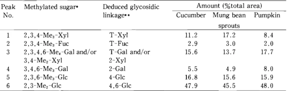
15)Each HC-II-1 fraction was methylated and hydrolyzed, and the sugar linkage composi- tion was analyzed by gas liquid chromatography of the resulting alditol acetates. 2,3,4- Tri-O-methyl-Xyl, 2,3,4-tri-0-methyl-Fuc, 2,3,4,6-tetra-0-methyl-Gal (and/or 3,4- di-O-methyl-Xyl), 3,4,6-tri-0-methyl-Gal, 2, 3, 6-tri-0-methyl-G1c, and 2,3-di-0- methyl-G1c were detected as major structural units in each methylated HC-II-1 fraction (Table 1), which suggests the possibility that each xyloglucan had a linear chain of fJ-l, 4 -D-G1c residues in the backbone, about 75% of which were branched at the 0-6 positions, with single
a-D-Xyl, fJ-D-Gal-(l
~2)-a-D-Xyl, and
a-L-Fuc-(l
~2)-fJ-D-Gal-(l~2)-a-DXyl residues. From these preliminary experimental results, the major polysaccharide in the
respective HC-II-1 fraction was found to be a xyloglucan.
Table
1.Sugar-linkage composition of the HC-II-1 fractions obtained from the cell walls of cucum·
ber, mung bean sprouts, and pumpkin.
Peak Methylated sugaf* Deduced glycosidic Amount (%total area)
No. linkage** Cucumber Mung bean Pumpkin
sprouts
1 2,3, 4-Me3-Xyl T-Xyl 11.2 17.2 8.4
2 2,3,4-Me3-Fuc T-Fuc 2.9 3.0 2.0
3 2,3,4 ,6-Me
4-Gal and/or T -Gal and/or 15.6 13.7 17.7
3,4-Mez-Xyl 2-Xyl
4 3,4,6-Me3 -Gal 2-Gal 5.5 4.9 8.0
5 2,3,6- Me3-Glc 4-Glc 16.8 15.6 15.9
6 2,3- Mez-Glc 4,6-Glc 47.9 45.5 48.0
* 2,3, 4-Me3-Xyl = 2,3, 4-tri-O-methyl-1, 4-di-O-acetyl-xylitol, etc.
** The numerical prefixes represent the carbon atoms involved in glycosidic linkages in the original polysaccharides. Prefix T indicates sugars linked through C-1 only.
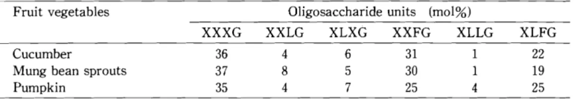
Comparative analysis of the oligosaccharide units of xyloglucans from cucumber, mung bean sprouts, and pumpkin - - Each HC- II -1 fraction (xyloglucan) was hydrolyzed with Penicillium sp. M451 xyloglucanase. A portion of xyloglucanase-treated xyloglucan was subjected to HPAEC- PAD. The residual xyloglucanase-treated xyloglucan were hydrol- yzed with Eupenicillium sp. M9 IPase, and the resultant reaction mixture was subjected to HPAEC- PAD analysis. Figure 1 shows the case of cucumber xyloglucan. Peaks 1,2,3,4, and 5 found in Fig.l-A were identified to be XXXG, XXFG, XLXG and/or XLFG, XXLG, and XLLG. The approximate ratio of XXXG, XXFG, XLXG and/or XLFG, XXLG, and XLLG are 35.5 : 31.4 : 28. 7 : 3.5 : 0.9, which were calculated from the peak area. Figure 1- B shows peaks 1 to 7 corresponding to Glc, IP, FG, LG, LXG, LFG, and LLG. It is well known that the IPase digestion of XXXG, XXLG, XLXG, XXFG, XLLG, and XLFG results in the production of IP and Glc (in the molar ratio of 3 : 1), IP and LG (2 : 1), IP and LXG (1 : 1), IP and FG (2 : 1), IP and LLG (1 : 1), and IP and LFG (l : 1).23) The oligosaccharides found in Fig.l- B are certainly derived from xyloglucan oligosaccharides (XXXG"'-'XLLG) found in Fig.l-A. LXG and LFG, which are characteristic IPase-digestion products of XLXG and XLFG, are found to be present in a ratio of 22 : 78. Cucumber xyloglucan was shown to be made up from XXXG:XXLG:XLXG:XXFG:XLLG:XLFG = 36 : 4 : 6 : 31 : 1 : 22.
In the same manner, the molar ratio of oligosaccharide units of other xyloglucans was calculated as shown in Table 2. The ratios of the six oligosaccharide units are slightly different among cucumber, mung bean sprouts, and pumpkin. However, the xyloglucans of cell walls of these vegetables are considered to be similar to each other in basic structure.
Tamarind seed xyloglucan has been shown to consist of four repeating oligosaccharide
units (XXXG:XLXG:XXLG:XLLG= 13 : 9 : 28 : 50, mol%).24) The presence of XLLG in
fucose-containing xyloglucans has not been reported. Most recently, we have reported that
cabbage, Chinese cabbage, spinach, chingentsuai, lettuce, turnip, Japanese radish, edible
burdock, carrot, and East Indian lotus xyloglucans are composed of XXXG, XXLG, XLXG,
XXFG, XLLG, and XLFG in 35 : 6 : 5 : 22 : 3 : 29, 35 : 9 : 5 : 24 : 2 : 25, 38 : 6 : 5 : 22 : 5 : 24, 35: 14 : 6 : 19 : 4 : 22, 33: 7 : 4 : 31 : 3 : 22, 35: 3 : 8 : 17 : 4 : 33, 34: 2 : 10 : 13 : 3 : 38, 34: 3 : 4 : 27 : 4 : 28, 33: 7 : 4 : 29 : 1 : 26, and 41 : 2 : 5 : 37 : 2 : 13, respec- tively.lOl Thus most fucose-containing xyloglucans may have essentially a small quantity of XLLG, XLXG, and XXLG besides XXXG, XXFG, and XLFG as major structural oligosaccharide units.
Fig.
1.HPAEC of xyloglucanase-hydrolyzate of cucumber xyloglucan (A) and of IPase-hydrolyzate of xyloglucanase-treated cucumber xyloglucan (B).
The retention times of peaks 1 to 5 of xyloglucanase-hydrolyzate of cucumber xyloglucan and of peaks 1 to 7 of IPase-hydrolyzate of xyloglucanase-treated cucumber xyloglucan were compared with the retention times of standard xyloglucan oligosaccharides.13.19-w The arrows in the figures indicate the elution positions of standard sugars, G1c. IP, FG (a), LG (b), LXG (c), LFG (d), LLG (e), XXXG (f), XXFG (g), XLFG and XLXG (h), XXLG
(i)and XLLG
0).0.04
A
f g h i j
~~ ~ ~ J
0.03
1
uC 0.02
2
0.01
5
0.000 10 20 30 40 50 60
Minutes
0.4
Glc IP a b cd e B
t t ~ ~ ~+ t
0.3
2
uC 0.2
O. 1
1 3
7
0.010 20 30 40 50 60
Minutes
Table 2. Structural oligosaccharide units of xyloglucans obtained from the cell walls of cucumber, mung bean sprouts, and pumpkin.
Fruit vegetables Oligosaccharide units (mol%)
XXXG XXLG XLXG XXFG XLLG XLFG
Cucumber 36 4 6 31 1 22
Mung bean sprouts 37 8 5 30 1 19
Pumpkin 35 4 7 25 4 25
REFERENCES
1) Fry, S. c., York, W. S., Albersheim, P., Darvill, A., Hayashi, T., Joseleau, 1. P., Kato, Y., Lorences, E. P., Maclachlan, G. A., McNeil, M., Mort, A. J., Reid, 1. S. G., Seitz, H. U., Selvendran, R R, Voragen, A. G. J., and White, A. R: An unambiguous nomenclature for xyloglucan-derived oligosaccharides. Physiol. Plant., 89, 1-3 (1993).
2) Hayashi, T.: Xyloglucans in the primary cell wall. Annu. Rev. Plant Physiol. Mol. Biol., 40, 139-168 (1989).
3) Kato, Y. and Matsuda, K: Examination of the fine structures of xyloglucans using endo-(1
~4)-