T h r e e D i m e n s i o n a l C e l l C u l t u r e
U s i n g N a n o p i l l a r P l a t e
R y o s u k e T a k a h a s h i
審査終了 •1 3.12.11
W 夫
- ¥ 1T h is th esis is b ased on the fo llo w in g publication s.
R. T akahashi, H . Sonoda, Y . T abata , A . H isada.
“Form ation o f h epatocyte spheroids w ith structural polarity and functional b ile canaliculi u sin g nanopillar sh eets.”
T issu e E ng. Part A ., 16:1983 (2 0 1 0 ).
d o i : 10.108 9 /ten .tea .2 00 9 .0 6 62 R. T akahashi, H . S on od a , A . H isada.
“H igher E xp ression s o f C ytochrom e P 4 5 0 , U D P -G lucuronosyltransferase, and Transporter G enes in N anopillar-C ultured Rat H epatocyte S p heroid s”
Journal o f D rug M etab olism and T o x ico lo g y , 4: article num ber 146 (p p a g es) (2 0 1 3) doi: 1 0 .4 1 7 2 /2 1 5 7 -7 6 0 9 .1 0 0 0 1 4 6
R. T akahashi, Y . Z hou, Y . H origuchi, H . S h ik u , H. Sonoda, N , Itabashi , J. Y am am oto, T_ Saito, T_ M atsu e , A . H isada
“N o n in v a siv ely m easuring respiratory activ ity o f rat prim ary h ep atocyte spheroids b y scann in g electroch em ical m icro sco p y ”
Journal o f B io scien ce and B io en gin eerin g, in p ress (2 0 1 3)
In the course o f drug d evelop m en t today, w h o le anim als are u sed to evaluate drug m etab olism and to x icity in p reclin ical stages. H ow ever,stud ies u sin g anim als are n o t alw ays su ccessfu l in predicting th e m etab olic fates o f drug candidates in the hum an bod y. In recent years, o n ly a sm all fraction (-1 1 % ) o f drug candidates that w ere selected for clin ical trials has reached m arkets. It is h op ed that alternative assay m o d els u sin g hum an cells in v itr o w ill b e better able to predict in v iv o pharm acokinetics and h elp reduce this h ig h attrition rate. C ells in such m od els m u st m aintain their differentiated fu nctions and v ia b ility , and, h e n ce,cell culture m eth ods that can fu lfill th is requirem ent are b ein g sought. T h is stu d y tested and sh o w ed th e utility o f n an o-scaled pillars (nanopillars) in culturing h ep ato cytes,cells o f the prim ary organ o f drug m etab olism ,the liver. Prim ary h ep atocytes cultured in nanopillar plates, ce ll culture plates w ith nanopillars in w ells, form ed th ree-d im ension al aggregates, or spheroids, w ith a structural polarity and fu nctional b ile canaliculi. T h ey w ere m ore sim ilar to fresh ly isolated h ep atocytes than th o se cultured in m on olayer w ith resp ect to g en e exp ression . T h is study also sh o w ed that scann in g electroch em ical m icroscop y can n o n -in v a siv ely evalu ate the respiratory activ ity o f the cultured cells and thus has a potential in validatin g the quality o f cultured c e lls and in m easuring th e resp on ses o f the cells to drugs.
H ep atocytes in the liver h ave a cu boidal shape w ith tw o or three basal surfaces
fa cin g the sin u soid and adjacent cells form b ile canaliculi. Prim ary hepatocytes cultured
in m on olayer lo se their structural and fu nctional characteristics and are no lon ger
suitable as a cell m od el o f the liver. In contrast,hepatocytes ind uced to form spheroids
m ain tain th e characteristics and h a v e p rom ise for drug screen ing. I en visaged that
nanopillars w o u ld b e a suitable substratum for form ation o f h ep atocyte spheroids,as
H eL a cells had b een sh o w n to w ea k ly adhere to the substratum and to take a round
sh ap e,w h en cultured in plates w ith nanopillars. W hen rat h ep atocytes w ere cultured on
found to b e optim al in that the num ber o f obtained sph eroids w ith diam eter o f 5 0 -1 0 0 jam in w h ich cells located centrally is not necrotic, starting from the sam e num ber o f cells, w a s the largest. P lates pre-coated w ith typ e I co lla g en at 100 n g/m L yield ed better results than un coated plates or th ose coated at 10 (ig/m L . T h e spheroids resem b led h ep atocytes in the liver, w h en im m un e-stain ed for actin and E -cadherin and ob served under a flu orescen ce m icroscop e. T ran sm ission electron m icro sco p y sh ow ed that th e spheroids had b ile can alicu li, so m e w ith w ell-d ev elo p ed m icrovilli. T h e w ell-d ev elo p ed m icrov illi had not b een ob served for h ep atocytes grow n in san d w ich culture sy stem s , w h ich h ave b een u sed to evalu ate h ep atocyte fu nctions. T h e fu n ction ality o f the b ile can alicu li such as biliary excretion w as confirm ed m icro sco p ica lly w ith C D F D A in th e spheroid. T h ese results estab lish ed h isto log ica l and functional sim ilarity o f the spheroid in nanopillar plates to the liver.
Sem i-q uan titative real-tim e P C R sh ow ed that exp ression o f A b cc2 (A T P -b in d in g cassette transporter, sup erfam ily C ,m em b er 2 ) , album in, and C y p 3 a 2 3 /3 a l (cytoch rom e P 4 5 0 -3 a 2 3 /3 a l) g en es,w h ich are k n ow n to b e h ig h ly exp ressed in the liver,w as higher in the spheroids than in h ep atocytes cultured in m onolayer. In order to co m p reh en sively exam in e g en e exp ression , D N A m icroarray an alysis u sin g A g ilen t W h o le R at G en om e M icroarray 4 x 4 4 K w as carried out for h ep atocytes cultured in nanopillar plates, in a san d w ich system , and in m onolayer, and for fresh ly isolated hepatocytes. P rincipal com p on en t an alysis o f all v a lid data sh o w ed that g en e exp ression w as sim ilar in h ep atocytes cultured in nanopillar plates and in th ose in a san d w ich system . T he cells in th ese tw o system s w ere closer to fresh ly isolated h ep atocytes alon g the prim ary com p on en t a x is than th ose in m onolayer. E xp ression o f gen es related to drug m etab olism and ph arm acokinetics, esp e cia lly th ose co d in g for cytoch rom es P 4 50 , U D P -glu cu ron osyltran sferase,and transporters, sh ow ed the sam e trend: the exp ression w a s h igh est in fresh ly isolated h ep atocytes and lo w est in m on olayer hepatocytes; it w a s interm ediate and sim ilar in h ep atocytes in nanopillar p lates and th ose in a san d w ich system . T h us, th e sph eroids obtained w ith nanopillar plates w ere sh ow n to resem b le fresh ly isola ted h ep atocytes m ore than the m on olayer culture o f hepatocytes w ith resp ect to g en e exp ression as w ell.
S can n in g electroch em ical m icrosco p y w a s tested for its u tility in n on -in v a siv e evalu ation o f the quality o f each spheroid through m easuring their respiratory activity.
In order to obtain sm aller spheroids for this purpose, nanopillar culture plates o f a n e w
d esig n w ere tested , in w h ich each m acroscop ic culture w ell had m icroscop ic h o llo w s
round nanopillar area w ith a diam eter o f 80 \im in the m iddle. W ith the n ew d esign , called h o llo w nanopillar plate, sm aller sph eroids w ith less siz e h eterogen eity w ere obtained. E ach spheroid w as m o v ed to co n e-sh ap ed m icro w ells and the o x y g en concentration w as m easured a lon g the vertical axis in the v icin ity o f each spheroid u sin g platinum m icroelectrod e. T he o x y g en con su m p tion rate d erived from th e m easurem ent sh o w ed an abrupt in crease in variab ility for spheroids larger than 70 (im in diam eter,su ggestin g that th e larger spheroids contained cells perform ing anaerobic respiration. T his interpretation w as supported b y the results o f sem i-qu an titative real-tim e P C R sh o w in g that exp ression o f th e g en e for lactose dehydrogen ase A , an indicator o f anaerobic respiration, w as higher in larger spheroids. R eal-tim e P C R a lso sh ow ed that exp ression o f h ep atocyte nuclear factor 4 a , w h ich is essential for the exp ression o f liver fu nctions, w as higher in sm aller spheroids and that sm aller spheroids w ere m ore lik ely to resem b le h ep atocytes in th e liver. T h u s,th e results sh ow ed that scanning electroch em ical m icro sco p y can b e u sed to evaluate ind ividu al hepatocyte spheroids prior to drug screen in g and to con tin u o u sly m onitor their resp on ses to drugs.
In con clu sion , cell culture in m icroscop ic com partm ents w ith nanopillars, com b in ed
w ith contin uous m onitoring w ith scann in g electroch em ical m icroscop y, has a great
p rom ise as a m uch sou gh t cell-b a sed in v itr o screen ing system and also as a
w ell-con tro lled system for cell b io lo g y in general.
1 Introduction 8
1.1 B ack gro u n d ...8
1.2 3 -D spheroid culture o f h ep a to cy tes...10
1.3 N anopillar cell culture plate (N P p la te)...11
1.4 Scan nin g electroch em ical m icro sco p y ...13
1.5 O rganization o f this th e sis...14
2 Higher Structural Polarity and Hepatic Functions of Nanopillar-Cultured Spheroids 17 2.1 In trod uction...17
2 .2 M aterials and M eth o d s... 21
2.3 R esu lts... 23
2.3.1 Form ation o f rat hepatocyte sph eroids o n N P p la te... 23
2 .3 .2 E ffects o f M atrigel overlay on h ep atocytes sp h eroid s...2 9 2 .3 .3 H igher fu nctional nanopillar 3 -D sp h eroid ... 3 4 2 .4 D isc u ssio n ...36
2.5 C o n clu sio n ... 38
3 Global Gene Expression Analysis of Nanopillar-Cultured Spheroids 39 3 •1 In trod u ction ...39
3 .2 M aterials and M eth o d s...41 3.3 R e su lts... 4 4 3.3.1 T issu e m o rp h o log y under three different culture conditions: N P , SW ,
and M L cu ltu res... 4 4
analysis (H C A )...4 4 3 .3 .3 C lo se a sso c ia tio n b e tw e e n N P and fr e sh ly iso la te d h e p a to c y te s
in vestigated b y principal com p on en t an alysis (P C A )... 45
3 .3 .4 H ig h er h e p a to c e llu la r fu n c tio n s rela ted to drug m e ta b o lism and excretion in th e N P -cultu red 3 -D sp h eroid ...4 7 3.4 D isc u ssio n ...53
3.5 C o n clu sio n ...57
4 Respiratory Activity Measurement of Nanopillar-Cultured Spheroids 58 4.1 Introd uction...58
4 .2 M aterials and M eth o d s... 60
4.3 R esu lts...6 4 4.3.1 Form ation o f rat hepatocyte sph eroids w ith various diam eters b y u sin g th e h o llo w and n o n -h o llow N P p la tes... 64
4 .3 .2 M easuring respiratory activity b y S E C M ...66
4.3 .3 R espiratory a ctivity and gen e ex p ressio n a n a ly sis...69
4 .4 D isc u ssio n ... 70 4.5 C o n clu sio n ...7 4
5 Concluding remarks and future prospect 75
References 78
Acknowledgements 88
I n t r o d u c t i o n
1.1 Background
D rug d evelop m en t is a lon g p rocess that gen erally spans 10 to 15 years. T h e standard p rocess o f n ew drug d evelop m en t gen erally co n sists o f the fo llo w in g stages (Figure 1.1).
A t the first stage, desired pharm aceutical candidates are screened. In other w ords, th e target d isea se for drug d evelo p m en t is selected after h avin g in vestigated pharm aceutical n eed s and effective lead candidates for the d isea se are op tim ized . A t the secon d and third stages, effica cy and safety o f a pharm aceutical candidate in the b o d y are p roved through n on -clin ical and clin ica l trial su ccessiv ely. A n d the m anufacturer files a n e w drug application based o n the evalu ation resu lts o f th ese studies to acquire the approval o f the country at the fourth stage. F o llo w in g accep tance, the m anufacturer can b e requested to cond uct additional post-m arketing study (1,2).
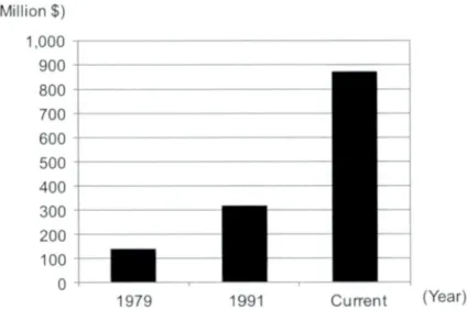
In recent years, the co st o f n ew drug d evelop m en t is sign ifican tly increasing (F igure 1.2) (1,3). T h e reasons for th e increasing co st co u ld b e accoun ted for b y a num ber o f explanation: th e pharm aceutical industry is currently attacking d isea se o f great com p lexity; the entry bar for n ew drugs is h igh er b ecau se th ey are o ften com p etin g w ith en hanced standard o f care; and/or the regulatory authorities are m ore dem anding (3).
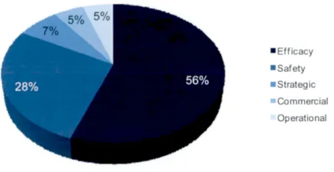
H ow ever, th e m ain reason for th e escalatin g d evelop m en tal cost is lo w clin ical su ccess rate (-1 1 % ) (3 ) b eca u se o f un accep table clin ical effica cy (56% ) and hepatocellular to x ic ity (28% ) (F igure 1.3) (4 ),w h ich su g g ests pre-clin ical anim al pharm acokinetics (P K ) stud y m a y not b e su fficien t for predicting th e fate o f drug candidate in hum an (5 ,6 ).
In other w ord s, great num ber o f attrition o f p re-clin ical and clin ical tests are con d u cted ,
as a resu lt,brings escalatin g d evelop m en tal cost. For this reason, the alternative in v itr o
Target discovery
lead and optimization
Pre-clinical study
2-5 years
Phase
Clinical stud Phase
II Phase
III 5-6 years
2-3 years
Figure 1.1 New drug development process (reproduced from reference 1 and 2).
(Millions) 1,000 900 800 600 700 500 400 300 200
1979 1991 Current (Year)
required from pharm aceutical com p an ies in recent years (6).
Today, m ost pharm aceutical com p an ies try to use a set o f sp ecific in v itro assays u sin g hum an cells as the initial step in drug lead discovery. N um erous cell-b ased assays h ave been establish ed o ver the past 5 to 10 years, incorporating a broad range o f tech n olo gical and instrum ent advances. T his p rom isin g p rocess is still on goin g, but the valu e o f any individual cell-b a sed assay to predict in v iv o effica cy has not been firm ly establish ed in the drug d isco v ery p rocess (7).
p o c^r
m a r CkDt
l n urva
的
Clll3ncD
(I
)
Figure 1.2 Temporal increase in developmental cost per approved new drug
(reproduced from reference 1 and 3).
■Efficacy
■Safety
■ Strategic
■ Commercial Operational
Figure 1.3 Cause of failure. The majority of failure s were due to lack of efficacy (56%) or to safety issues (28%) in Phase I/ll studies (reproduced from reference 4).
1.2 3-D spheroid culture of hepatocytes
To replace the in v iv o screen ing system w ith an in v itro high-throughput on e, prim ary h epatocytes are particularly interested as the cell source b ecau se the hepatocytes are m etab olic fundam ental elem en ts in the body. T h ey play critical roles in syn th esizin g m olecu les that are u tilized elsew h ere to support h om eostasis, and in con vertin g m olecu les o f on e type to another.
T he lack o f predictability o f in v iv o effica cy are reported to attribute to co m m o n ly em p loyed tw o-d im en sion al (2 -D ) cell culture system w h ich d oes not m im ic the resp on se o f cell o f in v iv o m icroenvironm ent (7 ). Traditional 2 -D m onolayer culture o f prim ary hepatocytes on tissu e culture plastic dish is problem atic and has been associated w ith a rapid lo ss o f differentiated function (8). T herefore, n ovel culture sy stem s are needed to facilitate short and long-term culture o f h epatocyte for diagn ostic, therapeutic, and drug d iscovery applications.
In order to o verco m e this draw back, three-dim ensional (3 -D ) m ulticellular
aggregates, spheroids, have been focu sed on. O riginal observations o f tissu e-lik e
aggregate form ation from isolated cells w as reported by M oscon a in 1961, using fetal
liver cells and a rotational technique (9 ). T he d escrip tive term “sph eroid” w as coin ed
years later b y Landry in 1985 w h en m ulticellular aggregates w ere form ed from isolated
rat h ep atocytes after 2 -5 days o f culture on non-adherent plastic surfaces precoated w ith
p oly(2-h yd roxy eth yl m ethacrylate) (p oly -H E M A ) (F igure 1.4A ) (10 ). S in ce th en,a great num ber o f 3 -D spheroid culture m eth od s or d evices other than th ose above h ave b een d evelop ed such as p roteoglycan (1 1 ), alginate scaffold (1 2 ,1 3 ), titanium d io x id e(1 4 ),h o llo w fibers (1 5 ),polyurethane form (1 6 ),and spinner flask (17 ,1 8).
R eports o f structural polarity and b ile can alicu li form ation b y prim ary h ep atocytes in spheroid aggregate provide further ev id en ce that hepatic spheroids m im ic th e hepatocellular m icroan atom y o f the liver (F igu re 1.4B ) (1 2 ). M oreover,the spheroid allo w s recapitulation o f the cu boidal g eom etry o f prim ary h ep atocytes w ith relatively stable long-term differentiated fu n ction such as album in secretion (F igure 1.4C ) (1 4 ).
T h e spheroid culture system is accord in gly an alternative culturing technique for evalu ating drug m etab olism and to x ic ity in th e n ew drug develop m en t. In resp on se to th ese issu es,w e h ave b een studied culture d e v ice to construct 3 -D tissu e in v itro . W e therefore m ade a start on d evelo p in g a n ovel culture d evice w ith aim in g at con trollin g spheroid form ation b y op tim izin g the loca l geom etric architecture o f th e substratum , w h ich is the “N anopillar C ell C ulture P late (N P p late)”.
1.3 Nanopillar cell culture plate ( N P plate)
Ingber’s group origin ally reported that m ech an ical forces serve as im portant regulators at the cell and m olecu lar lev els, and that th ey are eq u ally potent as ch em ical cu es in
1991 (19). P h y siolo g ist and clin ician h ave b e en recogn izin g the im portance o f th is m ech a n o b io log y for the d evelop m en t and fu n ction o f the cell over the p ast tw o d ecad es (2 0 ). N o w that,th e geom etric architecture o f th e culture substratum that brings cellu lar p roliferation,m o tility to tissu e m orp h ogen esis reach es n an oscale lev el.
N a n o tech n o lo g y has attracted the m u ch interest o f researchers not o n ly in the field
o f m aterial scien ce but also in that o f m olecu lar and cellular b iology. In order to
fabricate nanostructures or n an oscale pattern for culture d ev ice efficien tly and h ig h ly
throughput, nanoim printing is the k ey tech n o lo g y (21). C onven tion al nanostructures
h a v e b een fabricated u sin g large-scale integration (L SI) p rocesses b y ph otolith ograp hy
w ith m aterials lim ited to silico n (S i)-b ased . H ow ever,K uw abara e t a l have b een
su cceed ed in form ing n an o-scaled structures (nanopillars) w ith diam eters o f 8 0 -1 ,0 0 0
n m and h eigh ts o f 1-3 (im u sin g nanoim printing tech n ology, w h ich is nan o-scaled 3 -D
culture d e v ice (2 2 ). T h e exam p les are sh ow n in Figure 1.5.
0 0 3 6 9 12 15
Culture time (day)Figure 1.4 (A) Phase-contrast photograph of the hepatocyte spheroid on day 2. Bar, 50 lim (reproduced from reference 10). (B) Ultrastructural feature of the hepatocyte spheroid by transmission electron microscopy (TEM) on day 3. The spheroid exhibits tight junction (Tj) between adjacent cells. Rough endothelial reticulum (rER) indicates hepatic metabolism in active. Microvillus-lined channels (Me) and Bile canaliculi (B) show structural polarity. Bar, 200 nm (reproduces from reference 13). (C) Time course of albumin secretion rate (reproduced from reference 15).
A B
Tj rKR
Balaban et al. reported the elastic micropattemed substrates with nanopillar structures was applied as a measurement tool of local force added by cells to the substrates (23). Nomura et al. has reported the possible application of a nanopillar device as a new type of cell culture dish (24). In these studies, they reported using HeLa cells that the localization of cell adhesion-related molecule such as actin and vinculin commonly limited to the top of the nanopillars. This phenomenon means the adhesion to the substratum is weaker than that to the conventional flat cell culture dish. They also reported that the cell morphology on the nanopillar device was round-shape, however, that on the conventional flat petri dish were flattened 2-D manner.
From this point of view, we considered practical use of the nanopillar culture device, NP plate, for 3-D spheroid formation with aiming at utilization in the course of new drug development.
Figure 1.5 Scanning electron microscope (SEM) im ages of the NP plate surface structure. Bars, 3.0 jam.
1.4 Scanning electrochemical microscopy
When we apply 3-D spheroids as in vitro cell-based assay models for preclinical testing in the new drug development process, we need to establish noninvasive measuring method that can evaluate the cellular activity and viability. Noninvasive analysis is essential requirement in order to make use of spheroid itself which will be tested.
Furthermore, noninvasive measurement provides a benefit that the drug response or
metabolic process can be monitored continuously using the same spheroid throughout
evaluating. Based on this background, we focused attention on scanning electrochemical
microscopy (SECM). SECM is ideally noninvasive; oxygen consumption around cells
can be monitored continuously.
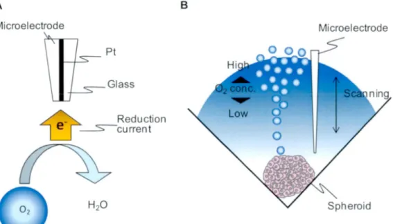
SECM w as originally developed in 1986 (25),w hich is the technology that the current that flow s through the very small electrode tip (generally an ultramicroelectrode w ith a tip diameter o f 10 jam or less) can be measured near a conductive, sem i-conductive, or insulating substrate im m ersed in solution (26). The features o f the SECM are the ability to induce a chem ical reaction in the local region and to measure in situ the chem ical reaction quantitatively in the local area o f biological sample.
Application to the biological sample w as firstly reported in 1990 in which the oxygen reduction current o f leaves produced by photosynthesis was measured (27).
Then,the target object has been m oved to m am m alian cells. SECM w as applied to measure the oxygen consum ption o f single bovine embryos to evaluate the viability for an effective artificial insem ination (28,29). In this case, m icroelectrode measures the reduction current with a negative charge that flow s w hen the dissolved oxygen in the m edium is reduced to water (Figure 1.6). The amount o f reduction current is proportional to the amount o f dissolved oxygen. In the very close region o f the cell surface, the amount o f reduction current is sm all because o f low concentration o f dissolved oxygen due to vigorous cellular respiration. H owever,at the bulk,a point aw ay from the cell surface, the amount o f reduction current increases because o f high concentration o f dissolved oxygen (Figure 1.6). B y scanning the m icroelectrode along vertical direction,w e measure the oxygen concentration difference (AC) betw een the cell surface and the bulk, and calculate the oxygen consum ption rate o f sample.
B y applying this m easuring principle, w e challenged to measure the respiratory activity o f single spheroids,which were formed on a N P plate in this study.
1.5 Organization of this thesis
A s described in Section 1.1, m ost pharmaceutical com panies demand a set o f specific in
vitro high-throughput cell-based screening system that can predict the fate o f drug
candidates in human. Primary hepatocytes w ould firstly be favored candidates for the
cell source. H owever, traditional 2-D m onolayer culture has a problem o f rapid loss o f
differentiated function. Therefore,novel 3-D culture system s are needed for diagnostic,
therapeutic, and drug discovery applications. In addition,a noninvasive m easuring
system that can evaluate the cellular activity and viability is also needed. N oninvasive
m easurem ent provides a benefit that the cellular viability, drug response or m etabolic
process can be monitored continuously. The objective o f this study is to establish
Figure 1.6 Measuring principle of SECM. (A) Microelectrode m easures the reduction current with a negative charge that flows when the dissolved oxygen in the medium is reduced to water. The amount of reduction current is proportional to the amount of dissolved oxygen. (B) In the very close region of the spheroid surface, the amount of reduction current is small because of low concentration of dissolved oxygen due to the cellular respiration. However, at the bulk, a point away from the cell surface, the amount of reduction current increases because of high concentration of dissolved oxygen. By scanning the microelectrode along vertical direction, we measure the oxygen concentration difference (AC) between the cell surface and the bulk, and calculate the oxygen consumption rate of sample.
a culturing method for 3-D spheroid formation using NP plate and a non-invasive cellular activity m easuring method by applying the scanning electrochem ical m icroscopy. Furthermore, w e analyze the m echanism the 3-D spheroid formation on the pillar structure, and the w ay o f cellular respiration o f spheroid depended on the spheroid size. In this study, w e used rat primary hepatocytes as basic study before applying human ones.
Chapter 2 describes the 3-D spheroid culture with higher structural polarity and
hepatic functions using NP plate that w as developed in Hitachi. Chapter 3 describes the
com prehensive increase in the drug m etabolism and pharmacokinetics (DM PK)-related
gene expression o f NP-cultured 3-D spheroid using global gene expression analysis.
Chapter 4 describes the noninvasive m easuring o f cellular respiratory activity using
SECM and offers the new numerical insight into the w ay o f cellular respiration o f
spheroid. Finally, the thesis is summarized in chapter 5
H i g h e r S t r u c t u r a l P o l a r i t y a n d
H e p a t i c F u n c t i o n s o f N a n o p i l l a r -
C u l t u r e d S p h e r o i d s
2.1 Introduction
M ost m ethodologies used to evaluate drug m etabolism and toxicity in the drug developm ent process are performed using animals and animal body parts. However, it is difficult to fully predict how drug candidates work in human body with this in vivo animal m odel (5,6). To replace the in vivo screening system with an in vitro one, primary hepatocytes must first be explored as the cell source. Additionally, culture conditions for hepatocytes must be identified that maintain the structural and functional polarity o f the hepatocytes after isolation from the liver (12). Hepatocytes in their native environm ent have a structural polarity. They have a cuboidal shape with two to three basal surfaces facing the sinusoid w hile adjacent cells that form the bile canaliculi (12,17). After isolation from the liver, hepatocytes rapidly lose their polarized structure and their differentiated functions, such as albumin secretion, urea synthesis, and cytochrom e P450 (Cyp) activity (12,17).
C on ven tion ally cultured hepatocytes have a flat, m onolayer m orphology on
extracellular matrix (ECM )-coated surfaces with a rare, short-lived bile canalicular
structure, and their structure and function are lost or decreased and cannot be recovered
under such m onolayer culture conditions (11,14,18,30). Am ong such 2-D m onolayer
culture conditions, “sandwich (SW )” culture system s (Figure 2.1) have been reported to
support differentiated functions and polarity o f adult rat hepatocytes (31,32). In this
Figure 2.1 Schematic view of sandwich (SW) culture. Hepatocytes are cultured on a thin layer of type I collagen and then overlaid with Matrigel.
system , hepatocytes are cultured on a thin layer o f type I collagen and then overlaid with M atrigel,a basement membrane extract from Engelbreth-Holm-Swarm (EH S) m ouse sarcoma (33). This system is used to evaluate hepatocyte function such as biliary excretion activity in vitro (34).
In contrast, primary hepatocytes maintain differentiated hepatocellular functions when they are induced to self-assem ble into 3-D aggregates, spheroids by culturing on an extracellular matrix or substrate such as proteoglycan (11), alginate scaffold (12), poly (2-hydroxym ethyl methacrylate) (poly-H EM A ) (10), titanium dioxide (14), hollow fibers (15), polyurethane foam (PUF) (16), and spinner flask (35). Several investigators have reported that hepatocyte spheroids possess structural polarity and transporters sim ilar to those o f the bile canaliculi in the native tissue, which explains the enhanced hepatocellular activities o f spheroids (12,17,18). Accordingly, the spheroid culture system is expected to be an alternative culturing technique for evaluating drug m etabolism and toxicity in the course o f drug screening (12,13). A great number o f cell culture devices have been developed for form ing three-dimensional structures (such as spheroids), including NP plate (14,24,36-38). In one case, NP plates were fabricated with high aspect ratio structures with a pillar diameter o f 160-1,000 nm and a height o f 2 8 0 -1 ,0 0 0 nm using nanoimprinting technology (24).
In the previous research on the effect o f ECM patterns in the micrometer range
during the initial phases o f cell adhesion and spreading, Ingber’s group found that the
long-term effect o f cell shape on growth and viability depends on the local geom etric
architecture o f the substratum, synergistically with a consequence o f the type o f matrix
protein (39,40). Bastmeryer and co-workers analyzed the effect o f ECM patterns in the micrometer range by controlling the size o f ECM squares (dots) and the distance between them. They found that a dot distance o f around 2 -5 pm affected the cellular shape and functions (41).
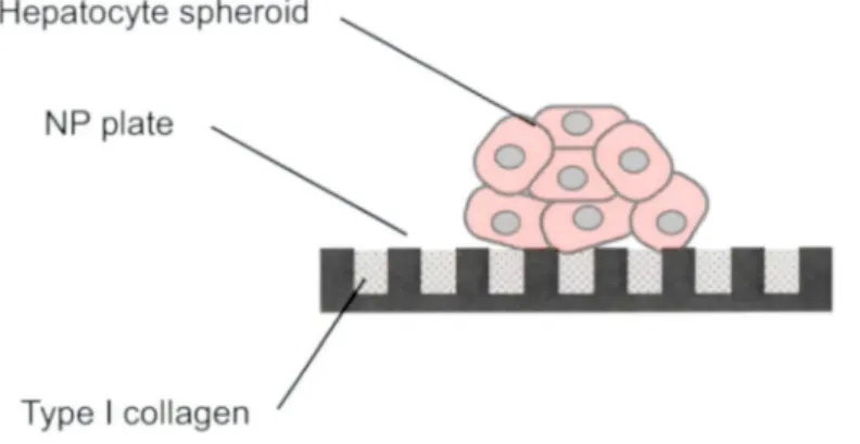
On the basis o f these findings, w e envisaged the 3-D spheroid would be formed on the NP plate (Figure 2.2), and investigated how the NP surface structure affects cell adhesion, migration, and consequent spheroid formation. We used the N P plates with pillar diameters o f 0.18, 0.5 , 1.0, 2.0, and 5.0 |im and with pillar pitches (pillar center-to-center distance) that were tw ice the pillar diameter (Figure 2.3). The adhesion area o f cells cultured on the N P plates w as reported to be restricted to the top o f the pillars (24). Accordingly, the m ode o f cell migration and the resultant m orphology o f tissues on the NP plate should differ from those on a conventional culture dish. From this point o f view, our objective is to identify the optimal pillar diameters for spheroid formation in terms o f cell adhesion and migration. We exam ined the structural polarity and biological functions o f spheroids formed on the NP plates and investigated the effects o f M atrigel-overlaying on the hepatocellular functions.
Figure 2.2 Schematic view of 3-D spheroid cultured by NP plate.
A
Nanomold T Heat-imprinting NP plate
Polystyrene
B 0.18NP
0.5NP
■■■■… ■■■■■■■■■■… …1.0NP 2.0NP 5.0NP Flat plate C Pillar diameter
i o r
Q Q R Pillar pitch
pi
「う : 令 ::Pillar height
Figure 2.3 (A) Fabrication process of the NP plate. Nanomold is made of silicon wafer
and fabricated using photolithography. By heat-imprinting the nanomold onto the
polystyrene film, nanopillar sheet is made. (B) A side views of surface patterns of NP
plates used in this study. B ar,1 \im. (C) Definitions of pillar diameter, pillar pitch and
pillar height.
2.2 Materials an d M et hod s 2.2.1 Nanopillar plate preparation
Polystyrene film with a thickness o f 1.0 m m w as spin-coated onto a glass substrate. A nanom old w as m ade o f silicon wafer and fabricated using photolithography. A n N P structure was formed by pressing the m old onto the film at 4 2 3 .K and then releasing it at room temperature (RT) (24). The surface patterns o f the fabricated N P plates are illustrated in Figure 2.3.
2.2.2 Hepatocyte culture
H epatocytes were isolated from 6- to 8-w eek-old m ale specific viral pathogen free W ister rats (Charles River Japan Inc., Japan) w eighing about 150-250 g by using a m odified two-step in situ collagenase perfusion method (42) and purified by isodensity Percoll centrifugation (43). The viability o f the hepatocytes w as determined by trypan blue exclusion, and hepatocytes with over 85% viability were used for culturing.
The hepatocytes were resuspended in W illiam ’s E m edium containing 10% fetal bovine serum, supplem ented with 8.6 nM insulin (Sigm a-Aldrich Corp. , M O ),255 nM dexam ethasone (Nacalai Tesque, Inc” Japan),50 ng/m l epidermal growth factor (Sigm a-Aldrich Corp.), 5 K lU /m l aprotinin (Wako Pure Chem ical Industries, Ltd”
Japan) and were seeded at a density o f 1 x 10⁵ cells/cm 2 onto prepared N P plates and into a 35-m m -diam eter culture dish. The plates were precoated with a solution containing 10 |ag or 100 ng/m l o f type I collagen. The dish that type I collagen w ere precoated w as purchased from AG C Techno G lass C o. , Ltd. (Japan). The procedure for the NP, SW ,and conventional m onolayer (M L) cultures is described below.
N P culture: after 24 hours o f post-seeding, the m edium was replaced w ith serum-free W illiam ’s E m edium containing the same supplem ents described above.
After 48 hours o f post-seeding, the m edium was replaced w ith a serum-free m edium containing M atrigel™ (BD B ioscience, M A ) w ith the sam e supplements described above. Subsequently,the culture m edium was changed daily. H epatocytes were cultured in total for 96 hours.
SW culture (31,32): after 24 hours o f post-seeding, the culture m edium w as replaced
w ith serum -free W illiam ’s E m edium containing M atrigel (BD ) w ith the sam e
supplem ents described above. Subsequently, the culture m edium without Matrigel w as
changed daily. H epatocytes were cultured in total for 96 hours.
M L culture: the culture m edium w as replaced with serum-free W illiam ’s E m edium every 24 hours. H epatocytes were cultured in total for 96 hours.
2.2.3 Immunohistochemistry
After a total o f 96 h o f culturing, the spheroids cultured on the N P plates and the hepatocytes cultured in the dish were w ashed with phosphate buffered saline (-) (PB S(-)) after rem oving the culture m edium , and then fixed in 4% paraformaldehyde in PB S(-) and perm eabilized in 0.05% Triton-X in PBS(-). After blocking by FBS in PB S(-), the sam ples were incubated in rabbit anti-human E-cadherin polyclonal antibody in a 100-fold dilution (SC -7870; Santa Cruz Biotechnology, Inc., CA), w ashed w ith 0.05% Tween 20 in PB S(-), and incubated in biotinylated anti-rabbit IgG antibody (B A -1000; Vector Laboratories, CA). The sam ples were then washed with 0.05% Tw een 20 in PBS(-) and incubated in Streptavidin-Fluorescein (NEL720; PerkinElmer Life Science, Inc., M A). They were then w ashed again with 0.05% Tween 20 in PBS(-) and incubated in rhodamine phalloidin (R415; Invitrogen Corp., CA). After that, the sam ples were washed w ith PBS(-) and incubated in H oechst 33258. After a final washing in deionized water (D W ), the sam ples were em bedded in glass slides. Fluorescence im ages were taken with an optical m icroscope (A X 70; Olym pus Optical, Japan).
2.2.4 Resin embedding and microscopy
Rat hepatocytes cultured on the NP plates were fixed with 2.5% glutaraldehyde in 0.1 M phosphate buffer,pH 7.4, incubated overnight at 4°C, and rinsed w ith 0.1 M phosphate buffer. Post-fixation w as performed w ith 1% osm ium tetroxide in 0.1 M phosphate buffer, pH 7.4. After being washed with 0.1 M phosphate buffer and DW, the hepatocytes on the N P plates were em bedded in 1.5% SeaPlaque® agarose (Lonza Ltd .,
Switzerland). Agar-containing hepatocytes w as dehydrated w ith a serial dilution o f ethanol, treated with propylene oxide,and em bedded in epon-araldite m ixed resin.
Vertical sections o f the sam ples were prepared using an ultramicrotome (Sorvall M T-6000; Du Pont Company , DE). Sem i-thin sections (500 nm ) were stained w ith toluidine blue and view ed with an optical m icroscope (AX70; Olym pus Optical, Japan).
Ultrathin sections (60 nm) were stained w ith uranyl acetate and lead citrate and
exam ined with a transm ission electron m icroscope (H-7100; Hitachi Ltd. , Japan).
2.2.5 Semi-quantitative gene expression analysis
Total RNA was extracted using an RN easy Tissue Kit (Qiagen GmbH, Germany) every 24 h from rat hepatocytes cultured on the NP plates with and without Matrigel, from ones conventionally cultured as a m onolayer, and from ones SW cultured. 2.0 jig o f the total RNA was reverse transcribed, and quantitative real-time PCR was conducted in a Thermal Cycler Dice Real Tim e System (TaKaRa Bio Inc., Japan) using a final reaction mixture o f the RT product, TaqMan Gene Expression A ssay (Applied Biosystem s, C A ), Premix Ex TaqrM (Takara Bio Inc.), and DW. The 2'AAC 1 method was used to calculate the relative change in the gene expression (44); TATA-binding protein (TBP) was used as an endogenous control. The relative changes in expression were determined toward the control sample that freshly isolated (tim e 0).
2.2.6 Biliary excretion analysis
Every 24 h after cell culture started, biliary excretion assay was performed. Before each assay, the medium w as washed by Hanks’ Balanced Salt Solution (HBSS;
Sigm a-Aldrich Corp.) with calcium and m agnesium . After that, it was replaced with a medium containing 5-carb oxy-2\ 7 ’-dichlorofluorescein diacetate (CDFDA; M olecular Probes, Inc., OR). After incubation at 37°C, the extracellular C D FDA was rinsed out by H BSS with calcium and m agnesium . Fluorescence and phase-contrast im ages o f the sam ples were acquired using an optical m icroscope (Axiovert 200; Carl Zeiss GmbH, Germany)
2.3 Results
2.3.1 Formation of rat hepatocyte spheroids on NP plates
To exam ine the effect o f the NP diameter on spheroid formation, hepatocytes were
seeded on N P plates with a pillar diameter o f 0.18, 0.5, 1.0, 2.0, or 5.0 (im, which are
referred to as 0.18NP, 0.5NP, 1.0NP, 2.0NP, and 5.0NP, respectively (Figure 2.3B). A
culture dish without NP (flat plate) w as used as a control (Figure 2.3B ). The pillar
diameter, pitch, and height were defined as show n in Figure 2.3C, and the dim ensions
are summarized in Table 2.1. To exam ine the effect o f the amount o f type I collagen in
the coating solution on spheroid formation, w e observed spheroid m orphology at 96 h post-seeding. Since hepatocytes are conventionally cultured in a culture dish precoated with a solution containing 1-1.5 mg/mL o f type I collagen (33,34),w e tested a sequential 100-fold dilution se r ie s:1 n g ,100 n g ,10 fig, and 1 m g/m l. We show here the results o f culture using 100 ng/ml and 10 |ig/m l o f type I collagen. In experim ents using six different kinds o f NP plates including flat plate and solutions with different concentrations o f type I collagen, w e observed spheroids with a compact m orphology in which individual cells constituting spheroid could barely be distinguished on the 0.18NP, 2.0NP, 5.0NP, and flat plates coated with a solution containing 100 ng/ml type I collagen (Figure 2.4B , K, N, and Q). Intermingled m onolayer hepatocytes and spheroids were observed on the 0.5N P and 1.0NP with the sam e concentration o f type I collagen (Figure 2.4E and H). Similar results were observed for the uncoated NP and flat plates.
However, unlike the spheroids on the NP plates coated with 100 ng/ml collagen solution, the spheroids on the uncoated 0.18NP, 2.0NP, 5.0NP, and flat plates (Figure 2.4A , J, M, and P) had weak adhesion to the substrate. The number o f spheroids decreased over tim e due to the changing o f the culture medium. Rat hepatocytes were spread on the N P plates and flat plate coated with 10 |ig/m l o f type I collagen solution in m onolayer configuration for all pillar diameters (Figure 2.4C , F, I, L, O, and R). This revealed that the hepatocyte and spheroid m orphologies depended on the pillar diameter and/or the concentration o f type I collagen. Spheroids with a compact m orphology adhered to the substratum were accom plished on 0.18NP, 2.0NP, 5.0NP, and flat plates coated with a solution containing 100 ng/ml o f type I collagen.
Table 2.1 Pillar diameter, pitch, and height of the NP plate
0 18NP 05N P 1 0NP 20N P 50N P
Diameter (^m) 0.18 0.5 1 0 2.0 5.0
Pitch (|im) 0 3 6 1 0 2 0 4.0 10.0
Height (卩 m) 0 2 1 0 1 0 1.0 1 0
Concentration of type I collagen
100 ng/ml 10 pg/ml
0 18NP
05NP 卜 ,、
公 ,霸
1 ONP
① TO Q- Z
20NP $
ベ
# ■
50NP
1
••
顧
flat plate ■ • ’
Figure 2.4 Phase-contrast micrographs of hepatocyte cultures on the NP plates 96 hr
after seeding. Hepatocytes were cultured on 0.18, 0.5,1.0, 2.0, and 5.ONP and on a flat
plate. NP plates were coated with a solution containing 100 ng/ml or 10 mg/ml of type I
collagen solution. Uncoated NP plates and flat plate were used as a control (-). Bars,
100 |im.
To determine the optimal size o f pillar diameter for spheroid formation, w e calculated the number o f spheroids and the distribution o f their diameters on the basis o f micrograph data for spheroids that formed on the NP plates coated with a solution containing 100 ng/ml o f type I collagen. M ost o f the spheroids on the 0.18NP, 2.0NP, and flat plates were 5 0 -1 0 0 |im in size, and m ost o f those on the other NP plates were 100-150 |im in size (Figure 2.5). As shown in Table 2.2,the 2.0N P had the m ost spheroids (153.6) and the sm allest standard deviation (±5.3), indicating that a number o f spheroids o f relatively uniform size formed on the 2.0NP. Since spheroids up to 100 |im in diameter have higher viability or effective cellular function (45), 2.0N P is suitable for formation o f spheroids because both the number (58.1 士 13.3) and ratio (37.8% ) o f spheroids on the 2.0NP ranging in size from 50-100 \xm were the highest am ong the NP plates (Figure 2.5) and because the number o f 1-100 jam spheroids on the 2.0N P versus the other NP plates was consistently larger with statistically significant differences (p<0.05) (Table 2.3). Consequently, 2.0NP coated with a solution containing 100 ng/ml o f type I collagen were used in the subsequent experiments.
The recruitment o f E-cadherin and actin to regions of intercellular contact is essential for the formation and stabilization o f adherens junctions, resulting in appropriate tissue structure (46). Accordingly, the localizations o f E-cadherin and actin were investigated using im m unohistochem istry to compare the structure o f hepatocyte spheroids with native liver tissue. Figure 2.6 show s double staining im ages for a frozen section o f rat native liver, an NP-cultured spheroid, and monolayer-cultured hepatocytes.
Rhodam ine-phalloidin was used to stain the actin (red), and anti E-cadherin antibody was used to stain the E-cadherin (green). The E-cadherin o f the rat liver and the spheroids formed on the NP plate were highly concentrated at the cell-cell junction (Figure 2.6D and E) as was the actin (Figure 2.6A and B). In contrast, the E-cadherin o f the conventional m onolayer culture was below the limit o f detection (Figure 2.6F), and actin stress fibers were observed (Figure 2.6C).
Table 2.2 Total number of spheroids formed on the nanopillar and flat plates;
mean±SD.
0.18NP 0.5NP 1.0NP 2.0NP 5.0NP flat sheet
120±21 3 1 2 0 4 H 3 8 1056±25 153 6±5 3 123 2±176 39 2±168
5.0NP
Flat plate 1 ONP
NP plate diameter of spheroids (um)
50-100 1-50 20N P 100-150
150-200 200- 50-100 1-50
0.5NP 100-150
150-200 200- 50-100 1-50 0 18NP 100-150
150-200 200-
Number of spheroids
0 10 20 30 40 50 60 70 80
B -
hl = E
卍
0
o
o
o
o 5
0
5
0
0 u
1 1
2
2 1
_
_
- o
o
o 5
0
5 1
o
o
o
o
o 5
0
5
0
0
J.
1 1
2 2
1 - - - o
o
o 5
0
5 o
o
o
o
o 5
0
5
0
0 u
1 1
2
2 1 - 1 1 - o
o
o 5
0
5
Figure 2.5 Size distributions of hepatocyte spheroids. Hepatocytes were cultured on
0.18, 0.5,1.0, 2.0, and 5.ONP and on a flat plate coated with a solution containing 100
ng/ml of type I collagen solution. Spheroids were counted 96 h post-seeding.
Table 2.3 Number of 1-100 fiin spheroids and p-value
20NP 0 18NP 05NP 1 ONP 50NP flat sheet
n=1 65 59 56 14 53 14
n=2 99 40 50 27 56 37
n=3 86 77 40 31 61 38
n=4 60 45 4 44 37 13
n=5 65 61 32 39 36 21
p-value
versus 2 ONP - <0.05 <0.01 <0.01 <0.01 <0.01
Actin
E-cadherin
Nuclear
Figure 2.6
cultured on 2.0NP and in conventional collagen-coated dish for 96 h. Frozen section of rat native liver, hepatocyte spheroids on the NP plates and hepatocyte monolayer cultured in dish were stained with rhodamine-phalloidin (red: A-C) and anti-E-cadherin antibody (green: D-F). Hoechst 33258 was used for nuclear counterstaining (blue: G-l). Bars, 50 \xrr\.
Native liver NP spheroid Monolayer culture A
SEE
Immunohistological staining of actin and E-cadherin for hepatocytes
2.3.2 Effects of Matrigel overlay on hepatocyte spheroids
Structural analysis. It is w ell established that an ECM can modulate gene expression in m any biological system s, and it has been suggested that a com plex ECM , but not its purified com ponents, is needed to maintain differentiated hepatocytes in vitro for a long period (47). Accordingly,w e expect the effect o f extracellular matrix, w hich sim ulates the environm ent o f a living organism by surrounding the cells, and overlaid M atrigel w hich includes com plex basem ent membrane extract but not purified com ponents on the spheroids formed by NP culturing.
To observe the m orphology and exam ine cellular viability before and after overlaying the M atrigel,w e cultured rat hepatocytes on the 2 .ONP coated with a solution containing 100 ng/m l o f type I collagen. Sem i-thin sections o f reconstituted spheroids were stained with toluidine blue. Rat native liver tissue and SW -cultured hepatocytes were also exam ined for comparison. In the SW culture system , monolayer-cultured hepatocytes are held betw een tw o kinds o f thin-layered ECM com ponents In the N P culture system with M atrigel, it is overlaid on spheroids formed on the N P plates. The hepatocytes cultured without the M atrigel overlay had a variety o f shapes but were spheroidal overall (Figure 2.7A and B) w hile those cultured with M atrigel were individually round polygons (Figure 2.7C), m uch like those o f native liver (Figure 2.7E).
The SW -cultured hepatocytes were uniform ly flat (Figure 2.7D ). These results indicate that the use o f the Matrigel overlaying onto spheroids is more effective than the use o f m onolayer culturing for recovering the native m orphology. The viability o f the hepatocytes w as then exam ined. The ratio o f dead hepatocytes represented by asterisks in the Figure 2.7 was increased tim e dependently under the condition o f without the M atrigel overlay 48 h and 96 h post-seeding (Figure 2.7A and B). A lm ost all the hepatocytes o f spheroids cultured with the Matrigel overlaid at 48 h were still alive at 96 h (Figure 2.7C ). The viability o f the hepatocytes was thus improved by the M atrigel overlay (Figure 2.7A -C ).
To investigate the bile canaliculi structures and their peripheral parts in more detail,
w e used a transm ission electron m icroscope (TEM ; H -7100; Hitachi Ltd., Japan). W e
found that bile canaliculi divided by a tight junction form ed in all the reconfigured
structures: NP-cultured spheroids (Figure 2.8A -C ) and SW -cultured tissues (Figure
2.8D ). B ile canaliculi with w ell-developed m icrovilli those were in native tissue (Figure
2.8E ) were observed in spheroids cultured with and without Matrigel (Figure 2.8A and
C). B ile canaliculi with and without w ell-developed m icrovilli were observed in
spheroids cultured with Matrigel (Figure 2.8B and C), but only bile canaliculi without w ell-developed m icrovilli could be observed in SW -cultured (Figure 2.8D).
A B C
Figure 2.7 Toluidine blue staining of hepatocyte spheroid sections on 2.ONP (A-C),
sandwich-cultured hepatocytes (D), and liver tissue (E). A: 48 h, B: 96 h, C: 48 h after
Matrigel overlay at 48 h (total 96 h culture), D: 72 h of sandwich culture (total 96 h
culture), E: rat liver tissue. Dead hepatocytes indicated by asterisks. B ars,10 |im.
Figure 2 . 8 「ransmission electron microscope (TEM) im ages of ultrathin section of hepatocyte spheroids cultured on 2.0NP for 96h without Matrigel overlay (A), hepatocyte spheroids cultured on 2.0NP for 96h with Matrigel overlay at 48 h post-seeding (B,C), sandwich-cultured hepatocytes for 96h (D), and rat liver tissue (E).
Tight junctions (TJ), bile canaliculus (BC), and microvilli (MV) are indicated. B a rs,1 |im
Semi-quantitative gene expression analysis. The real-tim e PCR was performed to m easure the relative amounts o f gene expression am ong four kinds o f culture conditions.
Freshly isolated hepatocytes were used as the calibrator sam ple,w hich is indicated as tim e 0 in Figure 2.9. The expression o f A bcc2 (ATP-binding cassette transporters superfam ily C ,member 2) tended to increase w ith tim e except for the M L culture,and those at 96 h for spheroids cultured on the N P plates w ith and without M atrigel were higher than those for the SW- and M L-cultured hepatocytes (p<0.05) (Figure 2.9A ). The expression at 72 h for spheroids cultured on the N P plate with M atrigel overlay at 48 h w as substantially higher than that for those cultured without M atrigel. However, the final values for these expressions at 96 h were virtually the sam e and were close to the initial level at tim e 0 (Figure 2.9A ). W hile a substantial increase w as also observed during the next 24 h after M atrigel overlay for the SW culture, a further increase over tim e w as not observed.
Under all conditions except for the m onolayer culture, the expression level o f C yp3a23/3al (cytochrom e P 450-3a23/3al) increased over tim e (Figure 2.9B ). Although the expression level for spheroids cultured on the N P plate with Matrigel overlay took 24 h after the overlay to substantially increase, that for the SW culture took 48 h. That is, there w as a tim e lag betw een these tw o culture conditions. Comparison o f the expressions at 96 h revealed that the one for spheroids cultured on the N P plate w ith M atrigel w as m uch higher than the others (p<0.05).
W hile the albumin expressions for spheroids cultured on the N P plates and the M L-cultured hepatocytes tended to decrease during the first 48 h,that for spheroids cultured on the N P plate with M atrigel increased after M atrigel overlay at 48 h (Figure 2.9C ). The expression for the SW -cultured hepatocytes trended upward for the first 72 h and then turned downward. Com parison o f the expressions at 96 h showed that the one for spheroids cultured on the N P plate with M atrigel w as m uch higher than that for the others (p<0.05). H owever, the expressions at 96 h were at m ost around 60% o f the initial level.
The expression patterns for talin differed from those for the three other genes. The expression for the ML-cultured hepatocytes increased over tim e, resulting in a 2.5-fold higher level at 96 h than the initial level (Figure 2.9D ). In contrast,for the other three conditions, the expression levels remained fairly constant,and the levels at 96 h w ere substantially low er than that for the M L culture (p<0.05).
The expression patterns for E-cadherin for spheroids cultured on the N P plate w ith
M atrigel show ed a substantial increase in a 24 h period after M atrigel overlay at 48 h
Figure 2.9 Semi-quantitative real-time PCR. Relative amounts of mRNA expression of Abcc2, Cyp3a23/3a1, albumin, talin, and E-cadherin were measured every 24 h under four culture conditions: culture on 2.0NP without Matrigel (MG -), culture on 2.0NP with Matrigel overlaid at 48 h post-seeding (MG +), monolayer culture in collagen-coat dish (ML culture),and sandwich culture (SW). Expression values of each gene were compared to freshly isolated hepatocytes (time 0 ,before seeding). Statistical comparison was performed for 96 h sam ples for each culture condition. A: p<0.05 for MG - (U) or MG + (*) versus SW ( す ) . B: p<0.05 for MG + (*) versus MG - (TJ), ML (t), or SW ( す ) . C: p<0.05 for MG + (*) versus MG - (H), ML ( 本 ),or SW ( す ) . D: p<0.05 for ML (t) versus MG - (H), MG + (*), or SW (t). E: Expressional levels have no statistically significant differences at p<0.05 for MG + (*) versus MG - (^|) or SW (|). The results are averages of three repetitions.
o
0 24487296 24487296 24487296 24487296 (h)spheroid spheroid
on NP on NP ML SW
(MG -) (MG +)
24487296 24487296(h) 0 24487296 spheroid
ML SW on NP
(MG -)
24487296 24487296 24487296 (h) spheroid
o n
NP ML SW (MG +)
Talin
24487296 s p h e r o i d
(MG +) onNP Albumin
24487296 spheroid
on NP (MG -) o
0 24487296 24487296 24487296 24487296(h) spheroid sphero»d
on
NP
o nNP ML SW (MG -) (MG +)
E E-cadhenn
3 0
0 24487296
spheroid on NP (MG-)
24487296 24487296
spheroid
on NP ML
(MG +)
24487296 (h)
SW
A Abcc2 B Cyp3a23/3a1
uolssoJdx
a) 0)>
= aJl9
J
5
0
5
0
5
0
5
3
3
2
2
1
d1
D
uolss
aJdx)
a>
a)>
一
ral a)
」
2 0 8 6 4 2 1 1
o o o
d
c
c s M( /) a)J d ax) a>
〜
ral a)J
u
.s co co a)J a x a) a)y y (lQl a>J
uolss
aJdx)
a>9
>
I CSI0
J