Toru SAWASAKI,
Yuji MORI and Yasuhiko
KANO
Stock Farm, Faculty of Agriculture, University of Tokyo Iwama, Ibaraki 319-02, Japan
(Received
for publication
: May 24, 1978)
The anatomical relations of visceral organs and other tissues in the thoracic,
abdominal and pelvic cavities of the male miniature •gShiba•h goat are illustrated
as thirteen semi-diagramatic representations of cross sections.
小 型 ヤ ギ,い
わ ゆ る シバ ヤ ギの躯 幹 の 断 面解 剖
沢 崎
徹 ・森
裕 司 ・加 納 康 彦
東京大学農学部附属牧場
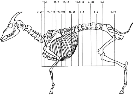
小型 ヤ ギ,い わ ゆ る シバ ヤ ギ の 実 験 動物 と し て の 有 用 性 は次 第 に評 価 され て来 て お り[4],ま た そ の基 礎 資 料 も整 いつ つ あ る[3,7]。 しか し実 験 動物 と して の有 用 性 を 更 に 高 め るた め には,生 理 学 的,形 態 学 的 な ど生 物 学 的 基 礎 資 料 を 整 備 す る こ とが 必 要 で あ る こ とは 言 うま で もな い 。 本 研 究 は,シ バ ヤ ギに 関 す る形 態 学 的 な資 料 が 特 に 少 い[5]こ とか ら,ま ず 内臓 諸 臓 器 の立 体 的 配 置, 相 互 関 係 等 を 明 らか に す る 目的 で,躯 幹 の 断 面 解 剖 学 的 検 討 を 行 な った もの で あ る。 材 料 お よ び 方 法 供 試 した シバ ヤ ギ は,本 場 で 生 産,育 成 した7ケ 月 令 の雄 で,体 重14kg,体 高42cm,胸 囲54cmの もの で あ る。 放 血 は抱 水 ク ロ ラー ル 静 脈 注 射 に よ る麻 酔 下 で 頸 動 脈 を 切 断 してお こ な った 。 そ の後,角,★ 甲,腰 角 を 支 点 と して,枠 内 に で き る限 り自然 な 駐立 姿 勢 で保 定 し,50% ホル マ リ ン 約400m2を 頸 動 脈 内 に 注 入 して 硬 化 させ, -20℃ の冷 凍 庫 内 で充 分 凍 結 した 後 ,大 型 の鋸を用 い て,水 平 面 に ほ ぼ 垂 直 に横 断 した 。 切 断 部 位 はFig.1 に 示 した 通 り,前 か ら順 に,第7頸 椎,第1,第3,第 5,第7,第9,第11,第13胸 椎,第1,第3,第5腰 椎,第1,第4仙 椎 で,部 位 の 選 択 は,Henri-Albert が ヒツ ジに用 い た方 法[2]を 参 考 と し,各 臓 器 の位 置 関 係 が 充分 に解 明 で き る よ うに 配慮 した[1,8]。 各 ブ ロ ッ クは 尾 側 よ り,切 断 面 の写 真 を撮 り,こ の写 真 と標 本 よ り解 剖 図 を描 き,断 面 に あ らわ れ た各 種 臓 器,筋,そ の 他 に 名 称 を 付 した 。学 名 はNonina Anatomica Veterinaria[6]に 従 った 。 さ らに 上 記 ヤ ギ とは 別 に10ケ 月 令,体 重19kg,お よび 12ケ 月 令,体 重22kgの 雄 シバ ヤ ギ2頭 を 局 所 解 剖 学 的 同 定 な らび に 比 較 の た め に 使 用 した 。 成 績 お よ び 考 察 シバ ヤ ギ に 関 す る形 態 学 的 資 料 の 一 つ と して,断 面 解 剖 を 行 な った 結 果 をFig.2∼14に と りま とめ,各 臓 器,筋 名 等 を付 記 した 。 シ バ ヤ ギ の椎 骨 数 は,Fig.1に み られ る よ うに頸 椎 7,胸 椎13,腰 椎6,仙 椎5,尾 椎9で,今 回使 用 した 他 の2頭 も 同様 の数 を構 成 し,シ バ ヤ ギ の椎 骨 数 に は 変 異 が 少 い こ とが 知 られ た 。 Fig.1の 破 線 は 横 隔 膜 の位 置を 図示 した も の で,胸
Fig. 1. The position
of cross sectioning.
The broken
line shows
Diaphragma
(median
section and costal line).
腔 と腹 腔 の境 界 を示 した 。 肺 は,第7頸 椎 に おけ る切 断 面(Fig.2)に は 認 め ら れ ず,第1胸 椎 の 断面(Fig.3)か ら,第9胸 椎 の 断 面 (Fig.7)ま で の 間に あ らわ れ るが,第11胸 椎 の断 面 (Fig.8)に は 見 られ な い 。 従 っ て肺 尖 は 第7頸 椎 と第 1胸 椎 に お け る 断面 間 に,後 縁 は,第9,第11胸 椎 に お け る 断面 間 に それ ぞれ 存 在 す る 。 肺 の位 置 を 外 側 か ら判 断 す る場 合,肺 尖 は 第1肋 骨 の,後 縁 は 第11肋 骨 の 位 置 に ほ ぼ一 致 す る 。 心 臓 は,第5胸 椎(Fig.5),お よび 第7胸 椎(Fig. 6)の 断 面 に あ らわ れ,腕 頭 動 脈幹,前 大 静 脈 は,第3 胸 椎 の 断 面(Fig.4)に 見 られ た 。 従 って,心 臓 の 前 縁 は ほ ぼ 第3肋 骨 の 線 に,後 縁 は ほ ぼ 第5肋 骨 の 線 に そ れ ぞ れ 一 致 し,心 臓 は この 間 に 存 在 す る。 心 底 の 中 心 と心 尖 を 結 ぶ 心 軸 は,後 方 よ り見 た 場 合 は ほ ぼ 垂 直 であ るが, 心 臓 全 体 は や や 左 側 に 偏 位 して い る。 また 側 方 よ り見 た 場 合,心 軸 の 腹 端 は や や 後 方 に 傾 い て い た 。 肝 臓 は,第7胸 椎 の 断 面(Fig.6)か ら第13胸 椎 の 断 面(Fig.9)に あ らわ れ,そ の 前 縁 は,第5,第6肋 骨 間 の 線 に,後 縁 は 第13肋 骨 の 線 よ り後 方 に あ る。 腎 臓 は,第13胸 椎,第1腰 椎,第3腰 椎 に お け る断 面 図(お の お のFi9.9,10,11)に 見 られ,Fig.9,10で は 右 腎を,Fig.11で は 左 腎 を示 して お り,左 腎が 遊 走 腎 であ る こ とは 明瞭 で あ る 。 複 胃は,第7,9,11,13胸 椎,第1,3,5腰 椎,お よ び 第1仙 椎 に おけ る断 面 に あ らわ れ,第II胃 は,第7, 9胸 椎 切 断面(Fig.6,7)に,第III胃 は 第11胸 椎切 断 面(Fig.8)に,第IV胃 は第9,11胸 椎 切 断 面 に 認 め ら れ る 。 第1胃 は,Fig.8か らFig.13(第1仙 椎切 断 面)ま で の 各 図 に 示 され る よ うに,そ の 容 積 は極 め て 大 きい 。 特 にFig.9以 後 の 各 図 に 見 られ る よ うに,第1 胃 は 腹腔 の 大 部 分 を 占 め て い る。 また 第1胃 の 後 縁 は, Fig.14に 示 した 断 面 図,す な わ ち,第4仙 椎 に お け る 断 面 よ りや や 前 方 に 存 在 し,骨 盤 腔 まで 達 してい た 。 第11胸 椎 切 断 面(Fig.8)に は 幽 門 お よび 第I,II胃 皺 が み られ る。 幽 門 は 第10肋 骨 の 線 の 下 部 に ほ ぼ 一 致 し, また 第I,II胃 皺(外 側 よ り見 た 場 合,第I,II胃 溝) は,こ の 断 面 よ り前 方 で,第8か ら第9肋 骨 の線 に あ る。 供 試 ヤ ギは で き る限 り自然 な 駐 立 時 の 姿 勢 の ま ま固 定 した の で,各 臓 器 の 配 置 は 生 体 の そ れ を お お む ね そ の ま まに 表 現 して い る とみ て 差 し支 え な か ろ う。 また,供 試 ヤ ギ を 選 出 した 本 場 の シバ ヤ ギは 導 入 以 来 約10年 間,閉 鎖 集 団 と して 繁 殖,飼 育 して お り,個 体 間 に 著 しい 体 型 の偏 りが 見 られ な い こ とを 合 せ 考 え る と,本 研 究 で示 さ
い 雌 に つ い て は,予 備 実 験 に お け る雄 との 比 較 か ら,雄 と大 差 の な い こ とが 知 られ て い るの で,本 研 究 の 結 果 は, 成 熟 シバ ヤ ギ全 個 体 を 代 表 す る と考 え て よい だ ろ う。 切 断 部 位 の 選 定 に 当 って は,で き る限 り少 ない 断 面 で 各 臓 器 の 相 互 関 係 を 良 く表 現 で き る よ うに,ま た,実 験 に 用 い る際 に 最 も有 用 で,か つ 使 用 頻 度 の 高 い部 位 を表 現 す る よ うに 配 慮 した 。 従 っ て,シ バ ヤ ギ の 内臓 諸 臓 器 の立 体 的 配 置は,Fig.2∼Fig.14に よ っ て,充 分 に 表 現 され て い る もの と考 え られ よ う。 要 約 1.シ バ ヤ ギ に 関 す る形 態学 的 資 料 を 整 備 す る 目的 で, で き る限 り自然 に 近 い 状 態 に 保 定 した 雄 を 用 い,断 面 解 剖 学 的 に 検 討 を 加 え た 。 2.胸 部,腹 部,骨 盤 腔 部 の 各 臓 器,お よび 筋 の解 剖 学 的 位 置 関 係 を 明 らか に した 。 3.こ れ ら の成 績 の詳 細 は,Fig.2∼Fig.14に 示 し た 。
Venzke, W. G. and Ghoshal, N. G. (1975). Ruminant. In Sisson and Grossman's Anatomy of the Domestic Animals. Getty, R., editor, W. B. Saunders, Phila-delphia, 737-1211.
[2] Henri-Albert, D. (1972). Contribution Iconographi-que a la Connaissance de la Topographie Vicerale des Ovins, Presentation de Coupes Transversales Seriees. Dissertation of Ecole Nationale Veterinaire de Toulouse. [3] 加 納 康 彦 ・沢 崎 徹 ・小 山徳 義 (1977).小 型 ヤ ギ い わ ゆ る シバ ヤ ギ の生 物 学 的 特 性 一 東 大 牧 場 コ ロ ニ ー6年 間 の記 録 一 実 験 動物, 26, 239-246. [4] 三 宅 仁 ・藤 正 巌 ・井 街 宏 ・面 坂 剛 ・大 道 久 ・岩 井 矩成 ・河 野 明 正 ・渥 美 和 彦 (1978).運 動 負 荷 時 に お け る人 工 心臓 の制 御 方法 に つ い て,人 工臓 器, 7, 145-148. [5] 西 田 隆雄 ・望 月公 子 (1978)。 山 羊 の 大 動 脈 弓 と背 肋 間 動 脈 に つ い て,第85回 日本 獣 医学 会 講 演 要 旨, 108.
[6] Nomina Anatomica Veterinaria (1973).International Committee on Veterinary Anatomical Nomenclature, World Association of Veterinary Anatomists, Vienna.
[7] 野 沢 謙 ・加 納 康 彦 ・沢 崎 徹 ・西 田 隆雄 ・阿 部 恒 夫 ・庄 武 孝 義 ・松 田洋 一 (1978).小 型 ヤ ギい わ ゆ る シバ ヤギ の 遺 伝 子 構 成,実 験 動 物, 27. 413-422.
[ 8] Popesko, P. (1968). Atlas of Topographical Anatomy of the Domestic Animals. W. B. Saunders. Philade-lphia, Vol. I 66-90, Vol. II 72-101, Vol. III 70-99 .
Fig. 2. Cross section through the neck at the level of the seventh cervical vertebra. View of the caudal surface of the section.
1. Funiculus
nuchae
2. Vertebra
cervicalis
VII
3. Medulla
spinalis
4. Pays cervicalis
mi. trapezii
5. M. rhomboideus
cervicis
6. M, splenius
7. M. semispinalis
capitis
8. M. spinalis
et semispinalis
dorsi et cervicis
9. M. serratus
ventyalis
ceyvicis
10. M. longissimus
capitis, ceyvicis, atlantis
11. M. multifidus
ceyvicis
12. M. brachiocephalicus
13. M. scalenus dorsalis
14. Mm. intertransversarii
15. Pays ventyalis
mi, scaleni
medii
16. M. longus
colli
17. M. sternomandibularis
18. M. sternomastoideus
19. Mm. steynohyoideus
et sternothyroideus
20. Esophagus
21. Trachea
22. A. carotis communis
sinistra
23. Tyuncus
vagosympathicus
24. V. jugularis
Fig. 3. Cross section through the thorax at the level of the first thoracic vertebra. View of the caudal surface of the section.
1. Lig. nuchae
2. Corpus vertebrae
thoracicae
I
3. Humerus
4. Acromion
5. Os costale 1
6. Cartilago
costalis 1
7. Sternum
8. M. trapezius
9. M. rhomboideus
10. M. semispinalis
capitis
11. M. spinalis
et semispinalis
dorsi et cervicis
12. M. serratus
ventralis
13. M. ion gissimus
capitis, cervicis, atlantis
14. M. multifidus
15. Mm. intertransversarii
16. M. longus colli
17. M. scalenus
dorsalis
18. Mm. intercostales
19. M. rectus thoracis
20. M. pectoralis
transversus
21. M. pectoralis
descendens
22. Mm. scaleni
medii (pans dorsalis
ventralis)
23. M. su praspinatus
24. M. brachiocephalicus
25. M. pectoralis
ascendens
26. Medulla
spinalis
27. Trachea
28. Esophagus
29. Ductus thoracis
30. T runcus brachiocephalicus
31. N. vagus sinister
32. N. vagus dexter
33. Vena cava cranialis
34. Pars cranialis
lobi cranialis
pulmonis
dextri
35. A. et v. axillaris
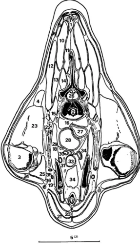
Fig. 4. Cross section through the thorax at the level of the third thoracic vertebra. View of the caudal surface of the section.
1. Lig, nuchae
2. Lig, interspanale
3. Corpus vertebrae
III
4. Ca put costae III
5. Scapula
6. Humerus
7. Sternum
8. M. spinalis
thoyacis
9. M. semispinalis
capitis
10. M. multifidus
dorsi
11. M. rotator
12. M. longissimus
capitis
13. M. longissimus
atlantis
14. M. longissimus
cervicis
15. M. rhomboideus
thoyacis
16. M. serratus
ventralis
17. M, trapezius
18. M. supraspinatus
19. M. infraspinatus
20. M. subscapularis
21. Ca put longum mi, tricipitis
bracii
22. M. deltoideus
23. M. latissimus
dorsi
24. Ca put mediale
mi. trici pitis bracii
25. M, extensor carpi radialis
26. Ca put laterale
mi. tricipitis
bracii
27. M. coracobrachialis
28. M. pectoralis
ascendens
29. M. biceps brachii
30. M. brachiocephalicus
31. M. pectoralis
descendens
32. M. pectoralis
transversus
33. Mm. intercostales
34. M. iliocostalis
35. M. intercartilagineus
36. M. transversus
thoyacis
37. M. longus colli
38. Pars cranialis
lobi cranialis
pulmonis
sinistri
39. Pars cranialis
lobi cranialis
pulmonis
dextri
40. N, vagus dexter
41. Esophagus
42. Ductus
thoracicus
43. Trachea
44. Truncus
brachiocephalicus
45. Vena cava cranialis
46. Truncus
ni. sympathici
47. A. axillaris
48. V. axillaris
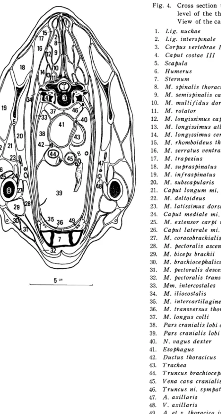
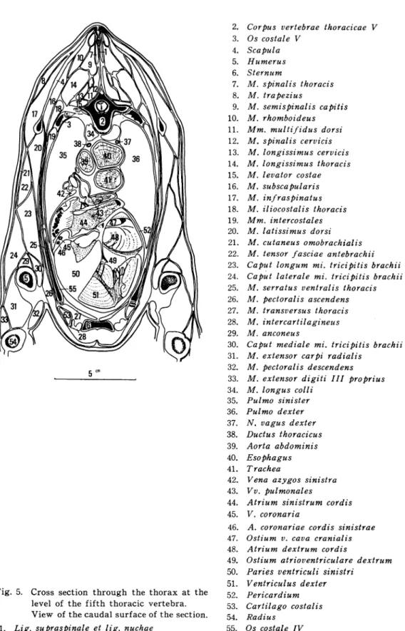
Fig. 5. Cross section through the thorax at the level of the fifth thoracic vertebra. View of the caudal surface of the section.
Fig. 6. Cross section through the thorax at the level of the seventh thoracic vertebra. View of the caudal surface of the section. 1. Lig, supraspinale et lig. nuchae
2. Processus spinosus vertebrae thoracicae 3. Corpus vertebrae thoracicae VII 4. Scapula et cartilago scapulae 5. Os costal e V I
6. Sternum 7. Olecranon
8. M. spinalis et semispinalis dorsi et cervicis 9. M. multi f idus dorsi
10. M. rhomboideus 11. M. tra pezius
12. M. ion gissimus dorsi 13. M. subscapularis 14. M. iliocostalis thoracis 15. M. levator costae
16. M. serratus ventralis thoracis 17. M. cutaneus omobrachialis 18. Mm. intercostales
19. M, transversus abdominis 20. M. intercartilagineus 21. M. pectoralis ascendens
22. Ca put longum mi, tricipitis brachii 23. Ca put laterale mi. tricipitis brachii 24. M, anconeus
25. M. flexor digitorum profundus 26. Lobus caudalis pulmonis sinistri 27. Lobus caudalis pulmonis dextri 28. Lobus accessories pulmonis 29. V. azygos sinistra
30. Aorta
31. T runcus vagalis dorsalis 32. Ductus thoracicus 33. Truncus vagalis dorsalis 34. Esophagus 35. V. cava caudalis 36. Dia phragma 37. He par 38. Reticulum 39. Apex cordis 40. Os costal e VII
1. Lig, supraspinale
2. Corpus vertebrae
thoracicae
IX
3. Os costale VIII
4. Os costale VII
5. Cartilago
costalis
VII
6. Sternum
7. M. multifidus
dorsi
8. M. spinalis
et semispinalis
doysi
9. M. longissimus
doysi
10. M. psoas minor
11. M. iliocostalis
12. M, latissimus
doysi
13. Mm. intercostales
14. M. serratus
ventralis
thoracis
15. Pars costalis
diaphragmatis
16. M, transversus
abdominis
17. M. rectus abdominis
18. M. pectoralis
ascendens
19. Pulmo sinister
20. Pulmo dexter
21. Aorta thoracica
22. V, azygos sinistea
23. Ductus thoracicus
24. Lien
25. Orificium
esophagi
26. Reticulum
27. Abomasum
28. He par
29. V. cava caudalis
30. V. hepatica
31. Ramus v, portae
32. Os costale V I
33. Os costale IX
Fig. 8. Cross section
through
the thorax
at the
level of the eleventh
thoracic
vertebra.
View of the caudal surface of the section.
1. Corpus vertebrae
thoracicae
XI
2. Os costale XI
3. Os costale X
4. M. spinalis
et semispinalis
dorsi et cervicis
5. M. multifidus
dorsi
6. M. longissimus
dorsi
7. M. levator
costae
8. M. psoas minor
9. M. iliocostalis
10. Mm. intercostales
11. M. serratus
dorsalis
12. M. latissimus
dorsi
13. M. serratus
ventralis
thoracis
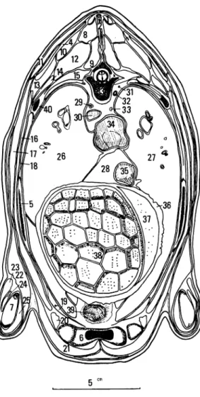
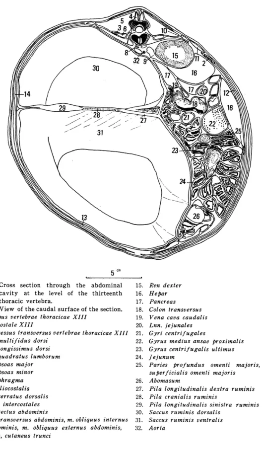
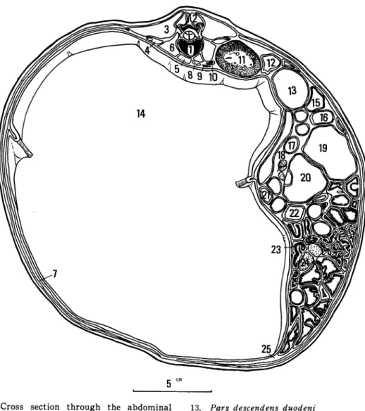
Fig. 9. Cross
section
through
the
abdominal
cavity
at
the
level
of the
thirteenth
thoracic
vertebra.
View of the caudal surface of the section.
1. Corpus vertebrae
thoracicae
XIII
2. Os costale XIII
3. Processus
transversus
vertebrae
thoracicae XIII
4. M. mul ti f ides dorsi
5. M. longissimus
dorsi
6. M. quadratus
lumborum
7. M. psoas major
8. M. psoas minor
9. Diaphragma
10. M, iliocostalis
11. M. serratus
dorsalis
12. Mm. intercostales
13. M. rectus abdominis
14. M. transversus
abdominis, m, obliquus
internus
abdominis,
m. obliquus
externus
abdominis,
et m, cutaneus
trunci
15. Ren dexter
16. He par
17. Pancreas
18. Colon transversus
19. Vena cava caudalis
20. Lnn, je junal es
21. Gyri centri f ugales
22. Gyrus medius ansae proximalis
23. Gyrus centrifugalis
ultimus
24. Jejunum
25. Panes
profundus
omenti
majoris,
Panes
superficialis
omenti majoris
26. Abomasum
27. Pila longitudinalis
dextra
ruminis
28. Pila cranialis
ruminis
29. Pila longitudinalis
sinistra
ruminis
30. Saccus ruminis
dorsalis
31. Saccus ruminis
ventralis
32. Aorta
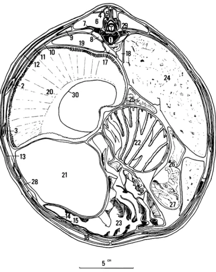
Fig. 10. Cross section through the abdominal cavity at the level of the first lumbar vertebra.
View of the caudal Surface of the section.
1. Corpus vertebrae
lumbalis
I
2. M. multifidus
dorsi
3. M. longissimus
dorsi
4. M. psoas major
5. M. quadratus
lumborum
6. M. psoas minor
7. M, transversus
abdominis,
m, rectos
abdomi-nis, m. obliquus
externus
abdominis,
et m.
cutaneus
trunci
8. Aorta abdominalis
et v. cava caudalis
9. Dia phragma
10. Ren dexter
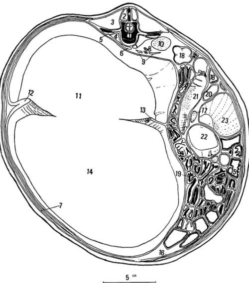
Fig. 11. Cross section through the abdominal cavity at the level of the third lumbar vertebra. View of the caudal surface of the section.
1. Corpus vertebrae
lumbalis
III
2. M. multifidus
dorsi
3. M. ion gissimus
dorsi
4. M. quadratus
lumborum
5. M. psoas minor
6. M, psoas major
7. M. transversus
abdominis,
m. rectus
nis, m. obliquus
externus
abdominis,
et m.
cutaneus
trunci
8. Aorta abdominalis
9. Diaphragma
10. V. cava caudalis
11. Ren sinister
(Ren migrans)
Fig. 12. Cross section through the abdominal cavity at the level of the fifth lumbar vertebra.
View of the caudal surface of the section.
1. Corpus vertebrae
lumbalis
V
2. Processus
transversus
vertebrae
lumbalis
V
3. M, longissimus
dorsi
4. M, multi f idus dorsi
5. M. psoas major
6. M. psoas minor
7. M, transversus
abdominis,
m. rectus
nis, m. obliquus
externus
abdominis,
et m.
cutaneus
trunci.
8. Saccus ruminis
dorsalis
9. Pila caudalis
10. Saccus ruminis
ventralis
Fig. 13. Cross section through the pelvis at the level of the first sacral vertebra. View of the caudal surface of the section.
1. Corpus vertebrae sacralis 1 2. Pars lateralis ossis sacri 3. Ala ossis ilium
4. Patella
5. Trochlea ossis femoris 6. Mm, erector spinae 7. M. gluteus medius 8. M. gluteus accessorius 9. M. gluteus profundus 10. M, tensor fasciae latae 11. M. psoas major 12. M. psoas minor
Fig. 14. Cross section through the pelvis at the level of the fourth sacral vertebra. View of the caudal surface of the section.