Bull. Natl. Mus. Nat. Sci., Ser. A, 40(1), pp. 15–51, February 22, 2014
Revision of Isoparorchis Southwell, 1913 (Digenea, Hemiuroidea, Isoparorchiidae), Parasites of the Air Bladder of Freshwater Catfishes:
a Molecular and Morphological Study
Takeshi Shimazu1, Thomas H. Cribb2, Terrence L. Miller3, Misako Urabe4, Nguyen Van Ha5, Tran Thi Binh5 and Marina B. Shedʼko6
1 10486–2 Hotaka-Ariake, Azumino, Nagano 399–8301, Japan E-mail: [email protected]
2 School of Biological Sciences, The University of Queensland, Brisbane, Queensland 4072, Australia
3 School of Marine and Tropical Biology, James Cook University, Cairns, Queensland 4870, Australia
4 Department of Ecosystem Studies, School of Environmental Sciences, The University of Shiga Prefecture, 2500 Hassaka-cho, Hikone, Shiga 522–8525, Japan
5 Department of Parasitology, Institute of Ecology and Biological Resources, Vietnamese Academy of Sciences and Technology, 18-Hoang Quoc Viet, Hanoi, Vietnam
6 Institution of Russian Academy of Sciences, Institute of Biological and Soil Sciences, Far Eastern Branch of Russian Academy of Sciences,
159 Stoletiya Street, Vladivostok 690022, Russian Federation (Received 5 November 2013; accepted 18 December 2013)
Abstract Isoparorchis Southwell, 1913 (Digenea, Hemiuroidea, Isoparorchiidae), parasites of the air bladder of freshwater catfishes (Osteichthyes, Siluriformes) from East, Southeast and South Asia and Australia, is revised based on a molecular and morphological study. Materials, including syntypes where available, were examined from Russia (Primorskiy Kray), Japan, Vietnam, Cam- bodia, Bangladesh, India and Australia. The entire second internal transcribed spacer region of the ribosomal DNA (ITS2 rDNA) was sequenced for 18 distinct samples. Four of the five previously described species are considered to be valid: Isoparorchis trisimilitubis Southwell, 1913 (type spe- cies) from India, I. hypselobagri (Billet, 1898) from Vietnam, I. eurytremum (Kobayashi, 1915) from Japan and Russia and I. tandani Johnston, 1927 from Australia. Isoparorchis pakistani Bilqees and Khatoon, 1972 from Pakistan is regarded as a species inquirenda. Not only syntypes of I. trisimilitubis but also other materials from India and Bangladesh consisted of specimens of two morphologically distinct species. One is identified as I. trisimilitubis, and the other remains an undescribed species, Isoparorchis sp. 3. Distinctions in the ITS2 sequences obtained for samples from India support the interpretation that there are two species there. One sequence is related to I.
trisimilitubis, but it remains undetermined whether the other sequence (Isoparorchis sp. 1) relates to Isoparorchis sp. 3. Some materials from East, Southeast and South Asia require further critical studying for definitive species identification. Each species is described and figured with a summa- rized life cycle where known. Molecular data are given. A key to the species is presented.
Key words : Isoparorchis, Digenea, revision, taxonomy, morphology, life cycle, sequences of ITS2 rDNA.
Introduction
Species of Isoparorchis Southwell, 1913 (Digenea, Hemiuroidea, Isoparorchiidae) are par- asitic in the air bladder of freshwater catfishes
(Osteichthyes, Siluriformes) in Asia and Australia (Gibson, 2002; this paper).
Billet (1898) described a new species, Disto- mum hypselobagri, from the fish “Hypselo- bagrus” (Siluriformes, Bagridae) collected in
Vietnam. Southwell (1913) established a new genus, Isoparorchis, with a new species, Isopar- orchis trisimilitubis, as the type species from Wallago attu (Bloch and Schneider, 1801) (Silu- riformes, Siluridae) collected in India. Kobayashi (1915a) created a new genus and species, Lepto- lecithum eurytremum, from Silurus asotus Lin- naeus, 1758 (syn. Parasilurus asotus (Linnaeus, 1758)) (Siluridae) collected in Japan. Travassos (1922a, b) recognized Leptolecithum as synony- mous with Isoparorchis and placed L. eurytremum in the latter genus as Isoparorchis eurytremum (Kobayashi, 1915) (see Shimazu et al., 2011).
Bhalerao (1926) considered that I. trisimilitubis and I. eurytremum are the same species. Johnston (1927) proposed a new species, Isoparorchis tandani, from Tandanus tandanus (Mitchell, 1838) (Silurifomes, Plotosidae) collected in Aus- tralia. Ejsmont (1932) transferred D. hypselo- bagri to Isoparorchis as Isoparorchis hypselo- bagri (Billet, 1898) and synonymized I.
trisimilitubis, I. eurytremum and I. tandani with I. hypselobagri without reexamining any of the type materials. Bilqees and Khatoon (1972) added a new species, Isoparorchis pakistani, from Wallago attu collected in Pakistan. Bhutta and Khan (1975) and Zaidi and Khan (1977) reduced this species to a synonym of I. hypselobagri. It has thus far been believed that I. hypselobagri alone is valid in the genus with I. trisimilitubis, I.
eurytremum, I. tandani and I. pakistani as its synonyms.
Shimazu et al. (2011) gave a full account of the morphology of material from Japan under the species name of I. hypselobagri. They pointed out that the sinus organ was much smaller in their material than in the specimens of I. eurytremum described by Odhner (1927) and of Isoparorchis described by Gibson and Bray (1979). The sinus organ is actually much smaller in their material than in those of I. trisimilitubis and I. tandani as originally described by Southwell (1913) and Johnston (1927), respectively. We thus decided to make a molecular and morphological study of materials of Isoparorchis as broad a range of collections as possible to revise the genus. In this
paper, we present the results attained and sum- marize the life cycle where known.
Materials and Methods Molecular study
Samples sequenced. Table 1 lists samples for which the entire second internal transcribed spacer region of the ribosomal DNA (ITS2 rDNA) was sequenced and GenBank accession numbers of their ITS2 sequences.
Molecular sample processing. Total genomic DNA was extracted from ethanol preserved worms by the extraction method using a QIAgen DNeasy Kit (QIAGEN INC., Valencia, Califor- nia) according to the manufacturerʼs instructions.
The ITS2 rDNA was amplified via Polymerase Chain Reaction (PCR) amplifications (20 μl) using 1.6 μl of MgCl2 (25 mM), 2 μl of PCR reac- tion buffer (PROMEGA) (10x), 0.8 μl of dNTPs (5 mM), 0.75 μl each of forward primer 3S (5′- GGT ACC GGT TCA CGT GGC TAG TG-3′) and reverse primer, ITS2.2 (5′-CCT GGT TAG TTT CTT TTC CTC CGC-3′) (10 pmol), 0.25 μl Taq polymerase (PROMEGA, Madison, USA) (5 units/μl), 1–2 μl of DNA template (5–100 ng), made up to 20 μl with millipore H2O and run on a Minicycler (MJ Research, supplied by Bresatec, Watertown, USA). The following thermocycling conditions were used: 4 min denaturation hold at 95°C, 2 min at 45°C, 90 sec at 72°C, 4 cycles of 45 sec at 95°C, 45 sec at 50°C and 90 sec at 72°C;
then 25 cycles of 20 sec at 95°C, 20 sec at 52°C and 90 sec at 72°C and 5 min extension hold at 72°C.
Amplified DNA was purified using QIAGEN® QIAquick™ PCR purification kit according to manufacturerʼs protocol. Cycle sequencing was conducted using the same primers utilized for PCR amplification on the purified DNA products at the Australian Genome Research Facility (AGRF) in Brisbane, Australia. The resulting sequences were edited and contigs constructed using Geneious Pro™ version 5.4 software (Bio- matters Ltd.). They have been registered at Gen- Bank (Table 1).
Revision of Isoparorchis (Digenea, Hemiuroidea, Isoparorchiidae) 17
Comparative molecular analysis. The ITS2 data generated were aligned using MUSCLE ver- sion 3.7 (Edgar, 2004) with ClustalW sequence weighting and UPGMA clustering for iterations 1 and 2. The resultant alignments were refined by eye using MESQUITE v. 2.75 (Maddison and Maddison, 2009). After alignment of the ITS2 dataset was edited, the ends of each fragment were trimmed to match the shortest sequence in the alignment. Total nucleotide distance matrices were calculated using MEGA v.5 software (Tamura et al., 2011).
Minimum evolution analysis was conducted on the ITS2 dataset using MEGA v.5 software.
Nodal support for the minimum evolution analysis
of the ITS2 dataset was inferred by bootstrap analysis using a heuristic search of 10,000 repli- cates.
Morphological study
Material examined. Specimens examined are listed in Material examined of each species.
They had been prepared by various methods, either whole-mounted or serially sectioned.
Some were borrowed from the National Museum of Nature and Science (NMNS, collection name code NSMT-Pl), Tokyo then, Japan; the Meguro Parasitological Museum (MPM), Tokyo, Japan; the Swedish Museum of Natural History (SMNH), Stockholm, Sweden; the National Museum of
Table 1. Samples of the species of Isoparorchis used for sequencing the IST2 rDNA and GenBank accession numbers of their associated sequences.
Sample, species, developmental stage, site of infection, host, locality and date GenBank accession no.
1) ISO11 —I. hypselobagri, adult, air bladder, Silurus asotus, Ninh Co River, Vietnam, 10 May
2011 KJ188114
2) ISO22 —I. hypselobagri, adult, air bladder, S. asotus, Hong River, Vietnam, 23–25 November
2011 KJ188115
3) ISO2 —I. eurytremum, juvenile, body cavity, Gnathopogon elongatus elongatus, near
Daido River, Lake Biwa basin, Japan, 16 October 2010 KJ188116
4) ISO33 —I. eurytremum, juvenile, body cavity, Sarcocheilichthys variegatus microoculus,
Lake Biwa, Japan, 14 December 2011 KJ188117
5) ISO3 —I. eurytremum, adult, air bladder, S. asotus, Lake Biwa, Japan, 30 November 2010 KJ188118 6) ISO4 —I. eurytremum, adult, air bladder, S. asotus, Lake Biwa, Japan, 30 November 2010 KJ188119 7) RUS1262 —I. eurytremum, juvenile, wall of esophagus, Huso dauricus, Amur River, Primorskiy
Kray, Russia, 7 September 2010 KJ188120
8) RUS1297 —I. eurytremum, juvenile, wall of esophagus, Lota lota, Amur River, Primorskiy
Kray, Russia, 11 September 2010 KJ188121
9) RUS1385 —I. eurytremum, adult, air bladder, Silurus soldatovi, Amur River, Primorskiy Kray,
Russia, 25 September 2010 KJ188122
10) RUS1712 —I. eurytremum, adult, air bladder, S. asotus, Lake Khanka, Primorskiy Kray, Russia,
28 July 2011 KJ188123
11) IND375* —Isoparorchis sp. 1, juvenile, body cavity, Ompok pabo, Mukutmanipur dam lake,
West Bengal, India, 18 October 2010 KJ188124
12) ISO1 —Isoparorchis sp. 1, juvenile, body cavity, Mastacembelus favus, India, date unknown KJ188125 13) 55A† —I. trisimilitubis, adult, air bladder, Wallago attu, Rishra near Kolkata, West Bengal,
India, 15 November 2007 KJ188126
14) 61A† —I. trisimilitubis, adult, air bladder, W. attu, Balurghat, West Bengal, India, 13
December 2007 KJ188127
15) ISO44† —I. trisimilitubis, adult, air bladder, W. attu, Balurghat, West Bengal, India, 9
December 2007 KJ188128
16) ISO5† —I. trisimilitubis, adult, air bladder, W. attu, Balurghat, West Bengal, India, 9
December 2007 KJ188129
17) VNT353# —Isoparorchis sp. 2, juvenile, body cavity, W. attu, Phnom Penh-market, Cambodia,
18 October 2010 KJ188130
18) ISOAU1 —I. tandani, adult, air bladder, Tandanus tandanus, Moggill Creek, Brisbane,
Australia, 20 August 2012 KJ188131
*Collected by T. Scholz and A. Ash; † collected by A. Ash; # collected by T. Scholz and R. Kuchta.
Natural History (MNHN), Paris, France; the Department of Life Sciences, The Natural History Museum (NHM, collection name code NHMUK), London, UK; the South Australian Museum (SAM, collection name code AHC), Adelaide, Australia; and the Institute of Parasitology (IPCAS), Biology Centre, Czech Academy of Sciences, Branišovská, Česke Budějovice, Czech Republic.
Methods. Measurements (length by width) are given in millimeters unless otherwise stated.
Drawings were made with the aid of a camera lucida. Most of the present newly collected spec- imens have been deposited in the NMNS, Tsukuba;
the NMNS; the NHM; the IPCAS; and the Queensland Museum (QM), Brisbane, Queensland, Australia.
Abbreviations used in the figures. csd, com- mon sperm duct; dm, Drüsenmagen; e, esopha- gus; ed, ejaculatory duct; ep, excretory pore; ev, excretory vesicle; ga, genital atrium; gp, genital pore; hd, hermaphroditic duct; ic, intestinal cecum; Lc, Laurerʼs canal; m, metraterm; md, male duct; Mg, Mehlisʼ gland; o, ovary; os, oral sucker; ot, ootype; p, pharynx; pc, prostatic cells;
pac, parturient canal; pp, pars prostatica; rsr, rudimentary seminal receptacle; sd, sperm duct;
so, sinus organ; ss, sinus sac; sv, seminal vesicle;
t, testis; tnc, transverse nerve commissure; u, uterus; v, vitellarium; vd, vitelline duct; vs, ven- tral sucker.
Results
Genus Isoparorchis Southwell, 1913
Isoparorchis Southwell, 1913: 91–92.
Leptolecithum Kobayashi, 1915a: 50–52; Kobayashi, 1921: 396–397.
Diagnosis. Digenea, Hemiuroidea, Isoparor- chiidae sensu Gibson (2002). Body broad-ovate, large, dorsoventrally flat. Ecsoma absent. Preoral lobe present. Oral sucker subterminal. Prephar- ynx absent. Pharynx posterodorsal to oral sucker.
Esophagus inverted T- or Y-shaped, short. Drüsen- magen present at commencement of intestinal cecum, with several internal longitudinal
grooves. Intestinal ceca undulating, ending blindly near posterior extremity of body. Ventral sucker in anterior half of body. Testes two, entire, almost symmetrical, posterolateral to ventral sucker. Sperm ducts long; common sperm duct short, anterior to ventral sucker. Seminal vesicle tubular, sinuous, anterior to ventral sucker, free in parenchyma. Male duct lacking prostatic cells, long, connecting seminal vesicle and pars pros- tatica. Pars prostatica tubular, long, surrounded by prostatic cells, posterodorsal to sinus sac, free in parenchyma. Sinus sac snifter-shaped, globu- lar or elliptical, large, composed of thick outer layer of diffuse musculature and thin inner layer of dense musculature, median, between ventral sucker and esophagus; its base protractile with sinus organ on it; small thin-walled tubular por- tion present between sinus sac and genital pore.
Sinus organ large or small, complex, with diffuse musculature, arising from base of sinus sac.
Ejaculatory duct long, convoluted in wall of sinus sac, joining to middle of hermaphroditic duct dorsally. Genital atrium consisting of ante- rior lumen of thin-walled tubular portion and posterior lumen of thick-walled sinus sac. Geni- tal pore large, median, immediately posterior to esophagus. Ovary tubular, dextral or sinistral, lying transversely between ootype and relevant intestinal cecum, well posterior to testes in hind- body. Oviduct and ovovitelline duct very short.
Laurerʼs canal opening dorsally, forming small rudimentary seminal receptacle proximally. Ootype small, median; Mehlisʼ gland well developed.
Uterus long, preovarian, embracing ovary on ovarian side of body, folding transversely in intercecal field between vitellaria and sinus sac, in dorsal parenchyma; uterine seminal receptacle present; metraterm thin, long, convoluted in wall of sinus sac, terminally forming hermaphroditic duct in sinus organ. Hermaphroditic duct not eversible, clavate, thickened, short, opening on apex of sinus organ. Eggs numerous, ovate, oper- culate, not filamented, fully embryonated. Vitel- laria two, dendritic (may be compound follicular glands with many small follicles), large, median, diagonal; anterior vitellarium located on anti-
Revision of Isoparorchis (Digenea, Hemiuroidea, Isoparorchiidae) 19
ovarian side of body, in dorsal parenchyma;
vitelline area mainly between ovary and cecal ends, extending a little anteriorly to ovary on antiovarian side of body, overlapping ceca.
Excretory vesicle Y-shaped, in ventral paren- chyma; stem swollen in median longitudinal pos- terior portion, folding transversely in intercercal field, bifurcating in hindbody; arms running for- ward, turning backward laterally to pharynx, sep- arate there; excretory pore postero-ventral, -ter- minal or -dorsal. Parasitic in air bladder of freshwater catfishes (East, Southeast and South Asia and Australia). Type species: Isoparorchis trisimilitubis Southwell, 1913.
Discussion. The above diagnosis is based on the present study. Some other details will be added in the description for I. trisimilitubis.
The short esophagus bifurcates into an inverted Y- or T-shape with a short stem (Kobayashi, 1915a, c, 1921; Shimazu et al., 2011; this paper).
The Drüsenmagen is well differentiated at the commencement of the intestinal cecum on either side of the body (Kobayashi, 1915a, c, 1921;
Shimazu et al., 2011; this paper). The anatomy of the terminal genitalia is complex (Kobayashi, 1915a, b, 1921; Bovien, 1927; Johnston, 1927;
Odhner, 1927; Gibson and Bray, 1979; Shimazu et al., 2011; this paper). The anatomy reported in this paper is essentially the same as that described by Bovien (1927, fig. 3) and Shimazu et al. (2011). Bovien (1927) referred to the male duct connecting the seminal vesicle and pars prostatica as the first section of the pars prostat- ica lacking the prostatic cells. The male duct and pars prostatica are lined with a single layer of epithelial cells. The terminal thickening of the metraterm dorsally receives the ejaculatory duct at about the middle and then becomes a true uneversible hermaphroditic duct (Fig. 17).
Ko bayashi (1915a) erroneously stated that the metraterm joined to the ejaculatory duct laterally.
The hermaphroditic duct should be interpreted as a continuation of the terminal thickening of the metraterm, because the posterior terminal thick- ening and anterior hermaphroditic duct are sub- stantially the same in the thickness and histology.
The small, thin-walled tubular portion following the sinus sac may be a true genital atrium (see Figs. 13–15). In this paper, for convenience sake, the hermaphroditic duct means a combination of the terminal thickening of the metraterm and the true hermaphroditic duct; and the genital atrium means a combination of the anterior lumen of the thin-walled tubular portion and posterior lumen of the thick-walled sinus sac (Figs. 13–15).
Molecular study
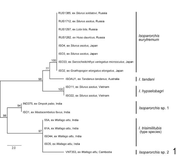
ITS2 Analysis. Alignment of the ITS2 dataset yielded 468 characters (including 54 nucleotides of the 5′ flanking 5.8S region and 56 nucleotides of the 3′ flanking 28S region) for analysis. The total pairwise nucleotide differences observed between the species of Isoparorchis examined are shown in Table 2. No intraspecific variation was observed in any of the species sequenced.
Interspecific variation observed among these taxa ranged from 3–18 nucleotides over the ITS2 region. Minimum evolution analysis of the ITS2 dataset resulted in a phylogram with the species I. eurytremum, I. hypselobagri and I. tandani forming a well-supported clade sister to Isopar- orchis sp. 1, I. trisimilitubis and Isoparorchis sp.
2 (Fig. 1). Each of the species clades resolved in the analysis was resolved with strong bootstrap support.
Morphological study
Isoparorchis trisimilitubis Southwell, 1913
(Figs. 2–7)
Isoparorchis trisimilitubis Southwell, 1913, in part (?):
92–94 (?), pl. 8, figs. 9 (?) and 11 (?), pl. 9, fig. 12 (?).
Isoparorchis hypselobagri: Ejsmont, 1932: 456.
Host and localities. Wallago attu (Bloch and Schneider, 1801) (Siluriformes, Siluridae) (type host) from a freshwater tank (type locality) at Bankipur [in Patna, Bihar State (?)], India (Southwell, 1913); and Balurghat and Rishra near Kolkata, West Bengal, India (this paper).
Site of infection. Air bladder (Southwell, 1913; this paper).
Fig. 1. Relationships between the species of Isoparorchis examined here based on minimum evolution analysis of the ITS2 rDNA dataset. Bootstrap values based on 10,000 replicates are indicated at the nodes.
Table 2. Number of nucleotide differences observed in the ITS2 rDNA dataset between the species of Isoparor- chis examined.
Sample, species, and locality 1 2 3 4 5 6 7 8 9 10 11 12 13 14 15 16 17 18 1) ISO11 —I. hypselobagri, Vietnam
2) ISO22 —I. hypselobagri, Vietnam 0 3) ISO2 —I. eurytremum, Japan 5 5 4) ISO33 —I. eurytremum, Japan 5 5 0 5) ISO3 —I. eurytremum, Japan 5 5 0 0 6) ISO4 —I. eurytremum, Japan 5 5 0 0 0 7) RUS1262 —I. eurytremum, Russia 5 5 0 0 0 0 8) RUS1297 —I. eurytremum, Russia 5 5 0 0 0 0 0 9) RUS1385 —I. eurytremum, Russia 5 5 0 0 0 0 0 0 10) RUS1712 —I. eurytremum, Russia 5 5 0 0 0 0 0 0 0 11) IND375 —Isoparorchis sp. 1, India 12 12 11 11 11 11 11 11 11 11 12) ISO1 —Isoparorchis sp. 1, India 12 12 11 11 11 11 11 11 11 11 0 13) 55A —I. trisimilitubis, India 15 15 14 14 14 14 14 14 14 14 7 7 14) 61A —I. trisimilitubis, India 15 15 14 14 14 14 14 14 14 14 7 7 0 15) ISO44 —I. trisimilitubis, India 15 15 14 14 14 14 14 14 14 14 7 7 0 0 16) ISO5 —I. trisimilitubis, India 15 15 14 14 14 14 14 14 14 14 7 7 0 0 0 17) VNT353 —Isoparorchis sp. 2, Cambodia 18 18 17 17 17 17 17 17 17 17 10 10 3 3 3 3 18) ISOAU1 —I. tandani, Australia 6 6 3 3 3 3 3 3 3 3 12 12 15 15 15 15 18
Revision of Isoparorchis (Digenea, Hemiuroidea, Isoparorchiidae) 21
Material examined. (1) 2 specimens (NHMUK 2000.4.10.78–80, syntypes 3 and 4 of I. trisimili- tubis), adult, whole-mounted, ex air bladder of W. attu, freshwater tank at Bankipur, March 1912 (Southwell, 1913). (2) 11 (NSMT-Pl 5887–5888, 1 whole-mounted immature, 2 whole-mounted adult, 3 serially sectioned adult; IPCAS D-694, 2 whole-mounted adult; NHMUK 2013.7.16.1, 1 whole-mounted adult; QM G 234252, 1 whole-
mounted adult; Nguyen Van Haʼs personal collec- tion, 1 whole-mounted adult), hot formalin-fixed, ex air bladder of W. attu, Balurghat, 9 and 13 December 2007 (another 2 ethanol-fixed samples 61A and ISO44 sequenced) (supplied by T.
Scholz). The ceca and uterus were seriously damaged in the hot formalin-fixed specimens.
Description. 1) Based on 2 adult syntypes 3 and 4 (Fig. 2). Body torn in parts, wrinkled,
Figs. 2–3. Isoparorchis trisimilitubis, adult specimens ex air bladder of Wallago attu from India. — 2, syntype 3 (NHMUK 2000.4.10.78–80), entire body, ventral view; 3, adult (NSMT-Pl 5887) from Balurghat, entire body, ventral view. Scale bars: 5 mm in Figs. 2–3.
Figs. 4–7. Isoparorchis trisimilitubis (continued), adult specimens ex air bladder of Wallago attu from Balurghat, India. — 4, adult (NSMT-Pl 5888), terminal genitalia, ventral view; 5, adult (IPCAS D-694), terminal genitalia, showing sinus organ protruding through genital pore, ventral view; 6, adult (NSMT-Pl 5888), sagittal section, showing sinus sac and sinus organ, lateral view; 7, adult (IPCAS D-694), ovarian complex, ventral view. Scale bars: 1 mm in Figs. 4–6; 0.5 mm in Fig. 7.
Revision of Isoparorchis (Digenea, Hemiuroidea, Isoparorchiidae) 23
scratched, ovate, large, 20–29 by 8–13; forebody 4–7 long, occupying 20–24% of body length.
Oral sucker 1.00–1.50 by 1.06–1.67. Pharynx 0.56–0.76 by 0.44–0.67. Esophagus 0.16–0.19 by 0.63–0.95. Drüsenmagen 0.32–0.47 by 0.59–
0.68. Ventral sucker 1.47–1.90 by 1.51–1.90;
sucker width ratio 1 : 1.1–1.4 (sucker length ratio 1 : 1.3–1.5). Testes slightly smaller than ventral sucker, 1.03–2.06 by 0.95–1.59. Seminal vesicle anterior to ventral sucker. Male duct thick. Pars prostatica posterodorsal to sinus sac. Sinus sac globular, 0.79–1.74 by 0.84–1.58, occupying 20–25% of forebody length. Genital atrium glob- ular, 0.44–0.95 by 0.48–1.03, occupying 55–56%
of sinus sac length. Sinus organ not clearly observed, large, filling genital atrium. Hermaph- roditic duct 0.44 by 0.09 in syntype 3. Ovary 5.86–6.75 by 0.18–0.28, dextral or sinistral. Lau- rerʼs canal not clearly traced; rudimentary semi- nal receptacle 0.06–0.12 by 0.06–0.08. Uterine seminal receptacle seen. Eggs 51–59 by 24–27 μm in syntype 3 and 44–48 by 22 μm in syntype 4. Vitelline area 4.12–4.76 by 4.60–5.08.
Excretory vesicle bifurcating greatly anteriorly to ovary or slightly anteriorly to middle of hind- body in syntype 3.
2) Based on newly collected specimens, 7 adult whole-mounts measured (Figs. 3–7). Iso- parorchis. Body large, 19–27 by 10–15; forebody rapidly tapering anteriorly, with squaring shoul- ders at about middle, 5–6 long, occupying 20–26% of body length; hindbody round to ellip- tical. Oral sucker almost globular, 1.11–1.35 by 1.19–1.43. Pharynx barrel-shaped, 0.56–0.74 by 0.56–0.71. Esophagus 0.08–0.40 by 0.79–1.59, surrounded by small gland cells. Drüsenmagen globular, small, 0.49–0.68 by 0.24–0.48. Intesti- nal ceca undulating 5–9 times in hindbody. Ven- tral sucker almost globular, 1.43–1.90 in diame- ter, located at about junction of anterior and second fourths of body; sucker width ratio 1 : 1.2–1.4. Testes globular to elliptical, smaller than ventral sucker, 1.27–2.30 by 1.11–2.14, symmetrical, directly medial to or overlapping ceca. Sperm ducts long; common sperm duct short, anterior to ventral sucker. Seminal vesicle
anterior to or overlapping ventral sucker. Male duct thick, lined with large epithelial cells. Pars prostatica thick, curved, posterior or posterodor- sal to sinus sac. Ejaculatory duct surrounded by small gland cells. Sinus sac almost globular, 1.50–2.06 by 1.23–1.67, occupying 25–34% of forebody length. Genital atrium globular, 0.95–
1.27 by 0.89–1.27, occupying 58–74% of sinus sac length; inner layer grooved spirally, sur- rounded by small gland cells located in outer layer. Sinus organ fairly large, contracted like double volcano (Figs. 4, 6), or extended on pro- tracted base of sinus sac, protruding to outside through genital pore (Fig. 5), filling genital atrium. Hermaphroditic duct 0.48–0.66 by 0.22–
0.28, surrounded by tall gland cells. Genital pore with sphincter. Ovary straight or folded once or twice, 5.32–8.39 by 0.21–0.32, surrounded by tall gland cells, dextral or sinistral, at about junc- tion of middle and posterior thirds of hindbody.
Laurerʼs canal fairly long; rudimentary seminal receptacle almost globular, 0.08–0.12 by 0.07–
0.15 in 3 specimens, but not clearly observed in other specimens. Uterus surrounded by small gland cells, forming some 7 large transverse turns across ceca between ootype and testes and then some 5 small median transverse turns; metr- aterm surrounded by small gland cells; uterine seminal receptacle seen. Eggs light brown, 41–51 by 22–24 μm. Vitelline area 1.49–4.28 by 4.44–
5.71. Excretory vesicle bifurcating greatly anteri- orly to ovary or a little anteriorly to middle of hindbody; excretory pore postero-terminal to -ventral.
Remarks. Southwell (1913) established Iso- parorchis Southwell, 1913 and described the type species Isoparorchis trisimilitubis Southwell, 1913 on the basis of adult [and possibly juvenile]
specimens found in the air bladder of Wallago attu collected in a freshwater tank at Bankipur.
Odhner (1927) reexamined some of Southwellʼs original specimens received from the Indian Museum in Calcutta [now Kolkata] and gave a specific diagnosis of I. trisimilitubis. Ejsmont (1932) regarded I. trisimilitubis as a junior syn- onym of Isoparorchis hypselobagri (Billet, 1898)
Ejsmont, 1932 in spite of the fact that I. trisimili- tubis is the type species of the genus.
Some wet specimens (NHMUK 2000.4.10.78–
80) of Southwellʼs original material have been deposited in the Department of Life Sciences, NHM (Eileen Harris, Senior Curator, personal communication). Odhner deposited none of the specimens reexamined but a serially sectioned juvenile specimen (SMNH Register Nos. 121143 and 121145). Neither a holotype nor a lectotype has previously been fixed for I. trisimilitubis.
Accordingly, the wet and Odhnerʼs specimens are syntypes of I. trisimilitubis in accordance with Articles 72.4 and 73.2 of the International Code of Zoological Nomenclature (ICZN) (Interna- tional Commission on Zoological Nomenclature, 1999). As far as we know, nobody else has previ- ously reexamined or redescribed the type mate- rial.
We borrowed four wet syntypes 1–4 from the NHM and made one (syntype 2) of them into serial sections and the others (syntypes 1, 3 and 4) into stained whole-mounts in Canada balsam.
The four syntypes reexamined were adults but poor preparations. However, the two suckers were clearly observed in them. The syntypes 3 and 4 had a low sucker width ratio of 1 : 1.1–1.4 (sucker length ratio 1 : 1.3–1.5). The syntype 1 had a high sucker width ratio of 1 : 1.9, and the syntype 2 had a high sucker length ratio of 1 : 2.0.
Furthermore, another difference was detected in the position of bifurcation of the Y-shaped excre- tory vesicle in the hindbody: either greatly or slightly anterior to the ovary. The excretory vesi- cle could be traced in places in the four syntypes.
It appeared that the excretory vesicle bifurcated greatly anteriorly to the ovary in the syntype 3 (Fig. 2) with the low sucker ratio and slightly anteriorly to the ovary in the syntype 1 (Fig. 30) with the high sucker ratio. These differences in the sucker ratio and position of bifurcation of the excretory vesicle were also clearly observed in the new specimens from India and Bangladesh.
The two above-mentioned morphological dif- ferences are correlated and considered to be of taxonomic importance at the species level. The
present specimens from India and Bangladesh are classified into two different species: (1) spe- cies 1 (syntypes 3 and 4), in which the sucker ratio is low, and the excretory vesicle bifurcates greatly anteriorly to the ovary; and (2) species 2 (syntypes 1 and 2), in which the sucker ratio is high, and the excretory vesicle bifurcates slightly anteriorly to the ovary. Since I. trisimilitubis is the type species of the genus, this species must be retained. Southwell (1913) gave the body of the 10 specimens as 10–35 mm long by 4.5–
20 mm wide, and the oral sucker as 0.7 mm in diameter and the ventral sucker as 1 mm in diam- eter in the largest adult specimen 35 mm long by 18 mm wide. Since the sucker width ratio is 1 : 1.4 (our calculation), this specimen probably belongs to species 1. On the other hand, he described the excretory vesicle as running for- ward a little distance from the more or less glob- ular or cylindrical contractile vesicle [the poste- rior longitudinal swollen part of the excretory vesicle in this paper]. He figured an adult speci- men (pl. 8, fig. 10) that had the sucker width ratio of 1 : 2.3 (our calculation) and the excretory vesicle bifurcating slightly anteriorly to the ovary. This specimen probably belongs to species 2. It seems likely that his original description for I. trisimilitubis was based on specimens of two different species, or our species 1 and 2. We here apply species 1 to I. trisimilitubis, because we have collected the 11 new better adult specimens of this species (described above). We abstain at present from designating a lectotype for I. trisi- militubis from the two syntypes 3 and 4, because they are poor whole-mounts. Species 2 will be described as Isoparorchis sp. 3 below.
Odhner (1927) described the oral sucker as 1.3–1.4 mm in diameter and the ventral sucker as 2.5–2.8 mm in diameter, with the sucker ratio of 1 : 2, in the specimens (19–35 mm long by 9–20 mm wide) reexamined. In the sagittal sec- tions of the juvenile syntype of I. trisimilitubis, the sucker length ratio was 1 : 2.0 (see Isoparor- chis sp. 3). These specimens evidently belong to species 2. Odhner stated that Southwellʼs mea- surements of the two suckers were completely
Revision of Isoparorchis (Digenea, Hemiuroidea, Isoparorchiidae) 25
incorrect. However, it seems that both Southwell and Odhner were correct. Odhner may have dealt, by chance, with only the adult and juvenile specimens of species 2.
In the present molecular study, the ITS2 sequences obtained for the samples from India also formed two different clades (Fig. 1). The samples 55A, 61A, ISO44 and ISO5 all from Wallago attu (I. trisimilitubis in Table 1 and Fig.
1) had been morphologically identified as I. trisi- militubis prior to DNA sequencing. The ITS2 sequences for these four samples were identical and they formed a well-supported clade with the specimen VNT353 (Isoparorchis sp. 2 in Table 1 and Fig. 1) from the same host (but different locality, Cambodia). The clade was sister to the clade formed from the samples IND375 (juve- nile) from Ompok pabo (Hamilton, 1822) (Siluri- dae) and the sample ISO1 (juvenile) from Masta- cembelus favus Hora, 1924 (Mastacembelidae) (Isoparorchis sp. 1 in Table 1 and Fig. 1). There- fore, the clade associated with the four samples from the type host (Wallago attu) collected in India is assigned to I. trisimilitubis. The samples IND375 and ISO1 had been sequenced before morphological observations for precise identifi- cation could be undertaken, because we sup- posed that they also belonged to I. trisimilitubis.
However, soon after that, we concluded that the syntypes of I. trisimilitubis consisted of two spe- cies as mentioned above. Since nothing is known at present about their morphology, the samples IND375 and ISO1 remain as an unidentified spe- cies, Isoparorchis sp. 1. Morphological observa- tion of adult specimens is necessary for species identification of Isoparorchis sp. 1.
Neither Southwell (1913) nor Odhner (1927) described the seminal vesicle, or the pars prostat- ica, or the anatomy of the terminal genitalia. The present study shows that the seminal vesicle and pars prostatica are present, posterior and pos- terodorsal to the sinus sac, respectively; a long thick male duct connects the seminal vesicle and pars prostatica; the sinus sac is globular and small (occupying 20–25% of the forebody length in the syntypes 3 and 4); the sinus organ is like a
double volcano and large, filling the genital atrium; the genital atrium is globular and small (occupying 55–56% of the sinus sac in the syn- types 3 and 4); the ejaculatory duct and metra- term are long and convoluted in the wall of the sinus sac; and the metraterm is differentiated into an uneversible clavate terminal thickening (the hermaphroditic duct interpreted in this paper), which receives the ejaculatory duct dorsally at about the middle and then becomes a true her- maphroditic duct (Figs. 4–6) (see also Figs.
16–17). Southwell (1913) did not say anything about eggs. The egg size differs in the two syn- types: 51–59 by 24–27 μm in the syntype 3 and 44–48 by 22 μm in the syntype 4. The syntype 3 has the largest eggs among the present specimens of I. trisimilitubis.
Gibson and Bray (1979) illustrated the anatomy of the sinus sac in Isoparorchis. Their specimens (NHMUK 1954.9.14.360–389) were part of the material collected by W. N. F. Woodland from the air bladder of W. attu collected in the Ganges River at Allahabad, India, in 1921 (D. I. Gibson, personal communication; see also Bhalerao, 1926).
In the sagittally sectioned specimen 29 mm long, the oral sucker was 0.94 mm long, and the ventral sucker was 1.46 mm long (D. I. Gibson, personal communication). The sucker length ratio is 1 : 1.5 (our calculation). Accordingly, this specimen is likely to belong to I. trisimilitubis.
Since Southwell (1913), many papers have been published on adult materials under the spe- cies name of either I. trisimilitubis or I. hypselo- bagri from India (e.g. Bhalerao, 1926; Simha and Rao, 1977; Srivastava, 1977), Bangladesh (e.g.
Bashirullah, 1972; Chandra and Banerjee, 1993), and Pakistan (e.g. Bilqees and Khatoon, 1972;
Bhutta and Khan, 1975; Zaidi and Khan, 1977).
Most of these materials lack details of the adult morphology, so that it is not possible at present for us to identify them definitely. In Bhutta and Khanʼs (1975) material, the sucker ratio was 1 : 1.5–3.0. We consider that this range is too wide to be consistent with a single species. Zaidi and Khan (1977, fig. 25) figured an adult, in which the sucker ratio is about 1 : 1.3, but the
excretory vesicle bifurcates posteriorly to the ovary. These materials need reexamination.
Life cycle. The final host is apparently con- fined to W. attu (this paper). According to Das and Manna (1993) and Manna and Das (2003), a first intermediate host of I. hypselobagri in India is the planorbid snail Indoplanorbis exustus (Deshayes, 1834). Chattopadhyay and Manna (1987) studied the chromosomes (karyotype) of adults of I.
hypselobagri from India. Immature worms have been recorded from the body cavity and other organs of freshwater fishes, a turtle and a croco- dile (e.g. Bhalerao, 1936; Simha, 1958; Dollfus, 1959; Srivastava, 1977). Accidental human infection has also been reported (Chandler, 1926;
Bhalerao, 1936). The morphology of the cercaria, juvenile and adult is quite unknown, because none of them has been described. It is not possi- ble for us to identify them.
Isoparorchis hypselobagri (Billet, 1898)
(Figs. 8–11)
Distomum hypselobagri Billet, 1898: 288–290, pl. 13, fig. 8.
Isoparorchis hypselobagri: Ejsmont, 1932: 456.
Hosts and localities. “Hypselobagrus” [most likely Hemibagrus sp.] (Siluriformes, Bagridae) (type host) from Bang Giang River (type local- ity) in Cao Bang Province (Billet, 1898; this paper); and Silurus asotus Linnaeus, 1758 (Silu- riformes, Siluridae) from Ha Bac water at Tu Son District, Bac Ninh Province (Kha Ki [Ha Ky], 1968; this paper); Thanh Tri District, Hanoi, Day River at Kim Son District, Ninh Binh Province, and Nhue River at Kim Bang District, Ha Nam Province (Nguyen Van Ha, 2003; this paper); and Ninh Co River at Hai Hau District and Hong River at Balat District, both in Nam Dinh Prov- ince (this paper), all in northern Vietnam.
Site of infection. Air bladder (Kha Ki [Ha
Ky], 1968; Nguyen Van Ha, 2003; this paper).
Material examined. (1) 2 specimens of I.
hypselobagri, adult, whole-mounted, ex air blad- der of S. asotus, Day River, 11 September 2001 (Nguyen Van Ha, 2003; his personal collection).
(2) 1 (NSMT-Pl 5884), juvenile, whole-mounted, ex air bladder of S. asotus, Ninh Co River, 10 May 2011 (another 1 sample ISO11 sequenced).
(3) 5 (NSMT-Pl 5885, 2 whole-mounted, 1 seri- ally sectioned; NHMUK 2013.7.16.2, 1 whole- mounted; IPCAS D-213, 1 whole-mounted), adult, killed in hot water, ex air bladder of S. aso- tus, Hong River, 23–25 November 2011 (another 1 ethanol-fixed sample ISO22 sequenced). The ceca and uterus were found seriously damaged in them. (4) 7 (NSMT-Pl 5886, 5 whole-mounted, 1 serially sectioned; QM G 234253, 1 whole- mounted), adult, hot formalin-fixed, ex air blad- der of S. asotus, Day River, 7 May 2012. The ceca and uterus were seriously damaged.
Description. Based on adult specimens (Figs.
8–11); juvenile specimen in parentheses. Body large, 12–23 by 6–16 (2.45 by 0.95); forebody with squaring shoulders, especially in large spec- imens, 3.25–5.95 (1.03) long, occupying 21–29%
of body length; hindbody broad-elliptical. Oral sucker 0.95–1.42 by 1.06–1.42 (0.27 by 0.29).
Pharynx 0.52–0.79 by 0.51–0.73 (0.18 in diame- ter). Esophagus 0.22–0.27 by 0.63–1.11 (0.05 by 0.19). Drüsenmagen 0.32–0.63 by 0.35–0.60 (0.08–0.09 by 0.06–0.12). Intestinal ceca undu- lating about 8 times in hindbody. Ventral sucker 1.30–2.06 by 1.31–2.11 (0.32 by 0.35); sucker width ratio 1 : 1.2–1.6 (1 : 1.2). Testes smaller than ventral sucker, 0.71–1.90 by 0.71–1.59.
Seminal vesicle anterodorsal to ventral sucker.
Male duct thin, lined with small epithelial cells.
Pars prostatica posterolateral to sinus sac. Sinus sac almost globular, small, 1.11–2.06 by 1.34–
2.02 (0.09 by 0.05), occupying 28–40 (8)% of forebody length; genital atrium globular, small,
Figs. 8–11. Isoparorchis hypselobagri, adult specimens (NSMT-Pl 5886) ex air bladder of Silurus asotus from northern Vietnam. — 8, entire body, ventral view; 9, terminal genitalia, ventral view; 10, forebody, sagittal section, reconstructed from several sections; 11, ovarian complex, ventral view. Scale bars: 5 mm in Fig. 8;
1 mm in Figs. 9–10; 0.5 mm in Fig. 11.
Revision of Isoparorchis (Digenea, Hemiuroidea, Isoparorchiidae) 27
0.71–1.35 by 0.79–1.42, occupying 45–69% of sinus sac length. Sinus organ large (small), con- tracted like double volcano or extended on pro- tracted base of sinus sac to protrude to outside through genital pore, filling genital atrium. Her- maphroditic duct 0.40–0.73 by 0.06–0.13 (in 9 specimens, but not clearly observed in 2 others).
Ovary dextral or sinistral, 3.18–7.12 by 0.21–
0.36. Rudimentary seminal receptacle 0.09–0.15 by 0.05–0.12. Eggs 38–51 by 21–25 μm. Vitelline area 2.22–4.76 by 2.36–6.66. Excretory vesicle bifurcating greatly anteriorly to ovary or a little anteriorly to middle of hindbody; excretory pore postero-ventral to -terminal.
Remarks. Billet (1898) briefly described Di- stomum hypselobagri Billet, 1898 on the basis of adult specimens found in the air bladder of the fish “Hypselobagrus” collected in “Song-Bang- Giang” [the Bang Giang River] in “Haut-Tonkin”
[Cao Bang Province, northern Vietnam]. The cur- rent scientific name of the fish “Hypselobagrus” is unknown. Hypselobagrus Bleeker, 1862 is a junior synonym of Mystus Scopoli, 1777 (Siluriformes, Bagridae) (Eschmeyer (ed.), 2013). Arthur and Bui Quang Te (2006) thus regarded the fish
“Hypselobagrus” as a species of Mystus. How- ever, this genus does not occur in Cao Bang Province. Instead, four species of Hemibagrus (Bagridae) are known there (Ministry of Fisher- ies, Vietnam, 1996). The original host may have been one of them. Odhner (1927) stated that D.
hypselobagri should be temporarily treated as a species inquirenda for lack of almost all mea- surements. Billet obtained many adult specimens but actually gave only the measurement of the body (about 25 by 7 mm) of one of them. Ejs- mont (1932) first placed D. hypselobagri in Iso- parorchis as I. hypselobagri.
Judging from our present knowledge of the genus, there are some problems to solve in Bil- letʼs description and figure (pl. 13, fig. 8) of the entire body. (1) The intestinal cecum has a small anterior diverticulum (d, d) on either side of the body. This diverticulum is interpreted as a longi- tudinal small fold of the intestine (this paper, Fig.
3; see also Dollfus, 1959, fig. 3). (2) The testes (t, t)
are anterolateral to the ventral sucker. We doubt if this is correct, because the testes are always posterolateral in all the present specimens. (3) The ovary (ger) is globular and median. It seems that the tubular ovary is misinterpreted as the seminal receptacle (rs); and Mehlisʼ gland, as the ovary (ger). (4) A globular organ (glc) of unknown nature is present anterior to Mehlisʼ gland. It is possible that this organ is the rudimentary semi- nal receptacle of Laurerʼs canal. (5) The excre- tory system is dorsal to the digestive system. The excretory system should be ventral. Billetʼs origi- nal specimens do need reexamination, but none of them were made available to Odhner (1927) or to us. It is believed that all of them were lost.
Therefore, the morphology of D. hypselobagri, or now I. hypselobagri, is vaguely understood at present. We attempted to obtain new specimens from Hemibagrus spp. and S. asotus in Cao Bang City, Cao Bang Province, in early August 2012 without success.
Kha Ki [Ha Ky] (1968) described adult speci- mens, under the species name of I. pseudobagri [sic, should be hypselobagri], found in the air bladder of S. asotus (syn. Parasilurus asotus) from the Kha Bak [Ha Bac] water [at several places near the Cau River at Tu Son District, Bac Ninh Province], northern Vietnam. Neither the sinus organ nor eggs nor the excretory vesicle was mentioned. None of the specimens were made available to us for reexamination.
Nguyen Van Ha (2003, fig. 3.14 b) also reported five adult specimens, under the species name of I. hypselobagri, from the air bladder of S. asotus collected in Thanh Tri District, Hanoi;
the Day River at Kim Son District, Ninh Binh Province; and the Nhue River at Kim Bang Dis- trict, Ha Nam Province, all in northern Vietnam.
Neither the sinus organ nor the excretory vesicle was mentioned. We reexamined two of the five specimens in the present study.
In the specimen of I. hypsleobagri figured by Billet (1898, fig. 10), the body is elongate; the sucker width ratio is 1 : 1.6 (our calculation); the testes are anterolateral to the ventral sucker; and the excretory vesicle bifurcates a little anteriorly
Revision of Isoparorchis (Digenea, Hemiuroidea, Isoparorchiidae) 29
to the ovary, or posteriorly to midway between the ventral sucker and ovary. We could not obtain any new specimens from Hemibagrus spp. col- lected in northern Vietnam. In the present speci- mens (Fig. 1), the body was broad-elliptical; the sucker width ratio was 1 : 1.2–1.6; the testes were posterolateral to the ventral sucker; and the excretory vesicle bifurcated greatly anteriorly to the ovary or a little anteriorly to the midlevel of the hindbody. We here tentatively identify Kha Kiʼs [Ha Kyʼs] (1968), Nguyen Van Haʼs (2003) and the present specimens from S. asotus in northern Vietnam as I. hypselobagri.
Morphologically, I. hypselobagri resembles I.
trisimilitubis (this paper) but differs from it mainly in having a broader body, a higher sucker ratio (1 : 1.2–1.6 instead of 1 : 1.1–1.4), a larger sinus sac (occupying 28–40% instead of 20–34%
of the forebody length), a smaller genital atrium (occupying 45–69% instead of 55–74% of the sinus sac length) and a thinner male duct. These differences are slight. However, I. hypselobagri is phylogenetically distinct from I. trisimilitubis (Fig. 1).
Life cycle. The entire life cycle is not known.
The final hosts are “Hypselobagrus” [most likely Hemibagrus sp.] (Billet, 1898) and Silurus aso- tus (Kha Ki [Ha Ky], 1968; Nguyen Van Ha, 2003; this paper). Juveniles [not larvae or meta- cercariae] were recorded in northern Vietnam:
from the intestine of Channa maculata (Lace- pède, 1801) (Perciformes, Channidae) (Moravec and Sey, 1989; IPCAS D-213/1; 5.39 by 1.63, forebody 2.22 long, sinus sac 0.26 by 0.23, sinus organ large, domelike, 0.11 by 0.12 in our reex- amination); the musculature of Ophicephalus maculatus (Kha Ki [Ha Ky], 1968); the intestine of Channa maculata and Hypophthalmichthys molitrix (Valenciennes, 1844) (Cypriniformes, Cyprinidae); and the body cavity of Anabas tes- tudineus (Bloch, 1792) (Perciformes, Anabanti- dae) (Nguyen Van Ha, 2003). Dollfus (1959) also recorded juveniles from the subcutaneous tissue, musculature, body cavity, etc. of fishes: Tylosurus annulatus (Valenciennes, 1846) (Beloniformes, Belonidae), Ophicephalus striatus, “Cà trèn” and
“Cà trê” from “Région de Càn-Tho (Cochi- nchine)” [Can Tho Province, southern Vietnam];
and Ophicephalus striatus from the La Nga River, central Vietnam, and Saigon City, southern Vietnam. The juveniles at least from northern Vietnam are likely to belong to I. hypselobagri.
Isoparorchis eurytremum (Kobayashi, 1915)
(Figs. 12–24)
Leptolecithum eurytremum Kobayashi, 1915a: 50–52, pl.
2, figs. 1–3; Kobayashi, 1921: 397–399, pl. 26, fig. 1.
Cercaria [U]: Ando, 1918: 619, fig. 14 b. Syn. nov.
Isoparorchis euritremen [sic, should be eurytremum]:
Travassos, 1922a: 20.
Isoparorchis eurytrema [sic, should be eurytremum]:
Travassos, 1922b: 230.
Cercaria introverta Faust, 1924: 294, table 1; Ito, 1953:
487–488, fig. 1; Ito, 1964: 478, fig. 97; Makita et al., 1996: 313, fig. 8. Syn. nov.
Isoparorchis eurytremum: Odhner, 1927: 2, fig. 1.
Isoparorchis hypselobagri: Ejsmont, 1932: 456; Shimazu et al., 2011: 24–26, figs. 30–33.
Isoparorchis trisimilitubis: Bhalerao, 1926: 248; Yama- guti, 1934: 502, fig. 129.
Hosts and localities. 1) Japan: Silurus asotus Linnaeus, 1758 (Siluridae) (type host) from Lake Kasumigaura (type locality) in Ibaraki Prefec- ture; Sawara (type locality), now in Katori City, Chiba Prefecture; Lake Biwa (type locality) in Shiga Prefecture; various places (not specified) (type localities) in Okayama Prefecture; Lake Kizaki in Oomachi City and Lake Suwa at Suwa City, Nagano Prefecture; Lake Biwa basin in Shiga Prefecture; Uji River at Uji City and Kizu River at Kasagi Town, Kyoto Prefectures; and Chikugo River at Hita City, Oita Prefecture (Kobayashi, 1915a, 1921; Yamaguti, 1934; Sawada and Osako, 1969; Shimazu, 2003, 2007; Shimazu et al., 2011; this paper); Silurus biwaensis (Tomoda, 1961) from Lake Biwa (Shimazu et al., 2011);
and “Pseudobagrus aurantiacus” ((?), vague) (Siluriformes, Bagridae) (type host) (locality unknown) (Kobayashi, 1915a, 1921; Shimazu et al., 2011).
2) Primorskiy Kray, Russia: S. asotus (Layman, 1930; Zmeev, 1936; Belous, 1952; Akhmerov,
Revision of Isoparorchis (Digenea, Hemiuroidea, Isoparorchiidae) 31
[1961]; Bykhovskaya-Pavlovskaya, 1962; Strel- kov, 1971; Bykhovskaya-Pavlovskaya and Kula- kova, 1987; Ermolenko and Besprozvannykh, 1987a, b; Ermolenko, 1992; Butorina and Ermo- lenko, 1998; this paper); and Silurus soldatovi Nikolskii and Soin, 1948 (Akhmerov, [1961];
Strelkov, 1971; this paper).
Site of infection. Air bladder (Kobayashi, 1915a; Shimazu et al., 2011; Bykhovskaya- Pavlovskaya and Kulakova, 1987; this paper).
Material examined. (1) 1 specimen (SMNH Register Nos. 121146–121147, a syntype of Lep- tolecithum eurytremum) of I. eurytremum, adult, serially sectioned (other data not given) (Odhner, 1927). (2) Specimens of I. hypselobagri, ex air bladder of Silurus asotus, Nagano Prefecture, Japan: 3 (NSMT-Pl 5407), adult, whole-mounted, Lake Kizaki in Oomachi City, 14 October 1980;
and 10 (NSMT-Pl 5408–5409), 1 juvenile, 9 adult, Lake Suwa at Suwa City, 19 May 1992 and 29 May 1993 (Shimazu, 2007). (3) 4 (Urabeʼs personal collection), adult, whole-mounted, ex air bladder of S. asotus, Chikugo River at Kobuchi Bridge, Miyoshikobuchi-machi, Hita City, Oita Prefecture, Japan, 25 August 2003. (4) Speci- mens, ex air bladder of S. asotus, Lake Biwa basin in Shiga Prefecture, Japan: 2 (NSMT-Pl 5861), adult, whole-mounted, Lake Biwa off Isakifudo, Shirao-cho, Omihachiman City, 30 November 2010; 10 (NSMT-Pl 5862–5864), adult, whole-mounted, Lake Biwa off Imazuhama, Takashima City, 2 and 5 December 2010; 7 (NSMT-Pl 5865, 6; NHMUK 2013.7.16.3, 1), adult, whole-mounted, Lake Biwa off mouth of Echi River, Higashiomi City, 17 December 2010;
11 (NSMT-Pl 5866, 6; IPCAS D-695, 2; QM G 234254, 1; Nguyen Van Haʼs personal collection, 2), adult, whole-mounted, Lake Biwa off Notogawa-cho, Higashiomi City, 18 December
2010; and 2 (NSMT-Pl 5867, 1; NHMUK 2013.7.16.4, 1), adult, whole-mounted, Iba-naiko (a lake connected to Lake Biwa by Daido River), Higashiomi City, 20 December 2010. (3) 20 (NSMT-Pl 5868, 17 whole-mounted, 2 serially sectioned; QM G 234255, 1 whole-mounted), adult, ex air bladder of S. asotus, Uji River at Uji City, Kyoto Prefecture, Japan, 20 May 2012. (5) 2 (NSMT-Pl 5869), juvenile, serially sectioned, ex body cavity of Gnathopogon elongatus elon- gatus (Temminck and Schlegel, 1846) (Cyprini- dae), irrigation canal closely connected to Yogo River at Nishiyama, Kinomoto-cho, Nagahama City, Shiga Prefecture, 27 May 2009. (6) 1 (NSMT- Pl 5870), adult, hot formalin-fixed, whole- mounted, ex body cavity of Sarcocheilichthys variegatus microoculus Mori, 1927 (Cyprinidae), Lake Biwa off Momose-gyoko Fishing Port, Chi- nai, Makino-cho, Takashima City, 14 December 2011 (another 1 ethanol-fixed sample ISO33 sequenced). (7) Specimens from Primorskiy Kray, Russia: 5 (NSMT-Pl 5871), adult, flattened, whole- mounted, ex air bladder of S. asotus, Razdolʼnaya River, 5 December 2004; 2 (NSMT-Pl 5872), adult, flattened, whole-mounted, ex air bladder of S. asotus, Lake Khanka, June, 2001; 6 (NSMT-Pl 5873), 1 juvenile, 5 adult, flattened, whole- mounted, ex air bladder of S. asotus, Amur River near Khabarovsk, 8 July 2011; 2 (NSMT-Pl 5874), adult, flattened, whole-mounted, ex air bladder of S. soldatovi, Amur River near Khabarovsk, 8 July 2011; and 1 (IPCAS D-695), adult, flattened, whole-mounted, ex air bladder of S. asotus, Ilistaya River of Lake Khanka basin near Chernigovka, 22 June 2011.
Description. 1) Based on Odhnerʼs serial sagittal sections of anterior half of body of 1 adult (Fig. 12; see also Odhner, 1927, fig. 1): oral sucker 1.00 long; ventral sucker 1.43 long;
Figs. 12–16. Isoparorchis eurytremum, adult specimens from Japan. — 12, syntype of Leptolecithum eurytremum (SMNH register nos. 121146–121147), forebody, sagittal section, reconstructed from several sections, neither host nor locality known; 13, small adult (NSMT-Pl 5866) ex air bladder of Silurus asotus, entire body, ventral view; 14, large adult (NSMT-Pl 5865) ex air bladder of S. asotus, forebody, ventral view; 15, large adult (NSMT-Pl 5868) ex air bladder of S. asotus, forebody, sagittal section, reconstructed from several sections; 16, adult (NSMT-Pl 5866) ex air bladder of S. asotus, part of terminal genitalia, ventral view. Scale bars: 5 mm in Figs. 13–14; 1 mm in Figs. 12 and 15; 0.5 mm in Fig. 16.
Revision of Isoparorchis (Digenea, Hemiuroidea, Isoparorchiidae) 33
sucker length ratio 1 : 1.4; sinus sac 2.30 long, occupying 63% of forebody length, its base pro- tracted like long thick column with small dome- like sinus organ 0.36 by 0.51 on it, genital atrium elongate, 2.11 long, occupying 89% of length of sinus sac; and hermaphroditic duct 0.49 long.
2) Based on other Japanese specimens (Figs.
13–18); 10 of adult specimens from Lake Biwa basin and Uji River measured. Body very large, 20–47 by 11–18; forebody without squaring shoulders, 7–15 long, occupying 27–37% of body length; hindbody elliptical. Oral sucker 1.26–2.46 by 1.39–2.22. Pharynx 0.63–1.27 in diameter. Esophagus 0.08–0.24 by 1.05–3.65.
Drüsenmagen 0.32–1.19 by 0.51–1.03. Intestinal ceca undulating about 7 times in hindbody. Ven- tral sucker 1.11–3.49 by 1.66–3.65; sucker width ratio 1 : 1.2–1.4. Testes smaller than ventral sucker, 1.26–4.44 by 1.19–3.96. Seminal vesicle posterior to sinus sac. Male duct thin, lined with small epithelial cells. Pars prostatica posterodor- sal to sinus sac. Sinus sac elliptical, large, 3.09–
11.11 by 2.06–7.93, occupying 43–74% of fore- body length, anterior to or overlapping ventral sucker (Figs. 13–14); genital atrium elliptical, elongated, 2.30–6.66 by 1.34–2.85, occupying 72–95% of sinus sac length. Sinus organ dome- like, small, 0.40–0.79 by 1.59 (Figs. 13–16).
Ejaculatory duct long. Genital pore with sphincter.
Ovary 3.28–10.92 by 0.14–0.21, dextral or sinis- tral. Rudimentary seminal receptacle 0.06–0.14 by 0.09–0.14. Uterus transversely folding about 7 times in intercecal field in hindbody; metraterm long; hermaphroditic duct 0.51–0.92 by 0.10–
0.22 (Figs. 16–17); uterine seminal receptacle
present. Eggs light brown, 41–54 by 24–32 μm.
Vitelline area 2.06–6.82 by 2.78–12.22. Excre- tory vesicle bifurcating greatly anteriorly to ovary or a little anterior to middle of hindbody;
excretory pore postero-terminal or -dorsal.
In adult specimen 13.25 by 7.30 from S. varie- gatus microoculus, base of sinus sac protracted like column with small domelike sinus organ on it (Fig. 19) as in Odhnerʼs specimen (Fig. 12).
Remarks. Kobayashi (1915a) established Leptolecithum eurytremum as a new genus and species in Japanese. Later, Kobayashi (1921) gave a generic diagnosis of the new genus and a description of the type species in English. He must have found several specimens in the air bladder of S. asotus (syn. Parasilurus asotus) and
“Pseudobagrus aurantiacus” (Japanese name:
“Gigi” of Kobayashi) from Japan, but he seems to have presented measurements of only a single adult specimen. He listed the localities (type localities) including Lake Kasumigaura in Ibaraki Prefecture; Sawara, now in Katori City, Chiba Prefecture; Lake Biwa in Shiga Prefecture;
and various places (not specified) in Okayama Prefecture. There is no doubt that S. asotus lived in all of these localities at that time as well as today. The question is where “Pseudobagrus aurantiacus” was really found infected, but he did not indicate it at all. The current species names of three related bagrids in Japan are:
Pseudobagrus tokiensis Döderlein, 1869 (Japa- nese name: Gibachi) in Ibaraki and Chiba prefec- tures, Tachysurus nudiceps (Sauvage, 1883) (syn.
Pelteobagrus nudiceps (Sauvage, 1883)) (Japa- nese name: Gigi) in Shiga and Okayama prefec-
Figs. 17–24. Isoparorchis eurytremum (continued), adult specimens from Japan and life cycle. — 17, adult (NSMT-Pl 5866) ex air bladder of Silurus asotus, terminal thickening (hermaphroditic duct) of metraterm and ejaculatory duct, ventral view; 18, adult (NSMT-Pl 5866) ex air bladder of S. asotus, ovarian complex, ventral view; 19, adult (NSMT-Pl 5870) ex body cavity of Sarcocheilichthys variegatus microoculus, sinus sac and sinus organ, ventral view; 20, fully embryonated egg, egg (A), miracidium (B); 21, [most likely daughter] spo- rocyst ex Juga sp. from Primorskiy Kray, Russia, scale not given; 22, daughter sporocyst (NSMT-Pl 5875) ex Semisulcospira libertina from Japan, anterior part of body; 23, Cercaria introverta, entire body ex Se. liber- tina from Japan (A), cercarial body proper (B), scale not given; 24, metacercaria ex an experimental second intermediate host, 21 days after infection, excretory vesicle should be ventral to intestinal ceca. Fig. 23A redrawn from Ito (1953); Figs. 21, 23B and 24 redrawn from Besprozvannykh (2000). Scale bars: 0.5 mm in Figs. 18, 19 and 22; 0.2 mm in Fig. 17; 0.1 mm in Figs. 23A and 24; 0.05 mm in Fig. 20.
tures, and Pseudobagrus aurantiacus (Temminck and Schlegel, 1846) (Japanese name: Ariake- gibachi) in western Kyushu (Eschmeyer (ed.), 2013; Nakabo (ed.), 2013). No records of adult worms of I. eurytremum have appeared from bagrids in Japan since Kobayashi (1915a, 1921) (see Hosts and localities). Kobayashi did not designate a holotype nor a type host nor a type locality (see also Shimazu et al., 2011).
Travassos (1922a, b) recognized Leptoleci- thum as a junior synonym of Isoparorchis and transferred L. eurytremum to Isoparorchis as I.
eurytremum (see Shimazu et al., 2011). Travas- sos (1922a) spelled the specific name euritremen [not euritrema in Shimazu et al., 2011]. Ejsmont (1932) synonymized I. eurytremum with I. hyps- elobagri.
Odhner (1927, fig. 1) examined two adult ones of Kobayashiʼs syntypes and provided a specific diagnosis of I. eurytremum and a photomicro- graph of a sagittal section through the genital pore of one of them. We reexamined this serially sectioned specimen (SMNH Register Nos.
121146–121147). Neither the host fish nor the locality of the specimen was indicated. Because it is believed that all the other syntypes were lost, Odhnerʼs specimen is considered to be the only existent syntype of L. eurytremum.
The present molecular and morphological study shows that the present specimens from Japan and Primorskiy Kray, Russia, belong to the same species. The present phylogenetic tree (Fig.
1) indicates that it forms, together with the most similar I. tandani, a clade sister to I. hypselo- bagri, being dissimilar to the clade formed by I.
trisimilitubis and Isoparorchis spp. 1 and 2.
Kobayashi (1915a, pl. 2, fig. 1; 1921, pl. 26, fig. 1) did not clearly describe the anatomy of the termi nal genitalia. In Odhnerʼs syntype of I. eurytrem um (Fig. 12), the sinus sac was large and ellip tical, and the sinus organ was small and domed as discussed above. The present adult specimens had a large elliptical sinus sac, a large elongated genital atrium and a small domelike sinus organ (Figs.
13–16). Kobayashi (1915a, 1921) described the excretory vesicle as bifurcating greatly anteriorly
to the ovary or a little anterior to the middle of the hindbody. This feature was also observed in the present specimens. Consequently, we identify the species in Japan and Primorskiy Kray as I.
eurytremum. Morphologically, I. eurytremum is distinct from I. trisimilitubis and I. hyselobagri (this paper) in the large elliptical sinus sac, large elongated genital atrium and small domelike sinus organ.
In Odhnerʼs specimen, the base of the sinus sac was protracted like a long column, on which a small domelike sinus organ lay, into the large elongate lumen of the sinus sac (Odhner, 1927, fig. 1, gp; this paper, Fig. 12). Odhner (1927) referred to this long column as “einige gewaltige Genitalpapille.” The terminal thickening of the metraterm stretched over the sinus organ and the anterior part of the column, and the ejaculatory duct and metraterm were longitudinally extended in the column. Obviously, the base of the sinus sac was protracted, but the sinus organ was not extended at all. The base of the sinus sac under- neath the sinus organ is protractile by an unknown mechanism. Such a protraction should be apparently very rare in I. eurytremum, because, among the present specimens, the protraction was observed only in the adult specimen (Fig.
19) from the body cavity of S. variegatus microoculus (see also Shimazu et al., 2011). On the other hand, it appears that the sinus organ itself is capable of contracting or extending in the other species (this paper; see also Gibson and Bray, 1979, fig. 2A, B).
In addition to Kobayashiʼs (1915a, 1921) records, I. eurytremum has previously been reported as I. trisimilitubis by Yamaguti (1934) (see Shimazu et al., 2011) and as I. hypselobagri by Sawada and Osako (1969), Kifune (1978), Shimazu (2007) and Shimazu et al. (2011) from Silurus asotus and S. biwaensis in Japan. The description and figures by Shimazu et al. (2011) for the terminal genitalia include some misinter- pretations: the metraterm in the wall of the sinus sac as the continuation of the uterus (figs. 31–32, u); the clavate terminal thickening of the metra- term as the hermaphroditic duct (figs. 31–32,
Revision of Isoparorchis (Digenea, Hemiuroidea, Isoparorchiidae) 35
hd); and the sinus sac as the posterior portion of the genital atrium (figs. 30–31, ga). Shimazu et al. (2011) stated that the uterine seminal recepta- cle was not seen, but the present study shows that it is present as in the other species.
In Primorskiy Kray, Russia, adult specimens have previously been recorded as L. eurytremum, I. trisimilitubis or I. pseudobagri [sic, should be hypselobagri] from S. asotus (syn. Parasilurus asotus) (Layman, 1930; Zmeev, 1936; Belous, 1952; Akhmerov, [1961]; Strelkov, 1971; Ermo- lenko and Besprozvannykh, 1987a, b; Ermo- lenko, 1992; Butorina and Ermolenko, 1998) and from S. soldatovi (Akhmerov, [1961]; Strelkov, 1971). The site of infection was the air bladder in all of these records except Laymanʼs (1930), in which the worm was obtained from the stomach of S. asotus (locality not given). None of these records were accompanied with a morphological description except Laymanʼs (1930), which merely presented a few measurements of his material. In addition, Bykhovskaya-Pavlovskaya (1962) and Bykhovskaya-Pavlovskaya and Kula- kova (1987) listed their hosts for I. pseudobagri or I. hypselobagri: S. asotus; Pseudobagrus fulvi- draco, Leiocassis ussuriensis and Leiocassis brashnikowi (Bagridae); and Esox reichertii (Esociformes, Esocidae). Little was mentioned of the site of infection, developmental stage, etc. of these records. Belous (1952) found juveniles of I.
trisimilitubis in the air bladder of Ps. fulvidraco.
Bykhovskaya-Pavlovskaya (1962, fig. 985) and Bykhovskaya-Pavlovskaya and Kulakova (1987, fig. 94-1) gave the same diagnosis and figure [original (?)] of an adult specimen. Ermolenko and Besprozvannykh (1987a) figured an adult specimen found in the body cavity of Leuciscus waleckii tumensis (Cyprinidae). An elliptical large sinus sac and a long genital atrium suggest that these specimens are referred to I. eurytremum, though the position of bifurcation of the excre- tory vesicle is not shown. Bykhovskaya-Pavlovs- kaya (1962, fig. 986) and Bykhovskaya-Pavlovs- kaya and Kulakova (1987, fig. 94-2) presented the same figure of the terminal genitalia. How- ever, this figure is a copy of the figure given by
Bovien (1927) for a barely mature specimen of I.
eurytremum found in the body cavity of Wallago attu from Java. The specimen is certainly not I. eurytremum as will be discussed below in Iso- parorchis sp. 4.
In China, Wu (1938, plate fig.) described adult specimens of I. hypselobagri from the air bladder of Silurus asotus (syn. Parasilurus asotus), Pel- teobagrus fulvidraco and Odontobutis obscura (Odontobutidae) from Hangchow [Hangzhou]. A large elongate organ that he referred to as the sinus sac is possibly the sinus sac lumen (or the genital atrium). The excretory vesicle bifurcates a little anterior to the middle of the hindbody.
Both the Institute of Hydrobiology, Hubei Prov- ince, China (chief ed.) (1973, pl. 53, fig. 8) and Song (1991, fig. 326) present the same figure of an adult that has a large globular sinus sac with a large elongated lumen, though the sinus organ was not mentioned. No adult specimens from China were made available to us for the present study. We reexamined the two juveniles (IPCAS D-213/2) of I. hypselobagri found in the body cavity of Leiocassis longirostris taken in the Yangtze River at Wuhan, Hubei Province (Moravec et al., 2003). One of them had a large elliptical sinus sac, a large elongated lumen and a small domelike sinus organ. The other was a hindbody part only. The anatomy of the sinus sac suggests that these Chinese adults and juveniles are not I. hypselobagri but I. eurytremum.
Life cycle. Live eggs (54–62 by 30–33 μm) were fully embryonated in the uterus of adults (Fig. 20A, B). They did not hatch in water. The epidermal plates appeared to be arranged in 3 : 3 : 1 in fully formed miracidia. The plates in the first row bore many apical spines (3–13 μm) in the anterior third and many cilia slightly longer than the miracidial body in the posterior two-thirds.
The other plates were naked. This naked part of the body was elastic. Two large cephalic gland cells, two flame cells, and posterior germ cells were observed.
Besprozvannykh and Ermolenko (1989) and Besprozvannykh (2000) studied the life cycle of I.
eurytremum, under the species name I. hypselo-