Clinical Outcomes after the endovascular treatments of pulmonary vein stenosis in patients with congenital heart disease
Authors
Yoshihiko Kurita1) Ohtsuki Shinichi1) Kenji Baba1) Maiko Kondo1) Takahiro Eitoku1) Hirokazu Tsukahara1) Shingo Kasahara2) Tatsuo Iwasaki3)
1) Department of Pediatrics, Okayama university Graduate School of Medicine, Dentistry and Pharmaceutical Sciences. Okayama-City, Okayama, Japan.
2) Department of Cardiovascular surgery, Okayama university Graduate School of Medicine, Dentistry and Pharmaceutical Sciences. Okayama- City, Okayama, Japan.
3) Department of Anesthesiology and Resuscitology, Okayama university
Graduate School of Medicine, Dentistry and Pharmaceutical Sciences.
Okayama-City, Okayama, Japan.
Corresponding author Kenji Baba, MD, Ph D.
Department of Pediatrics, Okayama university Graduate School of Medicine, Dentistry and Pharmaceutical Sciences. 2-5-1, Shikata-Cho, Kita-Ku, Okayama-City, Okayama, 700-8558, Japan
TEL +81-86-235-7249, FAX +81-86-221-4745 E-mail: [email protected]
Background
Pulmonary vein stenosis, which may develop following total anomalous pulmonary venous connection, and isolated congenital pulmonary vein stenosis are progressive diseases. While surgical or catheter treatments may be attempted, their effects are often temporary improvements and recurrence is common. Pulmonary vein stenosis is characterized by the transformation of a local lesion into a diffuse lesion encompassing the complete pulmonary vein. It may be accompanied by pulmonary hypertension and heart failure, and the prognosis is extremely poor1,2,3. In particular, patients intending to undergo the Fontan procedure are no longer considered eligible for the procedure and the mortality rate increases once pulmonary vein stenosis develops. Few reports are present that summarize the treatment of pulmonary vein stenosis in pediatric patients.
Pulmonary vein stenosis is treated with a combination of surgery and catheter treatment at our institution. This study aimed to evaluate the effect of endovascular therapy for pulmonary vein stenosis performed at our institution and to establish appropriate treatment strategies.
Methods
1、 Patients selection
The study included 31 patients who underwent balloon angioplasty and/or stent implantation for pulmonary vein stenosis associated with total anomalous
pulmonary venous connection or isolated congenital pulmonary vein stenosis at the Okayama University Hospital (Okayama, Japan) between January 2001 and December 2017. We retrospectively reviewed the medical records of the patients.
This study was approved by the research ethics committee of Okayama University Hospital.
2、 Hemodynamic and Vascular assessment, Cardiac Catheterization, and Surgery Evaluation and diagnosis of pulmonary vein stenosis were based on a combination of ultrasound, computed tomography (CT), and angiography. CT and angiography were used to measure pulmonary venous diameter, with the narrowest diameter set as the minimum diameter and the widest diameter in the upstream portion used as the reference diameter. If a bifurcation of the peripheral pulmonary vein was noted at this point, the reference diameter was considered as the diameter of the portion just before the bifurcation. Balloon sizes were determined based on the minimum and reference diameters. Stent size was determined on the basis of reference diameter; stent diameters were determined based on the distance between the ostium and the first bifurcation. Basically, we tried to use the stent size that is approximately 150% of reference diameter. The difference in terms of pressure prior to and after percutaneous catheter treatment was determined by measuring the pressure in all possible cases.
Cardiac catheterization was performed in all patients under general anesthesia managed by anesthesiologists. Percutaneous catheter evaluation and treatment were performed by a pediatric cardiologist in the catheterization laboratory.
Stent implantation was performed using two methods: percutaneous pulmonary vein stenting performed in the catheterization laboratory and hybrid pulmonary vein stenting, which is an open-heart surgery and performed in the operating theater. The indication of hybrid pulmonary vein stenting was discussed at an interdisciplinary conference. We determined a procedure assuming that anatomic problems such as difficulty reaching the stenotic site, difficult vascular access, and multiple closely spaced ostium, requirement of concomitant surgery. However, we have had a recent trend in which only hybrid pulmonary vein stenting was performed. Hybrid stenting is performed under cardiac arrest, and surgery is simultaneously performed at other sites if necessary.
Some patients undergo plasty of pulmonary veins prior to stent insertion into the site of pulmonary vein stenosis. All stents were directly inserted and dilated without the use of a sheath or guidewire. Following stent dilation, some veins were further dilated using a larger balloon. As shown in Figure 1, two stents were placed to perform simultaneous dilation if two sites of stenosis were in close proximity. Stent size was determined by the pediatric cardiologist based on the results of preoperative CT and angiography. In cases of hybrid stenting, the
protrude part of the stent to the atrium is treated by partial resection and flaring of
the proximal stent struts, and then by an anchoring suture to the atrial wall. These approaches were determined by the cardiothoracic surgeon.
In-stent restenosis following stent placement was diagnosed using CT or angiography. In this study, in-stent restenosis was defined as a 50% or higher stenosis of the stent size (≥50% luminal narrowing).
3、 Data and Statistical Analysis
The patients’ backgrounds and diagnoses are shown separately based on the methods used: balloon and stent cases. As the outcomes of this comparative investigation, overall survival rate was determined for all patients and occurrence of in-stent restenosis was determined among the stent cases. The patients were divided into two groups according to the survival rate: survival and mortality groups. Further, the selected parameters were compared between these groups.
Stent patency was compared between the patients that developed in-stent restenosis during follow-up and those that did not.
Statistical analysis was performed using IBM SPSS Statistics V25 Continuous variables are presented as mean ± standard deviation or median (range). Survival rates were investigated using the Kaplan–Meier curves, and between-group comparisons were performed using the log-rank test. Cut-off values for items in the log-rank test were determined using Receiver Operatorating Characterristic
curve analysis. Between-group comparisons were performed using the t-test, Mann–Whitney U-test, and chi-square test. P < 0.05 was considered statistically significant. Considering the small sample size of this study, only univariate analyses were performed.
Result
1、 Patients Demographics
Details of patients demographics are summarized in Table 1. In total, 31 patients (11 females) who underwent endovascular treatments were included in this study.
Pulmonary vein stenosis developed in 53 veins. A total of 40 stents were placed in 38 veins of 21 patients, and balloon angioplasty alone was performed 29 times in 15 veins of 10 patients. Pulmonary vein stenosis developed at a median of 7 months (3–20 months) from birth, and multiple stenosis developed in 17 cases.
Further, pulmonary vein stenosis associated with total anomalous pulmonary venous connection occurred in 24 cases, whereas isolated congenital pulmonary vein stenosis occurred in 7 cases. An intracardiac diagnosis of functionally univentricular heart was made in 23 cases, 8 of which were candidates for biventricular repair, including total anomalous pulmonary venous connection. The following surgical approaches were done: repair of total anomalous pulmonary venous connection (24 cases), modified Blalock–Taussig shunt (10 cases), pulmonary artery banding (13 cases), bidirectional Glenn shunt (6 cases), Fontan
procedure (2 cases), modified Norwood procedure (4 cases), atrioventricular valve plasty (7 cases), and release of pulmonary vein stenosis (25 cases). The median interval to the first intracardiac repair was 1 month (0.1–7 months) from birth.
2、 Patients of only balloon angioplasty and hemodynamic change
Among cases that underwent balloon angioplasty alone, the median follow-up period from PVS onset was 17 months (11–104 months). Balloon angioplasty was performed 29 times in 15 pulmonary veins of 10 patients, and 7 patients underwent treatment within 6 months following onset of pulmonary vein stenosis.
Details of the procedures are summarized in Table 2. Significant improvements were observed in terms of the stenosis site (2.0 ± 0.5 mm to 3.8 ± 1.0 mm, p = 0.02) and pressure difference (11.8 ± 3.5 mmHg to 5.7 ± 1.9 mmHg) following balloon angioplasty. In this group, eight veins (53%) presented complete vascular occlusion at the end of the follow-up.
3、 Patients and details of stenting
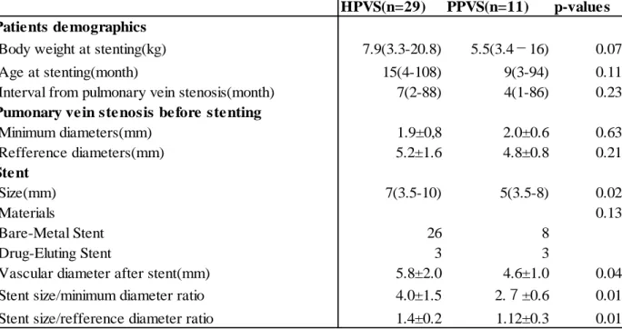
Table 3 presents a comparison of hybrid pulmonary vein stenting and percutaneous pulmonary vein stenting performed in the operating theater.
Significant differences were found in terms of stent size, vascular diameter after stenting, stent size/minimum diameter ratio, and stent size/reference diameter ratio. Two types of stents were used: bare-metal stent designed for peripheral
arteries and drug-eluting stent designed for coronary arteries. Thirty-four bare- metal stents were placed in 28 cases and 6 drug-eluting stents in 5 cases. Three types of bare-metal stents measuring 5–10 mm were used: Palmaz (Cordis, Miami Lakes, FL, USA), Express-Vascular (Boston Scientific, Natick, MA, USA), and Omnilink Elite (Abbott Vascular, Abbot Park, IL, USA). Two types of drug- eluting stents measuring 3.5 mm were used: Cypher sirolimus-eluting stent (Cordis, Miami Lakes, FL, USA) and Nobori biolimus-eluting stent (Terumo Corporation, Tokyo, Japan). Significant differences were observed between the bare-metal stent and drug-eluting stent groups in terms of the minimum and reference diameters of pulmonary vein stenosis prior to stent insertion and in terms of vascular diameter following stent insertion (table 4).
Among the 29 stents placed by hybrid procedure, 18 were subjected to anchoring suture to the atrial wall following stent insertion. Post-dilatation was performed in five stent cases. Simultaneous hybrid stenting at two stent sites in close proximity was performed in three cases.
4. Mortality and outcomes
The median overall follow-up period from pulmonary vein stenosis onset was 24 months (4–134 months). Fifteen patients died during the observation period owing to cardiac failure, pulmonary hypertension, or respiratory failure accompanying pulmonary vein stenosis. Figure 2 shows the Kaplan–Meier curve of survival rate.
The overall survival rate was 71% at 1 year and 49% at 5 years. As shown in Table 5, there were no significant differences except for the use of bare-metal stent between the survival and mortality groups. Although no significant differences were found between the cases of stent treatment and those of balloon angioplasty alone, the survival rate significantly differed between cases undergoing bare- metal stent and those undergoing other procedures (drug-eluting stent alone or balloon angioplasty alone). After treatment of pulmonary vein stenosis was initiated, biventricular repair, bidirectional Glenn shunt, and Fontan procedure were performed in two cases each. Takedown from bidirectional Glenn shunt to modified Blalock–Taussig shunt was performed in one case.
4、 In-stent restenosis
During the observation period, in-stent restenosis occurred in 27 stents (68%). The in-stent restenosis onset rate was 34% at 1 year and 70% at 5 years from stent implantation. Table 6 presents the comparison between the no in-stent restenosis and in-stent restenosis groups. The timing of pulmonary vein stenosis onset and stent implantation were significantly earlier and stent size was smaller in the in- stent restenosis group than in the no in-stent restenosis group. Regarding pulmonary vein stenosis before stent implantation, no significant difference was observed in terms of the minimum diameter, whereas the reference diameter before stent implantation and vascular diameter after stent implantation were
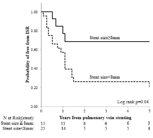
larger in the no in-stent restenosis group. Moreover, we found that stents placed by hybrid procedure were associated with freedom from significant in-stent stenosis than stents placed by percutaneous procedure. As shown in Figure 3 the in-stent restenosis onset rate during the observation period was significantly lower in the cases in which a stent of ≥8 mm was used.
5、 Re-intervention of stents
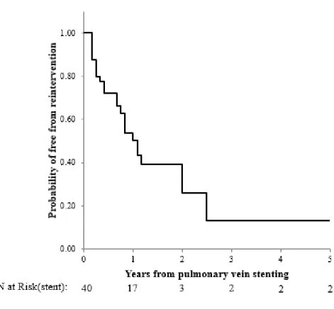
Re-intervention following stent implantation was performed for 24 stents (60%) in 16 cases, and the procedure was performed 48 times. Balloon dilation was performed 43 times. Stent-in-stent was performed in one case, whereas surgical dilation was performed in one case. Significant improvements were noted in terms of the narrowest diameter (2.2 ± 1.2 to 4.4 ± 1.7mm, p = 0.01) and stenosis site pressure difference (10.2 ± 3.1 to 4.1 ± 1.7 mmHg, p = 0.02) following balloon dilation. As shown in Figure4, re-intervention is commonly required following stent implantation. The median time of the first re-intervention was 5 months (1–
43 months) from stent implantation. Re-intervention was performed 31 times (65%) in the first year following stent insertion and 11 times (23%) in the following year. In 10 stents, multiple balloon dilations of stent were required.
Figure 5 shows the progress of a patient with isolated congenital pulmonary vein stenosis who received balloon angioplasty six times after stenting.
6、 Complication, obstruction, and malposition of stents
In our patients, we did not find any complications related to stent implantation other than a minor hemorrhage from the lung after a percutaneous procedure.
During follow-up, stent occlusion was noted in five stent cases (13%), among which four were recanalized with catheter intervention. Further, one stent migrated to the descending aorta and was retrieved with a percutaneous catheter.
Discussion
In this study, we present the results of balloon and stent treatment for pulmonary vein stenosis performed at our institution. The restenosis rate following surgery for the total anomalous pulmonary venous connection reported as 10%–15%2, 4, 5, and the reported risk factors include a small preoperative pulmonary veins6, preoperative stenotic lesions of pulmonary veins, and infra-cardiac total anomalous pulmonary venous connection7. Several studies have reported various effects of sutureless marsupialization on prognosis7,
8. Isolated congenital pulmonary vein stenosis is a rare disease with a poor prognosis, despite the existence of several treatments. The reported 3-year survival rate is 49% and risk factors for mortality include bilateral lesion, progression of diffuse stenosis, onset during early infancy, and restenosis following surgical treatment1, 9.
Regarding the pulmonary vein stenosis post pulmonary vein isolation for adult patients, the use of stents rather than balloon treatment alone has been reported to result in a lower risk of restenosis10,11, and stents of ≥10 mm have demonstrated good patency12.
Three types of stenosis may develop following surgical repair of total anomalous
pulmonary venous stenosis: stenosis of the atrial orifice following surgery, discrete stenosis in each inflow site of pulmonary vein, and long-segment stenosis from the inflow site to the upstream pulmonary vein. Our basic strategy is to perform rapid stenosis release, thus inhibiting the progression of the lesion to the upstream pulmonary vein13. While surgical release of the atrial orifice stenosis is effective, surgical and balloon angioplasty and/or stent implantations are considered for other types of stenosis. Although balloon angioplasty can be used to achieve stenosis release, the improvement is only temporary with a high likelihood of restenosis and many cases undergo the procedure multiple times.
Among our cases, following balloon angioplasty alone, eight of fifteen blood vessels (53%) became completely occluded during the follow-up period. Previous studies report the use of a cutting balloon for pulmonary vein stenosis, although only short-term improvement has been noted with a poor long-term prognosis14,15. Reported methods for stent implantation include bare-metal stent, drug-eluting stent, and covered stent.
Balasubramanian S et al. reported a significant difference in terms of the re-intervention rate for stents of ≥7 mm16, whereas Cory MJ et al. found that although no difference was found in the survival rate of drug-eluting stent and other types of stent, stent patency was superior in the group of drug-eluting stent17. As shown in Figure 2, our investigation revealed no significant differences in the survival rate between the group undergoing balloon angioplasty alone and that undergoing stent implantation. However, we did find a significant difference in terms of the survival rate between the bare-metal stent group
and other groups. The investigation based on stent type revealed that stent size, minimum diameter, reference diameter, and vascular diameter following stent implantation were all significantly larger in the bare-metal stent group than in the other groups (Table 4).
Therefore, we hypothesize that the placement of a large stent to secure a larger vascular diameter contributes to improved survival rates. On the other hand, our result show that it is difficult to place a large stent in an area where pulmonary stenosis is severe and progressive to upstream veins. Tomita H et al. found that good patency was achieved in blood vessels of patients who underwent dilatation with a stent of ≥5.6 mm and that repeated post-dilatation was required to maintain patency18. While the implantation of a large stent could contribute to prognosis and patency improvement, oversized stents could also contribute to in-stent restenosis development. Aggressive stent implantation using a high pressure or an oversized balloon is reportedly associated with the risk of in-stent restenosis, and damage to the intima could contribute to stenosis development19. Zamora CA et al. investigated in-stent restenosis in miniature pigs that underwent oversized stent insertion into the iliac artery and reported that the use of an oversized stent of 152% or greater resulted in a higher incidence of in-stent restenosis than smaller-sized stents20. Although we use stents of 150% or below the reference diameter, we have also inserted oversized stents larger than 150% into eight vessels for cases of hybrid pulmonary vein stenting.
According to a previous report by Furukawa T et al. on the use and effectiveness of drug-
eluting stent for pulmonary vein stenosis, comparison of similar-sized drug-eluting stent and bare-metal stent in pigs indicated that bare-metal stent was associated with more severe internal proliferation and stent stenosis and a significantly larger amount of granuloma formation than drug-eluting stent21. Drug-eluting stents used in our report were all of the size 3.5-mm. A drug-eluting stent has an advantage of pharmacological benefits; however, we selected a bare-metal stent because the larger stent is believed to be more important for good outcomes. Several reports have focused on the use of covered stent16, with Gordon BM et al. indicating that atrium expanded polytetrafluoroethylene covered stents are more effective for pulmonary vein stenosis than bare-metal stent22. Several reports have focused on hybrid stenting4, 23-26. We found that hybrid stenting resulted in no significant difference in terms of the minimum diameter of the stenosis site before stent implantation compared with percutaneous stenting; however, it did result in significantly larger stents being used and post-stent vascular diameter being significantly larger. Possible advantages of hybrid pulmonary vein stenting include the fact that it can be performed in cases with limited vascular access; stent insertion can be performed under direct vision; while the location must be predetermined, it can be easily adjusted; severe stenosis can be surgically dilatated prior to stent insertion; a larger stent can be inserted;
stent implantation is possible following plasty even for nearly occluded stenosis sites; any excessive stent length protruding to the atrium can be trimmed, flared and adjusted for future catheter treatment; and malposition can be prevented by an anchoring suture
connecting the stent tip to the atrial wall. Since 2010, our hospital has adopted the strategy of proactively performing only hybrid pulmonary vein stenting in patients. Although the physical burden associated with the use of a cardiopulmonary bypass (on-pump) and open-heart surgery is naturally greater than that associated with catheter treatment, it has several advantages. It has also been indicated that suturing of the atrial wall to prevent malposition can cause stent stenosis25. However, our strategy is to use stent suturing to prevent malposition. When suturing, the stent edge is slightly flared and sutured to the atrial wall, while ensuring that the thread of suturing does not contact the inside of stent.
Sullvian, P, M et al introduced the techniques of tailoring stents to fit the unique anatomical shaped vessels27. It may be possible to deploy trimmed stents to stenotic pulmonary vein using percutaneous procedure. However, advantages of hybrid pulmonary vein stenting are not only trimming, but also flaring, and suturing of stents.
These advantages are the primary reason why we selected hybrid stenting over percutaneous stenting in our hospital. Alternatively, avoiding cardiac surgery is an absolute merit for patients; hence, in the future, the feasibility of percutaneous stenting should be considered.
Our investigation of in-stent restenosis occurrence during the follow-up period indicated that the in-stent restenosis group had earlier onset of pulmonary vein stenosis and stent insertion timing and that the stent size tended to be smaller. As previously reported, early onset of pulmonary vein stenosis is a sign of poor prognosis, which may be due to severe
disease and stenosis in the upstream pulmonary vein, making it impossible to insert a larger stent9, 28. The tendency for stents of ≥8 mm to maintain patency was similar to those reported in previous studies.
During the observation period, stent re-intervention was performed 48 times. Quinonez LG investigated catheter treatment following stent insertion and reported that re- intervention became less common as the time since stent insertion increased. They hypothesized that disease progression might burn out28. In this study, the re-intervention- free rate was 50% at 1 year and 26% at 2 years, indicating that re-intervention was required by most stents. However, improvement was achieved in terms of vascular diameter and pressure difference in in-stent balloon dilation cases. Therefore, this procedure seems to be useful to maintain stent patency and is, therefore, strongly recommended. We believe that performing pulmonary vein stent implantation according to a long-term treatment plan, including balloon dilation of stent following insertion, can achieve better results. Cory MJ et al. reported that for in-stent balloon dilation, frequent re-intervention improved survival rates and vascular patency, suggesting that repeated performance of in-stent treatment to maintain patency contributes to achieving better outcomes17. Habara S et al. reported that use of a drug-coated balloon achieved a better clinical and vascular prognosis than a conventional balloon. However, they reported a maximum balloon size of 4 mm29. In the future, we hope to consider the use of drug- coated balloon also for the treatment of pulmonary vein stenosis and in-stent restenosis.
To date, we have not implemented chemotherapy to prevent stenosis nor have we conducted histological evaluation. The pathological mechanism of pulmonary vein stenosis is believed to be the occlusion of the pulmonary venous intima due to de novo intimal hyperplasia by myofibroblast proliferation30. Riedlinger WF et al. previously demonstrated that various types of receptor tyrosine kinase and ligands are expressed in the intima where stenosis has occurred31. Rehman M et al. found that the use of vincristine and methotrexate for inhibiting myofibroblast proliferation led to limited improvement32. Callahan R et al. reported that chemotherapy with imatinib mesylate and bevacizumab combination achieved a certain level of efficacy with few adverse events33. We believe that further investigation of such possibilities for the development of novel treatment of pulmonary vein stenosis needs to be conducted.
Study Limitations
This was a single-institution, retrospective study with a small sample size. It included the cases of pulmonary vein stenosis following surgery of total anomalous pulmonary venous connection and isolated congenital pulmonary vein stenosis with varying severity. While rare, there were some cases in which patients withdrew from the treatment prior to completion. Treatment approaches and devices that we can use have changed from 2001 to 2017. Our patients underwent balloon and stent treatments as well as various types of surgery; therefore, it was impossible to evaluate the effects of balloon and stent treatment
alone as well as each method’s issues. Further, it was impossible to investigate the effects of chemotherapy. Because pulmonary vein stenosis is a rare but often severe disease, comparison of different treatment selection is difficult. The conclusions derived from the present study are consistent with those of previous studies, indicating that insertion of a larger stent contributes to good vascular patency. However, we believe that to improve prognosis, proactive treatment is also required following stent implantation. Additional data are required to comprehensively evaluate surgical treatment, endovascular therapy, and chemotherapy, and further investigation is required regarding prevention of pulmonary vein stenosis.
Conclusion
We used balloon and stent treatments to aggressively treat pulmonary vein stenosis. The overall survival rates of 71% at 1 year and 49% at 5 years are not superior to those reported previously. The results suggest that the use of larger stents improves survival and vascular patency and that hybrid pulmonary vein stenting is suitable for the placement of larger stents. Treatment plans should consider post-stent re-interventions.
Acknowledgment
We would like to thank Dr Shunji Sano, Professor of surgery, Division of Pediatric cardiothoracic surgery of university California San Francisco for the surgery of congenital
heart disease in this research.
Financial Support
This research received no specific grant from any funding agency, commercial, or not- for-profit sectors.
Conflicts of interest None
Disclosure
The authors declare no conflict of interest.
Reference
1. Seale AN, Webber SA, Uemura H et al. Pulmonary vein stenosis: the UK, Ireland and Sweden collaborative study. Heart (British Cardiac Society). 2009;95:1944-9.
2. Seale AN, Uemura H, Webber SA et al. Total anomalous pulmonary venous connection: outcome of postoperative pulmonary venous obstruction. The Journal of thoracic and cardiovascular surgery. 2013;145:1255-62.
3. DiLorenzo MP, Ashley S, Faerber JA et al. Patient Characteristics and Mortality in Children With Pulmonary ein Stenosis. Circulation. 2018 136:A17454.
4. Mendelsohn AM, Bove EL, Lupinetti FM et al. Intraoperative and percutaneous stenting of congenital pulmonary artery and vein stenosis. Circulation. 1993;88:Ii210-7.
5. Hancock Friesen CL, Zurakowski D, Thiagarajan RR et al. Total anomalous pulmonary venous connection: an analysis of current management strategies in a single institution. The Annals of thoracic surgery. 2005;79:596-606; discussion 596-606.
6. Jenkins KJ, Sanders SP, Orav EJ, Coleman EA, Mayer JE Jr, Colan SD.
Individual pulmonary vein size and survival in infants with totally anomalous pulmonary venous connection. Journal of the American College of Cardiology. 1993;22:201-6.
7. Shi G, Zhu Z, Chen J et al. Total Anomalous Pulmonary Venous Connection: The
Current Management Strategies in a Pediatric Cohort of 768 Patients. Circulation.
2017;135:48-58.
8. Kanter KR, Kirshbom PM, Kogon BE. Surgical repair of pulmonary venous stenosis: a word of caution. The Annals of thoracic surgery. 2014;98:1687-91; discussion 1691-2.
9. Song MK, Bae EJ, Jeong SI et al. Clinical characteristics and prognostic factors of primary pulmonary vein stenosis or atresia in children. The Annals of thoracic surgery.
2013;95:229-34.
10. Fender EA, Widmer RJ, Hodge DO et al. Severe Pulmonary Vein Stenosis Resulting From Ablation for Atrial Fibrillation: Presentation, Management, and Clinical Outcomes. Circulation. 2016;134:1812-1821.
11. Buiatti A, von Olshausen G, Martens E et al. Balloon angioplasty versus stenting for pulmonary vein stenosis after pulmonary vein isolation for atrial fibrillation: A meta- analysis. International journal of cardiology. 2018;254:146-150.
12. Prieto LR, Schoenhagen P, Arruda MJ, Natale A, Worley SE. Comparison of stent versus balloon angioplasty for pulmonary vein stenosis complicating pulmonary vein isolation. Journal of cardiovascular electrophysiology. 2008;19:673-8.
13. Kato H, Fu YY, Zhu J et al. Pulmonary vein stenosis and the pathophysiology of
"upstream" pulmonary veins. The Journal of thoracic and cardiovascular surgery.
2014;148:245-53.
14. Seale AN, Daubeney PE, Magee AG, Rigby ML. Pulmonary vein stenosis: initial experience with cutting balloon angioplasty. Heart (British Cardiac Society).
2006;92:815-20.
15. McMahon CJ, McDermott M, Walsh KP. Failure of cutting balloon angioplasty to prevent restenosis in childhood pulmonary venous stenosis. Catheterization and cardiovascular interventions : official journal of the Society for Cardiac Angiography &
Interventions. 2006;68:763-6.
16. Balasubramanian S, Marshall AC, Gauvreau K et al. Outcomes after stent implantation for the treatment of congenital and postoperative pulmonary vein stenosis in children. Circulation Cardiovascular interventions. 2012;5:109-17.
17. Cory MJ, Ooi YK, Kelleman MS, Vincent RN, Kim DW, Petit CJ.
Reintervention Is Associated With Improved Survival in Pediatric Patients With Pulmonary Vein Stenosis. JACC Cardiovascular interventions. 2017;10:1788-1798.
18. Tomita H, Watanabe K, Yazaki S et al. Stent Implantation and Subsequent Dilatation for Pulmonary Vein Stenosis in Pediatric Patients-Maximizing Effectiveness-.
Circ J. 2003;67:187-190.
19. Klues HG, Radke PW, Hoffmann R, vom Dahl J. Pathophysiology and therapeutic concepts in coronary restenosis. Herz. 1997;22:322-34.
20. Zamora CA, Sugimoto K, Yamaguchi M, Sugimura K. Effect of stent oversizing on in-stent stenosis and lumen size in normal porcine veins. Journal of endovascular
therapy : an official journal of the International Society of Endovascular Specialists.
2005;12:495-502.
21. Furukawa T, Kishiro M, Fukunaga H et al. Drug-eluting stents ameliorate pulmonary vein stenotic changes in pigs in vivo. Pediatric cardiology. 2010;31:773-9.
22. Gordon BM, Moore JW. Treatment of pulmonary vein stenosis with expanded polytetrafluoroethylene covered stents. Catheterization and cardiovascular interventions : official journal of the Society for Cardiac Angiography & Interventions. 2010;75:263-7.
23. Ungerleider RM, Johnston TA, O'Laughlin MP, Jaggers JJ, Gaskin PR.
Intraoperative stents to rehabilitate severely stenotic pulmonary vessels. The Annals of thoracic surgery. 2001;71:476-81.
24. Gan C, Ji P, Lin K, Feng Y. A Novel Hybrid Approach for Balloon Dilation of Pulmonary Vein Stenosis Following Total Anomalous Pulmonary Venous Connection Repair with Atrial Septal Patching. Journal of cardiac surgery. 2015;30:608-10.
25. Shell KJ, Ebeid MR, Salazar JD, Dodge-Khatami A, Batlivala SP. "How to do it": hybrid stent placement for pulmonary vein stenosis. World journal for pediatric &
congenital heart surgery. 2015;6:284-7.
26. Yoon JK, Kim GB, Song MK et al. Hybrid Pulmonary Vein Stenting in Infants with Refractory to Surgical Pulmonary Vein Stenosis Repair. Pediatric cardiology. 2018.
27. Sullivan PM, Liou A, Takao C et al. Tailoring stents to fit the anatomy of unique vascular stenoses in congenital heart disease. Catheterization and cardiovascular interventions : official journal of the Society for Cardiac Angiography & Interventions.
2017;90:963-971.
28. Quinonez LG, Gauvreau K, Borisuk M et al. Outcomes of surgery for young children with multivessel pulmonary vein stenosis. The Journal of thoracic and cardiovascular surgery. 2015;150:911-7.
29. Habara S, Iwabuchi M, Inoue N et al. A multicenter randomized comparison of paclitaxel-coated balloon catheter with conventional balloon angioplasty in patients with bare-metal stent restenosis and drug-eluting stent restenosis. American heart journal.
2013;166:527-33.
30. Sadr IM, Tan PE, Kieran MW, Jenkins KJ. Mechanism of pulmonary vein stenosis in infants with normally connected veins. The American journal of cardiology.
2000;86:577-9, a10.
31. Riedlinger WF, Juraszek AL, Jenkins KJ et al. Pulmonary vein stenosis:
expression of receptor tyrosine kinases by lesional cells. Cardiovascular pathology : the official journal of the Society for Cardiovascular Pathology. 2006;15:91-9.
32. Rehman M, Jenkins KJ, Juraszek AL et al. A prospective phase II trial of vinblastine and methotrexate in multivessel intraluminal pulmonary vein stenosis in infants and children. Congenital heart disease. 2011;6:608-23.
33. Callahan R, Kieran MW, Baird CW et al. Adjunct Targeted Biologic Inhibition Agents to Treat Aggressive Multivessel Intraluminal Pediatric Pulmonary Vein Stenosis.
The Journal of pediatrics. 2018;198:29-35.e5.
Figure1. (A)Surgical view before hybrid pulmonary vein stenting, two orifices of pulmonary vein are very close(arrow). (B) After hybrid pulmonary vein stenting, two Express Vascular stents were implanted simultaneously. Both stent size had 5mm size and 15mm length. Cutting of extra stent strut and anchoring suture were placed to prevent migration.
Figure 2. (A)Kaplan-Meier analysis of patients treated for pulmonary vein stenosis. In overall, estimated survival is 71% and 49% at 1 years and 5 years. There is no significant difference between stent group and balloon angioplasty (BAP) group.
Figure 3. Kaplan-Meier analysis showing vascular outcome. Stent implanted with diameter≥8mm had a longer freedom from in-stent restenosis (ISR)(p=0.04).
Figure 4. Kaplan-Meier Analysis of re-intervention for pulmonary vein stents. Free from reintervention of pulmonary vein stent is 50% and 26% at 1 year and 2 years, respectively.
Figure 5. Angiogram and computed tomography of a patients who has isolated congenital pulmonary vein stenosis and common atrioventricular valve defect. Four pulmonary veins developed severe stenosis at early infantile period. (A) Right lower pulmonary vein stenosis of orifice before hybrid stenting at eleven months from birth. Two left stents were placed by percutaneous procedure. (B) Computed tomography of right upper and lower pulmonary vein stenosis. Two orifices of pulmonary vein were very close. (C) In-stent stenosis ten months after placement of 5mm bare-metal stent by hybrid procedure. (D) Follow-up catheter evaluation showed good patency except the occluded right upper
pulmonary vein. This is the recent angiogram five years after stenting. This patient underwent transcatheter re-interventions by balloon six times.
Table 1. Patients demographics and diagnostic data of pulmonary vein stenosis BAP group
(n=10)
Stent group (n=21)
Total (n=31) Patie nts
Body Weight at birth (g) 2881±276 2831±330 2874±324
IUGR 2 2 4
Preterm(<37weeks) 2 2 4
Female 5 6 11
Genetic malformation 0 2 2
Pulmonary ve in ste nosis
Onset age 8.5(1-20) 6(1-18) 7(3-20)
Stenotic veins>=2 veins 5 12 17
Bilateral pulmonary vein stenosis 5 12 17
Anticoagration the rapy
Warfarin 9 20 29
Aspirin 4 8 12
Ticlopidin 1 6 7
Pulmonary ve in de se ase
TAPVC 7 17 24
iCPVS 3 4 7
Additional cardiac le sions
None 0 6 6
CAVSD 0 1 1
Pulmonary Atresia with VSD 0 1 1
Heterotaxy syndrome 6 8 14
DORV 1 1 2
Hypoplastic Left Heart Syndrome 2 3 5
Tricupid Atresia 0 1 1
Mitral Atresia 1 0 1
BAP: Balloon Angioplasty, IUGR: Intra Uterine Growth Retardation, TAPVC: Total Anomalous Pulmonary Vein Connection, iCPVS: isolated Congenital Pulmonary Vein Stenosis, CAVSD: Complete Atrio-Ventricular Septum Defect, DORV: Double Outlet Right Ventricle
Table 2. Details about balloon angioplasty (BAP) of pulmonary vein stenosis Details of procedure
Balloon size (mm) 6(4-10)
Minimum diameter of stenosis(mm) 2.0±0.5
Refference diameter (mm) 5.1±1.1
Post BAP diameter(mm) 3.8±1.0
Balloon size/minimum diameter ratio 3.3±1.0
Balloon/refference diameter ratio 1.3±0.3
Pre BAP gradient of stenosis(mmHg) 11.8±3.5
Post BAP gradient of stenosis(mmHg) 5.7±1.9
Years from onset of pulmonary vein stenosis No. of interventions
~1y 16
~2y 8
~3y 2
Table 3. Difference between hybrid pulmonary vein stenting (HPVS) and percutaneous
pulmonary vein stenting (PPVS).
HPVS(n=29) PPVS(n=11) p-value s Patie nts de mographics
Body weight at stenting(kg) 7.9(3.3-20.8) 5.5(3.4-16) 0.07
Age at stenting(month) 15(4-108) 9(3-94) 0.11
Interval from pulmonary vein stenosis(month) 7(2-88) 4(1-86) 0.23 Pumonary ve in ste nosis be fore ste nting
Minimum diameters(mm) 1.9±0,8 2.0±0.6 0.63
Refference diameters(mm) 5.2±1.6 4.8±0.8 0.21
Ste nt
Size(mm) 7(3.5-10) 5(3.5-8) 0.02
Materials 0.13
Bare-Metal Stent 26 8
Drug-Eluting Stent 3 3
Vascular diameter after stent(mm) 5.8±2.0 4.6±1.0 0.04
Stent size/minimum diameter ratio 4.0±1.5 2.7±0.6 0.01
Stent size/refference diameter ratio 1.4±0.2 1.12±0.3 0.01
Table 4. Stent Types and Characteristics, comparison between Bare-Metal-Stents(BMS)
and Drug-Eluting-Stent(DES)
BMS(n=34) DES(n=6) p-value Patients demographics
Body Weight at stenting(kg) 6(3.3-20.8) 5.7(4.5-6.8) 0.53
Onset age of pulmonary vein stenosis(month) 6(1-20) 8(3-11) 0.65 Interval from pulmonary vein stenosis(month) 7(2-86) 4(1-7) 0.06
History of balloon dilation before stenting 20 1 0.06
Pumonary vein stenosis before stenting
Minimum diameter(mm) 2.1±0.7 1.3±0.4 0.02
Refference diameter(mm) 5.4±1.3 3.7±1.6 0.03
Stent
Size(mm) 7(5-10) 3.5(3.5) 0.01
Length(mm) 17(11-29) 18(8-24) 0.45
Vascular diameter after stenting(mm) 5.8±1.8 3.7±1.0 0.02
Table 5. Association with mortality of patients for pulmonary vein stenosis
Alive (n=16) Deceased (n=15) p-value Demographics
Body weight at birth(g) 2822±250 2874±397 0.81
Preterm(<37 weeks) 2 2 0.94
IUGR 2 2 0.94
Female 7 4 0.54
Genetic malformation 1 1 0.96
Pulmonary vein stenosis
Onset age of pulmonary vein stenosis(month) 9.5(1-20) 6(1-18) 0.22
Stenosic veins>=2veins 9 10 0.55
Bilateral pulmonary vein stenosis 10 10 0.81
Additional cardiac Issues
Single Ventricle 10 10 0.81
TAPVC 11 13 0.23
Heterotaxy Syndrome 7 7 0.87
History of intervention
First cardiac surgery 0.6(0.1-3) 1(0.1-7) 0.33
Sutureless repair 10 8 0.6
Use of Bare-Metal-Stent 12 5 0.02
IUGR: Intra Uterine Growth Retardation, TAPVC: Total Anomalous of Pulmonary Venous Connection
Table 6. Comparison of pulmonary vein stent with intra-stent restenosis (ISR) or not.
No ISR group (n=13)
ISR group
(n=27) p-value Follow up period from stenting(month) 19(4-63) 13(1-133) 0.24 Patie nts de mographics
Body weight at stenting (kg) 8.8(4.2-20.8) 5.5(3.3-15.6) 0.046 Onset age of pulmonary vein stenosis(month) 8(6-20) 4(1-18) 0.01
Age of stenting (month) 24(11-108) 10(3-72) 0.01
Ste nt
Size (mm) 8(5-10) 6(3.5-8) 0.01
Length (mm) 19(13-29) 17(8-24) 0.08
Pulmonary ve in ste nosis be fore ste nting
Minimum diameter (mm) 2.3±0.7 1.8±0.7 0.06
Refferece diameter(mm) 6.1±1.5 4.6±1.2 0.01
Vascular diameter after stenting(mm) 7.0±1.6 4.6±1.6 0.01
Proce dure of ste nting 0.03
HPVS 12 16
PPVS 1 11
HPVS: Hybrid Pulmonary Vein Stenting, PPVS: Percutaneous Pulmonary Vein Stenting