CASE REPORT
Spindle cell carcinoma of the breast – A case report –
Emiko Nozoe1, Tadahiro Nozoe1, Junko Tanaka2, Takefumi Ohga3, Aya Fujita4, and Katsuo Sueishi4Department of Breast Surgery1, Saiseikai Fukuoka General Hospital, Tanaka Junko Breast Clinic2, Department of Surgery3 and Pathology4,
Fukuoka Higashi Medical Center, Fukuoka, Japan
Abstract : Spindle cell carcinoma (SpCC) of the breast is quite a rare modality classified to the metaplastic car-cinoma of the breast. Regarding its biological behavior and the prognosis of the patients with this rare tumor, it has been remaining controversial. We herein report an 88 year-old woman who had a huge bleeding tumor on the right breast. She was a high-aged woman with low activities of daily life, even with some suspicion of distant organ metastasis. While the tumor proved to drastically bleed due to the tumor disintegration, a right simple mastectomy was performed. According to the histopathologic examinations, sarcomatoid spindle cells with severe atypia were observed. By an immunohistochemical examination, the tumor had proved to express neither estrogen receptor, progesterone receptor nor HER2 receptor. Moreover an immunohistochemical ex-pression of AE1/3 and CAM5.2, defining an epithelial neoplasm were observed in addition to an exex-pression of vimentin. From these findings, this bleeding tumor was diagnosed as spindle cell carcinoma of the breast. J. Med. Invest. 67 : 365-367, August, 2020
Keywords : spindle cell carcinoma, breast, malignant potential
INTRODUCTION
Spindle cell carcinoma (SpCC) of the breast is quite a rare tumor (1, 2) classified to the metaplastic carcinoma in general rules for clinical and pathological recording of breast cancer established by the Japanese Breast Cancer Society (3), and a mixture of sarcomatous spindle cells and epithelial tumor cells can account for the histopathologic characteristics of this tumor. Although the tumor often lacks the expression of ER, PgR, and HER2, it remains controversial whether its biological behavior is aggressive and the prognosis of the patients with this rare tumor is unfavorable (4-9).
Moreover while a study population for this tumor has been small, clinical features including the effectiveness derived from chemotherapy and radiotherapy has not been well appreciated.
We herein report an 88 year-old woman with SpCC of the breast who had undergone a surgical resection.
CASE REPORT
An 88-year old woman, who had a large tumor on the right breast came to see our institute.
The round-shaped tumor with a comparatively regular sur-face measuring 5.0cm in size was palpable in the lateral upper quarter area of the right breast. Moreover the tumor was found to partially invaded to the surface of the skin and a slight hemorrhage due to the disintegration of the tumor was also demonstrated.
In the laboratory examinations, the value of hemoglobin was found to be low (9.7g/dL), which was probably brought about by the hemorrhage of the breast tumor. There was no increase in the serum value of such tumor markers as CEA, CA15-3.
While she could not stand and stay still, a mammography and magnetic resonance imaging examination (MRI) could not be appreciated. Computed tomography demonstrated a breast tumor measuring 5 cm in size (Fig. 1). There existed no finding for distant metastasis and nodal metastasis in the axillary lymph nodes.
Preoperative histological examination demonstrated the histological diagnosis as ductal carcinoma, but the definitive histological type could not be determined.
While she was a high-aged woman with low activities of daily life, and the tumor proved to continuously bleed due to the tumor disintegration, a right mastectomy was performed (Fig. 2).
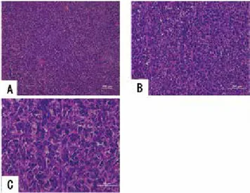
By the Histological examination, sarcomatoid spindle cells with severe atypia were observed (Fig. 3). An immunohistologi-cal examinations demonstrated that tumor was found to express
The Journal of Medical Investigation Vol. 67 2020
365
Received for publication January 28, 2020 ; accepted April 1, 2020. Address correspondence and reprint requests to Emiko Nozoe, Depart-ment of Surgery, Saiseikai Fukuoka General Hospital, 1-3-46, Tenjin, Chuo ward, Fukuoka, 810-0001, Japan and Fax : +81-92-716-0185.
Fig 1. Photo of computed tomography. A breast tumor measuring 5 cm in size with a comparably regular surface was demonstrated.
366
E. Nozoe, et al. Breast spindle cell carcinomaneither ER, PgR nor HER2 receptor. An immunoreactivity for Ki-67 proved to be 32% (Fig. 4). An expression of AE1/3 and CAM5.2, defining an evidence for epithelial neoplasm were observed (Fig. 5). Moreover a strong expression of vimentin was also observed (Fig. 6). These findings could compensate for the pathologic features of spindle cell carcinoma of the breast.
DISCUSSION
SpCC of the breast, a subtype classified as the carcinoma with metaplasia, spindle cell type in WHO classification, is quite a rare tumor, accounting for less than 0.5% among carcinoma of the breast (1, 2).
The most common and outstanding clinical features of this tumor is a formation of large palpable mass (10). High density tumor shadow with a unclear boarder on mammography, and tumor shadow with irregular shape, microlobulated margin, complex echogenicity, parallel orientation, and posterior acoustic enhancement on ultrasonography and higher intensity of the signal on T2 MRI imaging can account for the common features of breast SpCC in diagnostic images (11), but in our case mam-mography and MRI test could not be accomplished because of the physical condition.
While SpCC of the breast has been known to lack in expression of ER, PgR, and HER2 in many cases (8, 11), the malignant po-tential of this tumor has been reported to be more aggressive (6, 7). However, on the other hand, less frequent incidence of lymph node metastasis and more favorable prognosis of patients with SpCC of the breast, in spite of the large growth of the tumor, as is concomitant with our case, has been also reported (4, 5, 7).
Even when, she had distant metastasis that would prevent curative resection, surgical treatment might be required, even palliative resection. Because the hemorrhage owing to the dis-intergration of the tumor would definitively spoil the QOL of the patient. Indeed, the general condition of the patient remarkably improved after surgical resection.
Although chemotherapy other than hormonal therapy might be considered, no definitive anticancer drugs exerting a high efficacy for SpCC of the breast has been presented due to the small number of this modality. As our case is high-aged woman and activities of daily living (ADL) proved to be low, she desired to avoid chemotherapy.
Fig 2. Macroscopic finding.
Cut surface demonstrated the solid well-circumscribed tumor with central bleeding due to necrosis of the tumor.
Fig 3. Histopathologic finding.
Sarcomatoid spindle cells with atypia were shown.
A. Magnification ×10. B. Magnification ×20. C. magnification ×40.
Fig 4. Photo of immunohistochemical expression for Ki-67. An immunoreactivity was 32%.
A. Magnification ×20. B. magnification ×40.
Fig 5. Photo of immunohistochemical expression for keratin. Strong expression of AE1/3 (A. Magnification ×20.) and CAM5.2 (B. Magnification ×20.) in the cell membrane of the tumor cells was demonstrated, showing this tumor could be an epithelial neoplasm.
Fig 6. Photo of immunohistochemical expression for vimentin. Strong expression of vimentin was demonstrated.
367
The Journal of Medical Investigation Vol. 67 August 2020
There exists no clear understanding for the pathogenetic mechanism for the occurrence of SpCC of the breast. It remains still controversial whether this tumor originated from epithelial growth or derived from mesenchymal origin. Possible correlation of the concept of epithelial-mesenchymal transition (EMT) with the pathogenesis of SpCC of the breast has been demonstrated (12).
Pathologic investigation in this case demonstrated both a sarcomatoid features containing spindle shaped-cells with abun-dant mitoses and an epithelial proliferation.
Tumor cells in breast SpCC carcinoma has been derived from myoepithelial cells or differentiate to myoepithelial cells (13). In our case, immunohistochemical examination demonstrated an expression of both cytokeratin A1/A3 and vimentin, showing the correlation of myoepithelial cells with the pathogenesis of SpCC of the breast.
While the tumor of our case contained neither hormone recep-tors nor HER2 receptor, it was found to have strong expression of Ki-67, substantiating the malignant potential of the tumor. It has been reported that tumor cells in SpCC contained strong ex-pression of Ki-67 exex-pression, demonstrating an acquisition of the more aggressive cellular potential while epithelial components transit to the sarcomatoid lesion diagnosed as SpCC (14).
Nevertheless, meticulous follow-up for tumor recurrence should be mandatory.
REFERENCES
1. Ellis IO, Bell J, Roman JE, Elston CW, Blamey RW : Im-munocytochemical investigation of intermediate filament protein and epithelial membrane antigen in spindle cell tumours of the breast. J Pathol 154 : 157-165, 1988
2. Kaufman MW, Marti JR, Gallager HS, Hoehn JL : Car-cinoms of the breast with pseudosarcomatous metaplasia. Cancer 53 : 1908-1917, 1984
3. Japanese breast cancer society : General rules for clinical and pathological recording of breast cancer. Breast Cancer
12 : S12-14, 2005
4. Maemura M, Iino Y, Oyama T, Hikino T, Yokoe T, Takei H, Horiguchi J, Ohwada S, Nakajima T, Morishita Y : Spindle cell carcinoma of the breast. Jpn J Clin Oncol 27 : 46-50, 1997
5. Carter MR, Hornick JL, Lester S, Fletcher CD : Spindle cell (sarcomatoid) carcinoma of the breast : a clinicopathologic and immunohistochemical analysis of 29 cases. Am J Surg Pathol 30 : 300-309, 2006
7. Khan HN, Wyld L, Dunne B, Lee AH, Pinder SE, Evans AJ, Robertson JF : Spindle cell carcinoma of the breast : a case series of a rare histological subtype. Eur J Surg Oncol 29 : 600-603, 2003
8. Nahleh Z, Ebrahim V, Guerrero R, Gaur S, Ayyappan A, Padilla O : Spindle cell carcinoma of the breast : a case report and discussion. Breast Dis 33 : 115-119, 2011 9. Moten AS, Jayarajan SN, Willis AI : Spindle cell
carcino-ma of the breast : a comprehensive analysis. Am J Surg 211 : 716-721, 2016
10. Günhan-Bilgen I, Memiş A, Ustün EE, Zekioglu O, Ozdemir N : Metaplastic carcinoma of the breast : clinical, mam-mographic, and sonographic findings with histopathologic correlation. AJR Am J Roentgenol 178 : 1421-1425, 2002 11. Choi BB, Shu KS : Metaplastic carcinoma of the breast :
mul-timodality imaging and histopathologic assessment. Acta Radiol 53 : 5-11, 2012
12. Zhang Y, Toy KA, Kleer CG : Metaplastic breast carcino-mas are enriched in markers of tumor-initiating cells and epithelial to mesenchymal transition. Mod Pathol 25 : 178-184, 2012
13. Wargotz ES, Deos PH, Norris HJ : Metaplastic carcino-mas of the breast. II. Spindle cell carcinoma. Hum Pathol 20 : 732-740, 1989
14. Nakayama Y, Iwasaki H, Iwanaga S, Nakamura H, Shiroshita T, Kikuchi M, Jozaki H, Hashimoto M, Ikeda S : Spindle cell carcinoma of the breast : a case report and an immunohistochemical study including p53 and Ki-67 expression. Pathol Int 47 : 404-411, 1997