INTRODUCTION
Carcinomas of extrahepatic and intrahepatic bile duct account for a considerable amount of the cancer related death in Japan. As a group, intrahepatic and extrahepatic bile duct occurs with almost equal frequency in males and females and has a peak incidence during the sixth and seventh decade of life. All of these carcinomas have poor prognosis (6), although the prognosis of ampullary carcinoma and lower extrahepatic bile duct carcinoma is rela-tively good due to an earlier clinical presentation (6). There have been few studies regarding Ki-67 and p53 protein expressions of these tumors especially in extrahepatic carcinoma. The relationship between intrahepatic carcinoma and cell proliferative activity is unknown to the best of our knowledge. There have
been a few comprehensive studies done regarding the biological activity of bile duct carcinomas and prognosis after surgery (15, 18, 19). Therefore this study aims at studying the relationship, if any, between the cell proliferative markers Ki-67 and p53 in bile duct and gallbladder carcinomas and the prognosis of the patients as a whole.
Bile duct carcinoma persists as one of the carcinomas associated with a poorer prognosis. This poorer prog-nosis is attributed to the fact that characteristics of bile duct carcinomas are typically in an advanced stage at the time of diagnosis. The 1 year, 3 year and 5 year survival of intrahepatic cholangiocellular carcinoma in Japan is 19.9%, 3.4% and 1.1% (1) whereas in extrahepatic cholangiocelluar carcinoma (EHC), the survival rate is 30-50% in carcinoma of ampulla of Vater, 10-20% in pancreatic head cancer and 25% in lower bile duct carcinoma (2-4). In accordance with the advancement of Ultrasound scanning (US), Endoscopic retrograde cholangio-pancreatography (ERCP), Computed tomography (CT), Magnetic resonance image (MRI) and angiographic studies,
Prognostic significance of Ki-67 and p53 antigen
expres-sion in carcinomas of bile duct and gallbladder
Mitra Lal Shrestha, Hidenori Miyake, Toru Kikutsuji, and Seiki Tashiro
First Department of Surgery, The University of Tokushima School of Medicine, Tokushima, Japan
Abstract : Ki-67 and p53 protein expression was evaluated immunohistochemically in 32 patients with intrahepatic, extrahepatic bile duct and gallbladder carcinomas, who underwent surgery at First Department of Surgery, The University of Tokushima School of Medicine. p53 expression was found more in the well differentiated group than poorly differentiated group (p=0.007). MIB1 labelling index (MIB1 LI) was higher in EHC than in GBC (p=0.0061). MIB1 LI (T), (MIB1 LI in tumor) was higher in cases with lymph node metastasis than in those without lymph node metastasis (p=0.0189). Moreover, MIB1LI (L) (MIB1 LI in metastasized lymph node) was higher in poorly differentiated than in well differentiated carcinoma (p=0.0404). Prognostically, patients with high MIB1 LI (T) (>56.93) had a worse prognosis after surgery than those with low MIB1 LI (T) (p<0.05). There was no association between p53 positive tumors and MIB1 expression. These results suggest that cancer cell proliferative activity was markedly increased in cases with EHC compared to those with GBC and the poorly differentiated and lymph node metastasis group. MIB1 LI in tumor was found to be a good prognostic indicator whereas there was no association of p53 positive tumor with MIB1 expression and prognosis of the patients. J. Med. Invest. 45 : 95-102, 1998
Key words : Ki-67, MIB1, p53, cholangiocarcinoma, Gallbladder carcinoma
Received for publication June 15, 1998 ; accepted July 30, 1998.
1
Address correspondence and reprint requests to Seiki Tashiro, M.D., Ph.D., First Department of Surgery, The University of Tokushima School of Medicine, Kuramoto-cho, Tokushima 770-8503, Japan and Fax : +81-886-31-9698.
The Journal of Medical Investigation Vol.45 1998
the frequency of detection of bile duct carcinomas has been increased (5, 6). Similarly major hepatic resection combined with or without caudate lobectomy for carcinomas of hepatic duct confluence, with or without portal vein resection, has attributed the prolongation of survival in such patients (7).
To determine tumor growth and/or aggression, various techniques can be used. Carcinoembryonic antigen (CEA), cytokeratin, tumor grade and stage are reasonable predictors of tumor biological activity, but they do not reflect the exact proliferative rate of cancer. Precise proliferative activity of neoplasm can be assessed accurately using various immuno histochemical methods. One such method uses antibody to Ki-67 antigen, MIB-1, which is presented only in actively dividing cells, that is in G1, S, G2and M cell phasese (13, 14). Expression of this antigen in a tumor specimen approximates the cancer proliferative rate (11, 12). A major advantage of Ki-67 antigen labelling is the ability to apply this technique to paraffin embedded as well as to fresh specimens, allowing retrospective analysis of pathological mate-rial (14). The p53 suppressor gene is known to play an important role in cell proliferation, and its over-expression is related to prognosis (25, 26). Few reports to date have examined Ki-67 and p53 expression as a marker of tumor cell proliferation in extrahepatic and none in intrahepatic bile duct carcinomas to the best of our knowledge. In this study we have assessed the proliferative activity of EHC, IHC and gallbladder carcinomas (GBC) to specify the difference, if any.
MATERIALS AND METHODS
Patients and tumor materials :Histopathological studies were made on the surgical materials of 32 patients (19 males and 13 females). Formalin fixed paraffin embedded tissues from patients with EHC (n=11), IHC (n=12) and GBC (n=9) and non-neoplastic bile duct (n=18), who under-went surgery at First Department of Surgery, The University of Tokushima School of Medicine with verified follow up data from January, 1994 onwards were studied. The patients had undergone surgery according to the general rules of the surgical pathology study group of Japan in cancer of biliary tract and liver. Their mean age was 64.3±7.0 years. The range was 43-83 years. The medical records and surgical pathology reports were reviewed for each case. No patient had received preoperative cancer
chemotherapy and/or radiotherapy.
Bile duct carcinomas were further divided into well differentiated and poorly differentiated types. The well differentiated bile duct carcinomas were characterized by tumor cells that showed well defined glands where as the poorly differentiated ones had poorly defined glands characterized by tumor cells growing into cords, trabeculae, nests, nodules or sheet. Adequate clinical follow-up information was available for all bile duct and gallbladder cancer patients.
Immunohistochemical analysis :
The original tissue slides were reviewed for diagnostic confirmation and for adequacy of tumor. Immunohistochemical analysis was conducted using the avidin -biotin- peroxidase complex (ABC) method (Dako carpenteria, CA). Sections (4 mm thick) were cut from the paraffin embedded blocks and mounted on superfost glass slides. They were deparrafinized in Xylene and redehydrated in distilled water through a conventional ethanol scale. After washing through with phosphate-buffered saline (PBS) (pH 7.4), the slides for Ki-67 and p53 were heated to retrieve the antigenicity in a microwave oven at 600W (35 minute cycle) in 10mM sodium citrate buffer at pH6.0, and those for p53 were heated at 600 W (for 5 minutes) in deionized water. After heating, Ki-67 and p53 slides were left at room temperature for 2 hours. After endogenous peroxidase activity was blocked with 0.3% H2O2 in methanol for 15 minutes, the sections were washed in PBS and treated with normal rabbit serum for 10 minutes. The slides were then incubated for 20 minutes at room temperature with a mouse monoclonal antibody for p53 (DO-7, DAKO, Glostrup, Denmark) diluted at 1 : 50. The slides were incubated at 4℃ with a mouse monoclonal antibody for Ki-67 (MIB-1, Immunotech, Marcilles, France) diluted at 1 : 50. After being rinsed in PBS, they were treated with biotinylated rabbit antimouse immunogloulin G for 30 minutes, rinsed and treated with Streptavidin-bionylated peroxidase complex (SAB-PO : Nicheri) for 20 minutes. They were then visualized with 3,3’-diaminobenzidine containing 0.01% H2O2as chromogen and were counterstained with hematoxylin. For negative control, primary antibody was replaced with PBS. All slides were evaluated for immunostaining in a blinded fashion, without any clinicopathological information.
p53 expressions and MIB1 LI were evaluated under light microscopic observation at x 400 magnification. In the immunostaining analysis,‘positive’reddish brown tumor nuclei were detected and discriminated
M.L.Shrestha et al. Prognostic significance of Ki-67 and p53
from‘negative’blue hematoxylin nuclei. Calculations were based on 5 fields in each section within cancer cell areas. For p53 analysis, immunoreactive nuclei were counted out of 1000 tumor nuclei. Fields were not selected at random to avoid areas with stroma, but measurements were specifically no done in the most proliferative areas to obtain an average estimation of specimens.
p53 expression was classified into 3 grades accord-ing to the intensity of stainaccord-ing and proportion of positive tumor nuclei as follows ;
(++) > 10% immunoreactive tumor nuclei with intense staining
(+) 1-10% immunoreactive tumor nuclei with weak staining
( - ) < 1% immunoreactive tumor nuclei or the cases with no staining.
The extent of positivity in the case of p53 was determined using a cut off value of more than 10% based on previous reports by others. (22, 33, 34). These authors have reported a cut off value of p53 protein varying from 10-20% and a concordance rate between p53 expression and gene mutation of up to 73% (34). MIB1 indices were also determined by scoring the number of positively stained nuclei out of 1000 cancer cell nuclei at 5 places near the cancer cell areas as in p53 calculation. For Ki-67 immunostaining, the intensity of nuclear staining varied from weak to dense, and all slides were considered positive. The threshold high was defined as the mean value of nuclei stained positive when > 56.93% and high p53 index when 10% of the tumor nuclei were stained. The remainder were classified as low index. The value of 56.93% for the MIB1 index was used as a cut off value to divide the patients into 2 groups with different risks and was observed statistically to be the lowest allowing discrimination with a significant probability.
Histopathological examination
Histological classification, grade, depth of invasion, lymphatic and vascular permeation, lymph node involvement, and serosal involvement were examined histopathologically. The histological grading of cholangiocellular carcinoma was divided into well, moderate and poorly differentiated. The depth of invasion was divided into two ; invasion within the serosal layer and beyond the serosal layer. Similarly lymphatic permeation and vascular invasion also divided into positive and negative groups.
Statistical analysis ;
The frequency distribution was tested by the chi-square tests for pathological features and p53 expressions and the quantitative data on Ki-67 was examined by unpaired Mann-whitney test. The survival curve for the 32 patients was determined by Kaplan Meir and the log rank (Mantal-Cox) method. To assess statistical significance, a p value <0.05 was used.
RESULTS
MIB1 labelling index (MIB1 LI) and p53 expression was evaluated in 12 IHC, 11 EHC, 9 GBC and 18 non-neoplastic bile ducts.
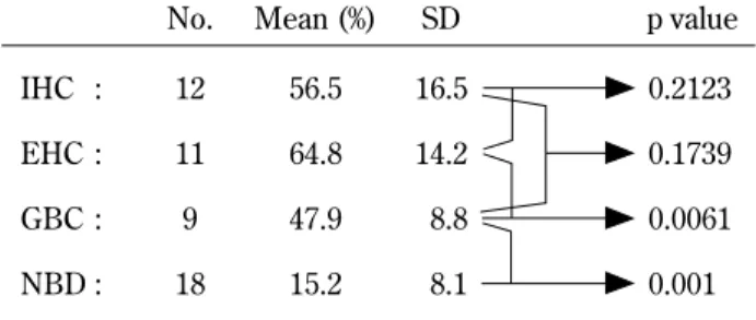
Immunohistochemistry of MIB 1 Expressions : The MIB1 LI ranged from 2-22% in non-neoplastic bile duct (mean 15.2±8.1%). In contrast, MIB1 LI of all grades in EHC were higher than those of GBC (p=0.0087). However there was no significant association between IHC and GBC, or IHC and EHC (Table 1). There was a significant difference in regard to MIB1 LI between poorly differentiated and well-differentiated carcinomas. The poorly differ-entiated carcinomas had an MIB 1 LI of 80±8.10% in comparison to well differentiated carcinoma which had a mean MIB1 LI of 60±16.2% (p=0.0404). Similarly, in 17 cases with lymph node metastasis group the mean MIB1 LI was significantly higher (Mean 62.7±14.2%) than in those without lymph node metastasis (mean 50.4±13.8%) (p=0.0189) (Table 2). Simple regressional analysis demonstrated a strong correlation between MIB1 LI in tumor and MIB1 LI in metastasized lymph node with a correlation coefficient of r2
of 0.957 (p<0.0001) (Fig.1).
Immunohistochemical analysis of p 53 expressions : Expression of p53 was detected in 6 (50%) of
Table 1. MIB-1 labelling index in biliary tract diseases
No. Mean (%) SD p value IHC : EHC : GBC : NBD : 12 11 9 18 56.5 64.8 47.9 15.2 16.5 14.2 8.8 8.1 0.2123 0.1739 0.0061 0.001
IHC : Intrahepatic cholangiocellular carcinomas EHC : Extrahepatic cholangiocellular carcinomas GBC : Gallbladder carcinomas
NBD : Non neoplastic bile duct
12 IHC, 8 (72.7%) of 11 EHC and 4 (44.4%) of 9 GBC (Table 3). p53 was expressed in 19 (79.2%) out of 24 well differentiated carcinoma while it was expressed only in 1 (12.5%) of 8 poorly differentiated carcinoma., which was significant (p=0.007). The relation between depth of cancer invasion particularly in reference to serosal involvement and p53 expressions, showed p53 was expressed in 8 (88%) of 9 patients with serosal involvement. On the other hand, 12 (52.2%) of 23 patients without serosal involvement showed p53 expressions. Though statistically not significant, a tendency to be expressed more in cases with serosal involvement than without serosal involvement was observed (p=0.0537) (Table 4). However there was no correlation between p53 expressions and lymphatic, vascular permeations and lymph node involvement
Table 2. Mean MIB-1 Labelling index and clinicopathological characteristics
No. MIB-1 LI(%) SD p value Remarks Grade Well 24 Poor 8 55.04 16.0 12.66 20.85 0.173 MIB 1 (T) NS Grade Well 13 Poor 4 60.0 80.0 16.22 8.10 0.0404 (MIB-1 L) Serosal involvement Absent 23 Present 9 54.82 62.33 14.43 15.15 0.2127 NS Venous invasion Absent 20 Present 12 53.70 62.33 14.83 14.58 0.1193 NS Lymphatic permeation Absent 18 Present 14 54.91 59.84 16.22 13.39 0.3765 NS Lymph node metastasis
Absent 15 Present 17 50.40 62.72 13.80 14.15 0.0189 Perineural invasion Absent 28 Present 14 52.58 64.53 14.92 13.23 0.308 NS p 53 Expression Absent 14 Present 18 55.83 58.26 18.52 13.14 0.6721 NS
MIB1 LI in tumor was higher in cases with lymph node metastasis than without (p=0.0189) Moreover MIB1 LI in metastasized lymph node was higher in poorly differentiated than in well differentiated carcinomas (p= 0.0404). There was no association between p53 positive tumors and MIB1 expression. (T=tumor, L=lymph node, NS=not significant)
Fig.1. Simple regressional analysis demonstrated a strong correlation between MIB1 LI in tumor and MIB1 LI in metas-tasis lymph node with a correlationn coefficient of r2=0.957 (p<
0.0001)
M.L.Shrestha et al. Prognostic significance of Ki-67 and p53
(Table 4) There was also no correlation between p53 expression and MIB1 LI.
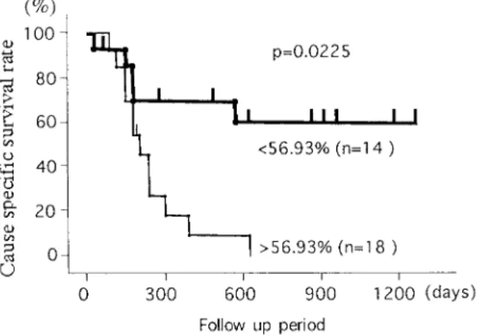
MIB1 and Cause Specific Survival :
MIB1 LI was found to be predictive of survival. The probability of 1200 days survival was 47% with tumors with low MIB1 indices (<56.93%). Virtually all patients died within 700 days with high MIB1 indices (Fig.2).
p53 and cause specific survival :
There was no significant correlation between
positive p53 expressions as per cut off value of 10% of low and high expressers and prognosis in our study (p=0.3077). (Fig.3)
DISCUSSION
Cell cycle activity and cell kinetics are important indicators of growth and behavior of various human tumors (8). The most sensitive method of assessing tumor cell proliferation is immunostaining of these sections with antibodies to Ki-67 (9). The antibody
Table 3. Incidence of p53 Expression in biliary tract diseases
p53 Total Expression Rate(%) ‐ + + + IHC EHC GBC NBD 6 3 5 18 1 1 3 0 5 7 1 0 12 11 9 18 50 72.7 44.4 0 NBD : Nonneoplastic bile duct
IHC : Intrahepatic cholangiocellular carcinoma EHC : Extrahepatic cholangiocellular carcinoma GBC : Gallbladder carcinoma
p53 (-) : No staining
p53 (+) : Weak staining or less than 10% positive nuclei p53 (++) : intense staining or more than 10% positive nuclei
Table 4. p53 expression vs clinicopathological chracteristics
p 53 expression
(-) (+) (+ +) Total Expression rate p value Grade Well Poor 5 7 5 1 14 0 24 8 79.20 12.50 0.007 Serosal involvement Absent Present 11 1 3 2 9 6 23 9 52.17 88.88 0.054 Venous invasion Absent Present 9 4 2 2 9 6 20 12 55.00 66.70 0.515 Lympatic permeation Absent Present 8 5 3 1 8 7 19 13 57.90 61.50 0.837 Lymph node involve.
Absent Present 7 5 3 2 5 10 15 17 50.00 57.90 0.314
p53 expressions were found more in the well differentiated than poorly differentiated group (p=0.007). p53 had a tendency to be expressed more in cases with serosal involvement than without (p=0.0537).
Fig. 2. Cause specific survival curves according to MIB1
Labeling index
to the Ki-67 antigen recognizes a nuclear protein of 345 and 395 kD, encoded by a gene on chromosome 10 (10-12). Ki-67 is present on cycling cells with peak concentrations during the G2and M phase of cell cycle and it is not expressed in the cells in the Go phase or early G2phase (12). MIB1 is the true equivalent of Ki-67 and provided an antigen retrieval process is used, it can be detected in formalin fixed, wax embedded material (13). MIB-1 expression correlates with semiconservative DNA synthesis but not DNA repair synthesis (14). Ki-67 has a short half-life and the level of Ki-67 in the cell declines rapidly after the mitotic phase. Ki-67 expression in the form of MIB-1 immunoreactivity is a more accurate indicator of cell proliferation in histologic material (14).
This study demonstrates MIB1 immunoreactivity in extrahepatic, intrahepatic bile duct and gallbladder carcinoma. The proliferative activity in benign and neoplastic lesions has been compared. MIB1 indices in EHC, IHC, and GBC were significantly higher than in non-neoplastic bile duct (p<0.001). Similar results were obtained in the study of Lee et al. (19). However MIB1 expressions in GBC were signifi-cantly lower than in EHC in our study. Moreover there was no significant association between GBC and IHC (Table 2).
It is not surprising that the patients with higher MIB1 LI tend to have a low survival rate. Several others have reported prognostic significance of Ki-67 in breast, ovary and gastric carcinoma (16, 17, 31). In our study, cases with lymph node metastasis had high MIB1 levels and these cases are also related to poorly differentiated component. Similar findings have been noticed by others (15, 17).
Irene OL et al . also reported better differentiated hepatocellular carcinoma had lower MIB1 labelling
index than poorly differentiated one but had no influence on venous permeation, liver invasion, and positive resection margin which was similar to our findings (20). Similarly a different study on invasive gastric carcinoma showed that poorly differentiated tumor had a much higher proportion of tumor cells labelled for MIB1 than well differentiated tumor (17). Simple regressional analysis demonstrated a signif-icant correlation between MIB1 LI in tumor and MIB1 LI in lymph node with a coefficient of deter-mination of r2
=0.957 (p<0.001). Our results further confirm that the MIB1 labelling index of the tumor is of prognostic significance.
The findings of a higher incidence of p53 in well differentiated carcinoma in our study are also reported by Ming et al (18). But in contrast to our study, the poorly differentiated gallbladder carcinoma in their study also showed a higher overall p53 positivity of 73%. Although statistically not significant because of the smaller number of cases in our group, our findings that well differentiated IHC and EHC tend to show increased p53 expressions in contrast to GBC support these could be of different genetic entities (data not shown), although more studies on p53 genetic sequence for specific point mutation would be certainly helpful in confirming these findings. We could not detect any association between p53 expressions and survival. Kastan MB et al . also examined MIB1 and p53 index in hepatocellular carcinoma and found a strong correlation in that p53 positive tumors had a significantly high Ki-67 in all grades of HCC (21). In contrast to his findings we could not detect any association between p53 and MIB1 expressions. Therefore HCC and bile duct as well a gallbladder carcinomas might have separate genetic events during initiation and progres-sion of carcinogenesis.
It has been regarded that p53 might act as tumor suppressor gene, located in the short arm of chro-mosome 17 (25), because its mutation or allelic deletion plays an important role in the development of many human cancers. Normal p53 induces the apoptosis in the detection of the damage of DNA, while mutant p53 and allelic deletion of p53 sup-presses the apoptosis. Immunohistochemical detection of p53 has been considered to be associated with p53 mutation in situ (22). Also the nuclear over-expression of p 53 has been reported in various malignancies such as lung, breast, esophageal, gastric, colon and extrahepatic bile duct cancer (27-30). The concordance rate of p53 expression and p53 gene mutation differs from study to study. Guldo Coggi et al . and
Fig. 3. Cause specific survival curves according to p53 expression
M.L.Shrestha et al. Prognostic significance of Ki-67 and p53
Salem et al . reported in less than 10% of cases of p53 protein expression, immunohistochemistry did not reveal any p53 gene mutation (33, 34). Coggi also reported p53 expression was found in esopaha-geal carcinoma in 68% of those who also showed p53 accumulation by immunohistochemisty (34). In pancreatic carcinomas, Dorandeu et al. reported nei-ther p53 nor Ki-67 had any effect on the prognosis of the patients by immunohistochemical analysis (35). They found that p53 had accumulated in 49% of their series. We found that p53 had accumulated in altogether (56%) 18 patients (6 of 12 IHC, 8 of 11 EHC and 4 of 9 GBC) in our study. This is similar to the rate reported by Ming et al. showing p53 positive results in 5 out of nine tumor of papilla of Vater and 66% of common bile duct tumors (18). However, he found that unlike tumors of gallbladder, there was no association between low degree of differentiation and p53 positivity, a finding which may be related to lack of prognostic value for p53. Until the present study, no report had examined over-expression of p53 and depth of cancer invasion in bile duct carcinomas. p53 expression had a tendency to be increased with the extent of tumor invasion. However, there was no correlation with lymphatic permeation, vascular permeation and lymph node metastasis.
CONCLUSION
In conclusion, the Ki-67 index correlated with the prognosis of the patient and with the poorly differentiated and lymph node metastasized group but did not correlate with other pathological findings. p53 expression did not show any association with prognosis and MIB1 expressions, though the expres-sion was seen in more well differentiated carcinoma. Thus analysis of Ki-67 of tumor and lymph node will provide useful prognostic information for patients with EHC, IHC and GBC who undergo surgery.
ACKNOWLEDGEMENTS
The authors would like to thank Keisuke Izumi, M.D., Ph.D., Nobuya Sano, M.D., Ph.D. (Second Department of Pathology), and Masashi Tsuji, M.D., Ph.D.(Department of Urology) for valuable contri-butions to this study.
REFERENCES
1. Okuda K. and the liver cancer study group of Japan : Cancer 45 : 2663-69., 1980
2. Yamaguchi K. Enjoji M : Ampullary carcinoma in patients with under 50 years of age with a poor prognosis : J Surg Onco 45 : 201-06, 1990 3. Forrest JF, Longmire WP : Carcinoma of pancreas and periampullary region. Ann Surg 89 : 129-38, 1979
4. Cameron JL, Crist WD, Sitzman V : Factors influe-ncing survival after pancreaticoduodenectomy for pancreatic cancer. Am J Surg 161 : 120-25 l, 1991
5. Tompkins RK, Saundrs K, Roslyn JJ, Longmire WP. Jr : Changing patterns in diagnosis and management of bile duct cancer. Ann Surg 211, 215 : 614-20, 1990
6. Neulaus H, Hoffman W : Hilar bile duct cancer diagnostic and therapeutic strategy. Bildgebung 60 (suppl. 1) : 51-6, 1993
7. Tashiro S, Uchiro R, Hiraoka T, Tsuji T, Kuwamoto S, Saito N, Yamasaki K, Miyauchi Y : Surgical indication of and significance of portal vein resection in biliary and pancreatic cancer. Surgery 109 : 481-87, 1991
8. Meyer JS. Cell kinetic measurement in human tumors : Human Pathol 13 : 847-7, 1982 9. Hall PA, Levison DA : Review, assessment of
cell proliferation in histologic material. J Clin Pathol 43 : 184-92, 1990
10. Gerdes J, Schwab U, Lemke H, Stein H : Pro-duction of mouse monoclonal antibody reactive with a human nuclear antigen associated with cell proliferation. Int J Cancer 31 : 3-20, 1983 11. Gerdes J, Lekme H, Stein h, Wascher H-H,
Schwab U, Stein H : Cell cycle analysis of a cell proliferation associated human nuclear antigen defined by the monoclonal antibody. J Immunology 133 : 1710-15, 1984
12. Hall PA, Woods AL : Immunohistochemical markers of cell proliferation, achievements, problems and prospects. Cell Tissue Kinet 23: 505-22, 1990
13. Cartottoretti G, Becker MHG, Key G, Duchow M, Schulter C, Galle J : Monoclonal antibody against recombinant parts of Ki-67 antigen (MIB-1 and MIB-3) detect proliferating cells in microwave processed formalin fixed paraffin embedded section. J Pathol 168 : 357-63, 1992 14. MC. Cormic D, Chong H, Hobbs C, Hall PA :
Detection of the Ki-67 antigen in fixed and
wax embedded sections with the monoclonal antibody MIB-1. Histopathopathology 22 : 355-60, 1993
15. Lee Soon C : Differences in cell proliferation and prognostic significance of proliferating cell nuclear antigen and Ki-67 antigen immunoreactivity in situ and invasive carcinomas of the extrahepatic biliary tract. : Cancer 78 : 1881-87, 1996 16. Kawamura T, Goseki N, Koike M, Takizawa
T, Endo M : Acceleration of proliferative activity of esophageal carcinoma with invasion beyond the mucosa. Cancer 77 : 843-49, 1996
17. Oya M, Yato T, Nagai E, Tsureyoshi M: Metastasizing intramural gastric carcinomas, well-differentiated type and proliferative activity using proliferative nuclear antigen and Ki-67. Cancer 75 : 926-35, 1995
18. Ming T, Wee A, Gangaraju CR : An immuno-histochemical study of p53 protein in gallbladder and extrahepatic bile duct/ampullary carcinomas. Cancer 74 : 1542-45, 1994
19. Lee YC, Song SY, Chung JB, Kang JK, Park IS : p53 expression in extrahepatic bile duct carcinomas. Yon Sei Medical Journal 37 : 112-7, 1996
20. Irene OL, Jia NA, Edward CS, Fan ST, Matthew NG : Ki-67 antigen expression in hepatocellular carcinoma using monoclonal antibody MIB1a comparison with proliferating cell nuclear antigen. Anatomic Pathology 104 : 313-318, 1995 21. Kastan MB, Okyekemere O, Sidransky D : Participation of p53 protein in cellular response to DNA. Cancer Research 51 : 6504-11, 1991 22. Milner J, Medceff EA : Co-translation of activated
mutant p53 with wild type drives the wild-type p53 protein into the mutant confirmation. Cell 65 : 765-74, 1991
23. Linder S, Parrado C, Falmer UG, Blasjo M, Sudelin P, Rosen A : Prognostic significant of Ki-67 antigen and p53 protein expression in pan-creatic duct carcinoma : a study of monoclonal antibodies MIB-1 and DO-7 in formalin fixed paraffin embedded tumor material. Br J Cancer 76 : 54-59, 1997
24. Leveine A J, Mohamad J, Finaly CA : The p53 tumor suppressor gene : Nature 351 : 453-6, 1991 25. Isobe M, Emmanuel BS, Givol D, Cren M, Croce CM : Location of the gene for human p53 tumor antigen to band 17 p 1. Nature 320 :
84-5, 1986
26. Banks L, Matlashewski G, Crawford L : Isolation of p53 specific antibodies and their use in the studies of human p 53 expression. Eur J Bio-chemistry 159 : 529-34, 1986
27. Iggo R, Gatter K, Bartec J, Harris AL : Increased expression of mutant form of p53 oncogene in primary lung cancer. Lancet 335 : 675-9, 1990 28. Barken J, Bartkova J, Vojtesek B, Staskwa Z,
Rejthar A, Kovario A : Pattern of expression of p53 tumor suppressor gene in human breast tissues in situ and in vitro. Int J Cancer 41 : 837-41, 1990
29. Martin HM, Flipe MI, Morris RW, Lane DP, Silvestre F : p53 expressions and prognosis in gastric cancer. Int J Cancer 50 : 859-62, 1992 30. Hurlimann J, Sagara EP : Expression of p53 in
gastric carcinomas association with histologic type and prognosis. Am J Surg Pathol 18 : 1247-53, 1994
31. Leaned E, Garland S, Seri G, Marui FA, Perinea G, Scampini S : PCNA and MIB1 in breast carcinoma correlation with clinical and bio-logical variables. J Clin Pathol 45 : 416-9, 1992 32. Derrico A, Garbisa S, Liotta LA, Castronovo G, Stelter-Stervon WG : Agumentation of type IV collagenease, lamin in receptor and Ki-67 proliferation antigen associated with human colon, gastric and breast carcinoma progression. Mod Pathol 97 (suppl. 1) : 521-528, 1991 33. Salem CE, Tomasic NA, Elmajian DA, Peter
DE, Nicholas WN : p53 protein expression and gene alterations in pathologic stage C prostate carcinoma. J Urology 158 : 510-514, 1997 34. Coggi G, Bosari S, Graziani B, Viale G, Buffa
R, Ferrero S, PIazza M, Blandamura S, Segalin A, Bonavia R, Perchhia A : p53 protein accu-mulation and p 53 gene mutation in esophageal carcinoma molecular and immunohistochmical study with clinicopathological correlation. Cancer 79 : 425-431, 1997
35. Dorandu A, Raol JL, Siriser F, Leclercq-Riox, Gosselin M, Martin ED, Ramee MP, Launoius B : Carcinoma of ampulla of Vater : prognostic factors after curative surgery : a series of 45 cases. Gut 40 : 350-355, 1997
36. Scarpa A, Capelli P, Zamboni G, Oda Y, Mukai K, Bonneti F : Neoplasia of ampulla of Vater. Am J Pathol 142 : 1163-72, 1993
M.L.Shrestha et al. Prognostic significance of Ki-67 and p53