Radiosynthesis of 11C-Labeled Single Chain
Antibody to HER2 by Cell-free Protein
Synthesis System
著者
Abe Y., Ishikawa Y., Iwata R., Higuchi K.,
Kigawa T., Yokoyama J., Furumoto S.
journal or
publication title
CYRIC annual report
volume
2016-2017
page range
122-125
year
2017
122
CYRIC Annual Report 2016-2017
VI. 5. Radiosynthesis of
11C-Labeled Single Chain Antibody to HER2 by
Cell-free Protein Synthesis System
Abe Y.1, Ishikawa Y. 1, Iwata R.1, Higuchi K.2, Kigawa T.2, Yokoyama J.3, and Furumoto S.1
1Cyclotron and Radioisotope Center, Tohoku University 2RIKEN Innovation Center, 3Taiyo Nippon Sanso
Introduction
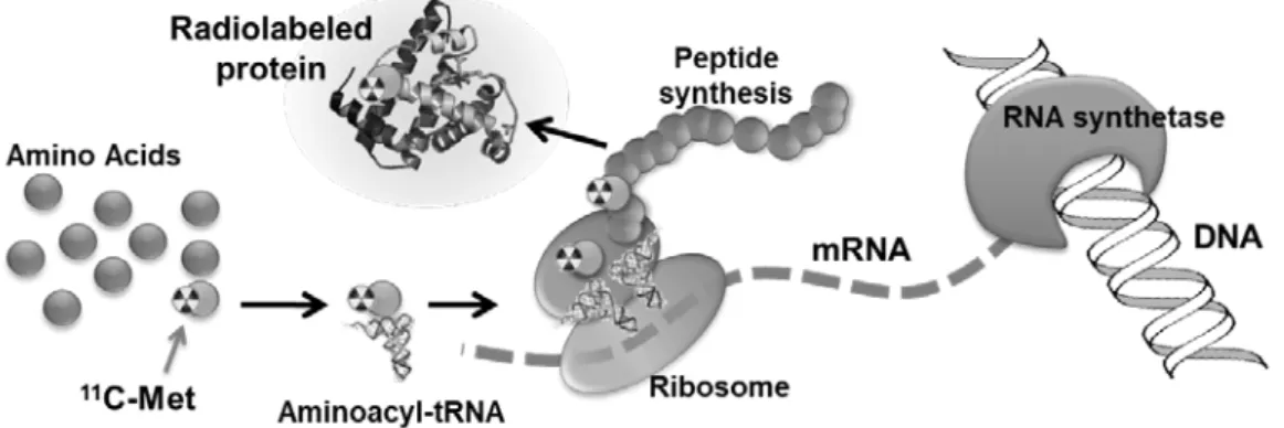
Recent advances in genetic engineering has made it possible to prepare large amounts of antibodies, to humanize them, and to make their size small. Since molecular biology of cancer rapidly progressed, cancer treatment is shifting to molecular targeted therapy with an antibody specific for antigen of cancer cells. As a result, development of a molecular imaging technique of the therapeutic target has become important. For example, an antibody type diagnostic probe can be used for monitoring in the course of treatment by the immunotherapy with an antibody. Therefore, we have tried to develop a novel method for preparation of positron-emitter labeled antibody by using cell-free protein synthesis (CFPS) system (1). The method is to synthesize an antibody in vitro by using enzymes, several factors, template mRNA, natural amino acids, and a positron-emitter labeled amino acid (Fig. 1). In this study, we prepared and biologically evaluated a 11C-labeled single chain antibody (scFv) to HER2 which is a transmembrane protein receptor with tyrosine kinase activity and overexpressed in breast and stomach cancers. HER2 is used as a prognostic factor for breast cancer diagnosis and a predictor of therapeutic effect. That is, HER2 positive breast cancer has poor prognosis and anti-HER2 therapy responds well. HER2-PET would be applicable to imaging biomarkers for diagnosis and therapy monitoring of breast cancer.
Methods
In this study, a single chain antibody of trastuzumab (tra-scFv) which bind strongly to HER2 was prepared by using CFPS system (Musaibo-Kun®) and 11C-methionine as a part of amino acid sources for protein synthesis. To the kit solution of CFPS system, template DNA (plasmid) and 11C-methionine (ca 1400 MBq/mL in saline) were added and incubated at 37℃.
123
After the incubation, 11C-tra-scFv was purified by immune-affinity chromatography. Radiochemical yields was determined by SDS-PAGE and autoradiography (ARG). Binding affinity of 11C-tra-scFv to HER2 was assessed by cell-biding assay using SK-OV-3 (human ovarian cancer cells with high expression of HER2) and MCF7 (human breast cancer cells with low expression of HER2). The tissue uptake of the tracer was evaluated by biodistribution study and small animal PET imaging.
Results and Discussion
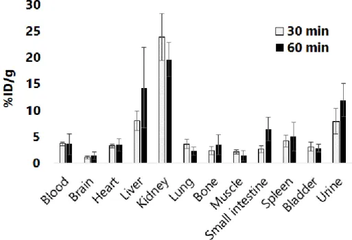
Decay-corrected radiochemical yields of 11C-tra-scFv were 8.6~13%. The binding rates (%ID/106 cells) of 11C-tra-scFv were 15.8 ± 0.6 %ID/106 cells and 2.2 ± 0.2 %ID/106 cells for SK-OV-3 and MCF7, respectively (Fig. 2). The binding to SK-OV-3 was strongly inhibited by addition of trastuzumab, suggesting the specific binding of 11C-tra-scFv to HER2 molecule. Biodistribution study in normal mice (ICR mice) demonstrated that 11C-tra-scFv accumulated mainly in the kidney at 30 min and 60 min post-injection (Fig. 3). This biodistribution was also confirmed by PET imaging. These data indicate that the injected 11 C-tra-scFv shows smooth clearance from the body through kidney. This feature is necessary to obtain PET images of the thoracic region with low radioactivity background. Actually, when 11C-tra-scFv was injected into a tumor bearing mouse made by implantation of SK-OV-3 cells to flank, the tumor was clearly visualized by PET (Fig. 4). Moreover, 11C-tra-scFv showed high binding to the tumor tissue sections of SK-OV-3 in vitro, and the binding was completely blocked by addition of trastuzumab. These findings indicate that 11C-tra-scFv can specifically bind to HER2 molecule expressed in the tumor tissue.
Conclusion
Using cell-free protein synthesis system and 11C-methionin, we succeeded to prepare 11C-labeled a single chain antibody which shows high binding to the antigen molecule. This study demonstrated that 11C-tra-scFv synthesized by this method could be useful as a PET tracer for HER2 positive tumor imaging.
References
1) Matsuda T, Furumoto S, Higuchi K, Yokoyama J, Zhang MR, Yanai K, Iwata R, Kigawa T., Bioorg. Med. Chem. 2012, 20, 6579-6582.
124
Figure 1. Radiosynthesis scheme of 11C-labeled protein by cell-free protein synthesis system and 11C-methionin (11C-Met).
Figure 2. Cell binding assay for evaluation of binding of 11C-tra-scFv to HER2
using SK-OV-3 (human ovarian cancer cells with high expression of HER2) and MCF7 (human breast cancer cells with low expression of HER2).
125
Figure 3. Biodistribution of 11C-tra-scFv in ICR mice.