Neurochemical studies of neuroprotection by
PTP1B inhibitors in the cerebral ischemia in
mice(マウスの脳虚血におけるPTP1B阻害剤による
神経保護に関する神経化学的研究)
著者
Sun Meiling
学位授与機関
Tohoku University
学位授与番号
11301甲第18624号
URL
http://hdl.handle.net/10097/00125883
Neurochemical studies of neuroprotection by PTP1B
inhibitors in the cerebral ischemia in mice
(マウスの脳虚血における PTP1B 阻害剤による神経保護に関する神
経化学的研究)
平成 30 年度
東北大学大学院薬学研究科
生命薬科学 薬理学分野
孫 美玲
LIST OF ABBREVIATION
4-HNE 4-hydroxy-2-nonenal
8-OHdG 8-hydroxy-2-deoxyguanosine
AD Alzheimer's disease
AIS acute ischemia stroke
Akt protein kinase B
bEnd.3 cell mouse brain microvascular endothelial cells
BBB blood-brain barrier
Bim Bcl-2-interacting mediator of cell death
CBF Cerebral blood flow
CREB cyclic AMP-responsive element binding protein
CML chronic myelogenous leukemia
CNS central nervous system
eNOS endothelial nitric oxide synthase
EGF epidermal growth factor
ERK1/2 extracellular signal-regulated kinase
FDA Food and Drug Administration
FoxO1 forkhead box protein O1
HIF-1α hypoxia-inducible factor-1α
HO-1 heme oxygenase-1
HT hemorrhagic transformation
iNOS inducible nitric oxide synthase
I/R ischemia/reperfusion
IR Insulin receptor
IRS Insulin receptor substrate
JAK Janus kinase 2
JAM junctional adhesion molecule
nyl) benzamide
LPS lipopolysaccharide
MAGUK membrane-associated guanylate kinase
MMP matrix metalloproteinases
MMP mitochondria membrane potential
mPTP mitochondrial permeability transition pore
nNOS neuronal nitric oxide synthase
Nrf2 nuclear factor-erythroid 2-related factor 2
PDGF-α platelet-derived growth factor alpha
PI3K phosphatidylinositol 3'-kinase
PTKs protein tyrosine kinases
PTP1B protein tyrosine phosphatase 1B
Rac1 Ras-related C3 botulinum toxin substrate 1
ROS reactive oxygen species
RT-PCR real-time polymerase chain reaction
STAT3 signal transducer and activator of transcription 3
tMCAO transient middle cerebral artery occlusion
tPA tissue plasminogen activator
TCA trichloroacetic acid
TEER transendothelial electrical resistance
TG triglyceride
TTC 2,3,5-Triphenyltetrazolium chloride
TJs tight junctions
TLR4 Toll-like receptor 4
VO(OPT) bis(1-oxy-2-pyridinethiolato)oxovanadium (IV)
Contents
Chapter 1: General introduction... 1
1. Cerebral ischemia... 1
1.1 Reactive oxygen species ... 3
1.2 Blood-brain barrier dysfunction ... 5
2. Protein tyrosine phosphatase 1B (PTP1B) ... 6
3. KY-226 ... 9
Chapter 2: KY-226 could protect from cerebral ischemic injury via AKT/eNOS and ERK signaling pathway ... 11
1. Introduction ... 11
2. Results ... 13
2.1 Pharmacokinetics of KY-226 in the blood and brain after I/R insult in vivo ... 13
2.2 KY-226 exhibits a neuroprotective effect on ischemia/reperfusion in vivo ... 15
2.3 KY-226 activates the Akt/eNOS signaling pathway in ischemic mice 17 2.4 KY-226 plays a vital role in ERK signaling pathway ... 19
2.5 KY-226 could attenuate the ROS stress induced by ischemia/reperfusion in vivo ... 21
3. Discussion ... 24
Chapter 3: KY-226 protects blood-brain barrier function through Akt/FoxO1 signaling pathway in brain ischemia ... 30
1. Introduction ... 30
2. Results ... 32
2.1 KY-226 ameliorated BBB breakdown in brain I/R insults ... 32
2.2 KY-226 attenuated the degradation of TJs in the ischemic brain ... 33
2.3 KY-226 ameliorated the loss of TJ proteins via Akt signaling and ERK pathways ... 38
2.5 KY-226 activated Akt/FoxO1 signaling in bEnd.3 cells stimulated with LPS ... 41 3. Discussion ... 43 Chapter 4: Summary ... 48 1. Conclusions ... 48 2. Future prospect... 51
Materials and Methods ... 56
Reference ... 69
Acknowledgements ... 104
Chapter 1: General introduction
1. Cerebral ischemia
Stroke is one of the primary causes of death and long-term disability worldwide, followed by cardiovascular disease (Wattanapitayakul and Bauer, 2001, Pendlebury and Rothwell, 2009, Krishnamurthi et al., 2013). According to the World Health Organization (WHO), every year 15 million people suffer stroke worldwide. Out of which, more than 6 million die and another 5 million are permanently disabled (Rodrigo et al., 2013). Over the last four decades, the stroke incidence in low- and middle-income countries has more than doubled. Moreover, stroke is a leading cause of dementia and depression (Owolabi et al., 2015).
According to its clinical symptoms, stroke is classically divided into ischemic stroke (~85%) and hemorrhagic stroke including subarachnoid hemorrhage, due to bleedings. Ischemic stroke is the most common type of stroke, resulting from the blockage of blood vessels thereby shortage of oxygen and glucose supplied to the brain. Restoration of the cerebral blood flow and the supply of oxygen and glucose, the damage caused by ischemia should be improved. However, studies have demonstrated that restoring blood circulation in a short period of time can improve cerebral ischemic injury, which was called “effective reperfusion time”. Rapid reperfusion beyond the effective time aggravates ischemic injuries because of the generation of reactive oxygen species (ROS), produced a so-called “reperfusion injury” (Moro et al., 2005). Both ischemia and reperfusion could result in the dysfunction of organs and
neuronal cell death.
Reperfusion injury has been demonstrated by accumulating clinical and experimental studies. For example, the infarct size significantly increased at 6-24 h after reperfusion compared with permanent occlusion in a rat middle cerebral artery occlusion (MCAO) model (Zhang et al., 1994). Multiple pathological processes are involved in ischemia/reperfusion (I/R) injury, including oxidative stress, mitochondrial dysfunction, inflammation, blood-brain-barrier (BBB) disruption and so on, consequently leading to brain edema or hemorrhagic transformation (HT) and eventually causing neuron death and neurological dysfunctions in the brain (L et al., 2016) (Fig.1).
Up to date, recombinant tissue plasminogen activator (r-tPA) administration intravenously is the only Food and Drug Administration (FDA)-approved therapy for acute ischemic stroke. Originally, tPA was demonstrated to be safe within 3 h after stroke onset. However, recent studies have demonstrated that the therapeutic window of tPA treatment was extended to 4.5 h after stroke onset (Del Zoppo et al., 2009). So far, many investigators are doing clinical and preclinical studies about neuroprotection in ischemic stroke. However, few studies have been success and shown any clinical benefit.
Fig.1 Pathological signaling pathways involved in the cerebral ischemic stroke. Ischemic stroke causes the release of the excitatory amino acid glutamate and increased calcium (Ca2+) influx, activating numerous cellular signaling pathways. Reperfusion could aggregate the ischemic damage by the formation of free radical. ROS, as well as Ca2+ influx and other factors, can also permeabilize the mitochondrial membrane, leading to the release of proapoptotic molecules, such as cytochrome c into the cytoplasm. In general, apoptosis and necrosis involve in the cell death following cerebral ischemia/reperfusion injury.
1.1 Reactive oxygen species
Reactive oxygen species have been implicated in brain injury after cerebral ischemia, resulting from the imbalance of excessive production of ROS and limited
antioxidant defense. These oxidants, such as superoxide (O2·−), nitric oxide (NO·) and
hydroxyl radical (OH·), can directly damage proteins, lipids, and DNA, leading to autophagy, apoptosis and necrosis (Love, 1999, Wong and Crack, 2008, Radermacher et al., 2013) (Fig.2). Generally, most of the ROS are produced in mitochondria after I/R with increased mitochondria membrane potential (MMP). In addition, NADPH oxidase (NOX) could generate free radicals by transferring one electron to molecular oxygen to induce ROS production (Lambeth, 2004). The initial formation for ROS production is the conversion of the oxygen (O2) to superoxide anion (O2·−) consumed by
mitochondria (Olmez and Ozyurt, 2012). O2·− is a precursor of other ROS (Fig.2).
ROS can regulate cellular growth, differentiation, proliferation and apoptosis at low levels (Poli et al., 2004). During cerebral reperfusion, ROS production is further stimulated. Unfortunately, tPA might increase ROS level and led to further damage (Green, 2008).
Increased ROS are major causes of tissue damage after cerebral ischemia through destructing cellular proteins, lipids, and DNA, or disrupting normal cellular signaling and gene regulation. Elevated ROS production in mitochondria can trigger the opening of the mitochondrial permeability transition pore (mPTP) and further induce the release of cytochrome c and other apoptotic proteins into the neuronal cytoplasm, causing apoptotic signaling cascade after stroke (Kirkland et al., 2002). In addition, during I/R injury, ROS promote tissue inflammation and activates NLRP3 inflammasome activation (Bryant and Fitzgerald, 2009). Therapeutics such as Radicut® (MCI-186, edaravone), an anti-oxidative radical scavenger, have been used in the clinical setting in
Japan (Tanaka, 2002). However, safe and long-acting compounds for inhibiting ROS are needed for ischemic stroke therapy.
Fig.2 Reactive oxygen species metabolism. ROS include free radicals, such as superoxide (O2·−), nitric oxide (NO·) and hydroxyl radical (OH·) and other molecular
species, such as hydrogen peroxide (H2O2) and peroxynitrite (ONOO −
). O2 ·−
is spontaneously converted to H2O2, or catalysed by SOD (Fridovich, 1995). H2O2 can
be fully reduced to water by GSH, or partially reduced to hydroxyl radicals (OH·) in the presence of reduced transition metals (Fe2+) (Liochev and Fridovich, 1994). In addition, O2·− can react with NO radicals to produce peroxynitrite (ONOO−), a highly
toxic molecule (Beckman and Koppenol, 1996).
1.2 Blood-brain barrier dysfunction
The BBB is a selective permeability barrier between the blood and the brain tissue, which mainly composed of endothelial cells, pericytes and astrocytes. BBB is
involved in the maintenance of central nervous system (CNS) microenvironment, regulation of influx and efflux transport, and protection from damage. BBB disruption is a common pathophysiological feature in cerebral I/R injury, and sometimes considered as an outcome of the processes such as oxidative stress and inflammation in I/R injury. NOX, which is highly expressed in the cerebrovascular endothelium, produces oxygen radicals during I/R, consequently leading to the loss of BBB integrity such as disrupting tight junctions and endothelial functions (Gursoy-Ozdemir et al., 2012). BBB disruption is associated with other neurodegenerative disorders including Alzheimer’s disease (AD) and stroke (Abraham et al., 2002, Bowman et al., 2007). Moreover, BBB dysfunction elicits induction of proinflammatory factors, matrix metalloproteinases (MMP), and aquaporins (Candelario-Jalil et al., 2009, Alam et al., 2011), thereby resulting in cerebral hemorrhage after thrombolysis (Kanazawa et al., 2017). MMPs induced during I/R injury could disrupt tight junction (TJ) proteins, promoting the BBB leakage and entrance of neurotoxic agents to the brain (Gu et al., 2012). TJs between adjacent endothelial cells are critical for maintenance of BBB structure and function by inhibiting the penetration of toxic substances into the brain (Mitic and Anderson, 1998).
2. Protein tyrosine phosphatase 1B (PTP1B)
Protein tyrosine phosphorylation is a key regulatory process in various cells and balanced by actions of protein tyrosine kinases (PTKs) and protein tyrosine phosphatases (PTPs): PTKs phosphorylate the tyrosine residues in a number of
proteins, while PTPs dephosphorylate tyrosine phosphorylation (He et al., 2014). Thus, dysregulation of protein tyrosine phosphorylation is involved in many diseases, such as diabetes and neurological diseases (He et al., 2014, Song et al., 2016). Protein tyrosine phosphatase 1B (PTP1B) is a member from PTP family and negatively regulates the insulin and leptin signaling pathways. In insulin signaling, PTP1B dephosphorylates the insulin receptors (IR) and insulin receptor substrate (IRS), while in leptin pathway, PTP1B dephosphorylates Janus kinase 2 (JAK2) and signal transducer and activator of transcription 3 (STAT3) (Tsou and Bence, 2012) (Fig.3).
PTP1B expression is upregulated in the brain after LPS injection, suggesting the role of PTP1B in neuroinflammation (Song et al., 2016). Moreover, PTP1B levels increased significantly in rat spinal cord following injury (Zhu et al., 2015). PTP1B activation significantly promotes ER stress-induced neuronal apoptosis (Jeon et al., 2017). PTP1B deletion in the hippocampus and cortex of mice showed significantly improved cognitive performance (Fuentes et al., 2012). Based on these findings, PTP1B may involve in brain damage and PTP1B inhibition would be a useful target for neurological disorders. In addition, inhibition of PTP1B could attenuate endothelial dysfunction (Wang et al., 2018b). In endothelial cells, PTP1B deletion could protect against chronic afterload-induced heart failure through improvements in cardiac VEGF-signaling and angiogenesis (Gogiraju et al., 2016).
Fig.3 PTP1B negatively regulates insulin and leptin signaling pathways. Insulin stimulation is followed by IR autophosphorylation and activation, which promoting tyrosine phosphorylation of IRS-1, thereby activating phosphatidylinositol 3'-kinase (PI3K) and Akt. MAPK/ERK signaling cascade is another essential signaling pathway branched from IR. PTP1B could dephosphorylate IR and IRS, negatively regulates the insulin signal transduction. Leptin binding to leptin receptor induces the recruitment and tyrosine phosphorylation of JAK2, subsequently leading to STAT3-mediated transcription. PTP1B attenuates the leptin signal transduction through dephosphorylation of JAK2 and STAT3.
Previously, we reported that sodium orthovanadate (Na3VO4), a PTP1B inhibitor,
could rescue neurons from delayed neuronal death following transient forebrain ischemia in gerbils. This implies that nonspecific inhibition of PTP1B may be a potential target for stroke therapy (Kawano et al., 2001). Sodium orthovanadate can
activate or maintain the activity of growth factor receptors by inhibiting dephosphorylation of tyrosine residues, which are activated by autophosphorylation of tyrosine residues (Sugano et al., 2009). Furthermore, in a mouse model of tMCAO, treatment with sodium orthovanadate before and after arterial occlusion decreases ischemic damage significantly (Shioda et al., 2007). Sodium orthovanadate also exhibits neuroprotective effects in rat models of MCAO through activation of both Akt and ERK activation (Hasegawa et al., 2003). However, orthovanadate treatment by peripheral administration resulted in severe diarrhea because of the inhibition of ATPase (Shioda et al., 2007).
3. KY-226
Recently, a novel series of benzoylsulfonamide derivatives were synthesized. 4-(biphenyl-4-ylmethylsulfanylmethyl)-N-(hexane-1-sulfonyl) benzamide (KY-226) was identified as a potent and selective inhibitor for PTP1B (IC50 = 0.25 µM) (Fig.4).
KY-226 was reported to bind to the allosteric sites of PTP1B to act as a non-competitive inhibitor (Ki = 0.35 µM) (Morishita et al., 2017).
Fig.4 The chemical structure of KY-226. 4-(biphenyl-4-ylmethylsulfanylmethyl)- N-(hexane-1-sulfonyl) benzamide
KY-226 exhibited high oral absorption in mice, which was enough for PTP1B inhibition (Morishita et al., 2017a). In db/db mice, KY-226 lowered the plasma glucose and triglyceride (TG) levels to exhibit anti-diabetic effects, without affecting body weight, possibly via improvement in insulin signaling (Ito et al., 2018). In high-fat diet-induced mice, anti-obesity effects of KY-226 through leptin signaling enhancement was demonstrated (Ito et al., 2018).
Chapter 2: KY-226 could protect from cerebral ischemic injury via
AKT/eNOS and ERK signaling pathway
1. Introduction
In recent years, the Akt signaling pathway, which is a serine/threonine protein kinase with demonstrated effects on BBB dysfunction, has been an increasing focus for stroke researchers. Specifically, the Akt pathway has been implicated in the stabilization of the BBB through decreased expression of VCAM-1, ICAM-1, and β-catenin, as well as increased expression of claudin 3 and 5 (Radisavljevic et al., 2000, Tsoyi et al., 2010, Krafft et al., 2013). Akt activity is upregulated by phosphorylation at Ser573 and
Thr308 by phosphoinositide-dependent protein 1 (PDK1) and
phosphoinositide-dependent protein 2 (PDK2), which is downstream from phosphatidylinositol 3'-kinase (PI3K) (Andjelkovic et al., 1997). Several reports demonstrate that Akt activity decreases after I/R injury, and its reduction promotes I/R-induced neuronal cell death and damage (Friguls et al., 2001, Janelidze et al., 2001, Yano et al., 2001, Li et al., 2017).
A number of compounds, including growth factors, estrogen, and free-radical scavengers have been shown to reduce ischemic damage, possibly (in part) by upregulating Akt phosphorylation (Zhao et al., 2006). Furthermore, activation of Akt in ischemic regions induces neuroprotection through regulation of eNOS (endothelial nitric oxide synthase) signaling. In response to various forms of cellular stimulation, eNOS is phosphorylated by Akt. Activation of Akt signaling by statins has also been
completely absent in the eNOS knockout (KO) mice), suggesting that eNOS is vital to the protective effect of the Akt signaling cascade (Robert M. Bell, 2003). Moreover, studies show that neurological deficits following cerebral ischemia are more severe in eNOS KO mice compared with wild-type mice (Chen et al., 2005). In addition, in 2013, Yabuki et al. reported that supplementation with l-citrulline was able to rescue cerebrovascular injury by restoring eNOS expression following brain ischemia (Yabuki et al., 2013). Together, these findings indicate that eNOS plays a crucial role in Akt-mediated neuroprotection in the brain following ischemic injury.
The p44/42 extracellular signal-regulated protein kinase (ERK) belongs to a family of mitogen-activated protein kinases (MAPK) (Kyriakis and Avruch, 2012), which are closely correlated with cerebral ischemia, like promoting neuronal survival after ischemia (Irving et al., 2000, Kilic et al., 2005, Zhu et al., 2013). ERK is normally activated in response to growth and differentiation factors, and its phosphorylation is increased in rodents with transient MCAO (Alessandrini et al., 1999). ERK activation has also been reported to be involved in increased tolerance to hippocampal ischemia (Choi et al., 2006), and exhibits neuroprotective effects via anti-oxidative actions (Wang et al., 2012). Activation of survival ERK and Akt signaling in rat brain neurons share a cooperative role after ischemic insults in rats (Li et al., 2003). In addition, erythropoietin has been shown to exert neuroprotective functions in brain hypoxia and ischemia by dual activation of ERK and Akt pathways (Kilic et al., 2005). Furthermore, ERK activation involved in the neuroprotective effects of calcitriol in rat models of global cerebral ischemia (Yuan et al., 2018). However, some studies have
found that activation of ERK can induce cell death during brain ischemia, although the mechanisms underlying this response remain unclear (Alessandrini et al., 1999, Namura et al., 2001, Zhuang and Schnellmann, 2006). In mice, treatment with U0126 (a specific inhibitor of MEK (MAPK/ERK kinase)) has been shown to protect brain tissue against damage after forebrain ischemia, as well as during focal cerebral ischemia (Namura et al., 2001). Similarly, the MEK1 inhibitor PD98059 has been shown to decrease infarct volume following focal cerebral ischemia (Alessandrini et al., 1999).
2. Results
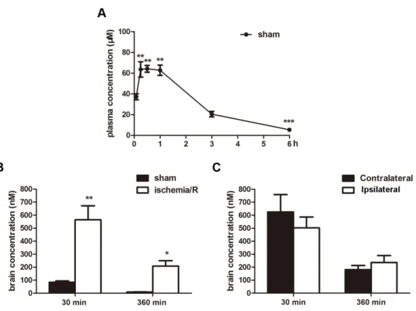
2.1 Pharmacokinetics of KY-226 in the blood and brain after I/R insult in vivo To determine if KY-226 could be useful for ischemic injury, we first determined if KY-266 could penetrate the BBB. To do this, we measured KY-226 concentrations in the plasma and brain tissues of mice after a 10 mg/kg intraperitoneal (i.p.) injection of KY-226 30 min following reperfusion. Results showed that KY-226 was able to enter the bloodstream, showing a slight increase in plasma concentrations in control mice 30 min after injection (tmax = 0.5 ± 0.14 h; Cmax = 71.64 ± 4.41 μM) (Fig.5 A). However, the concentration of KY-226 in brain tissues 30 min after i.p. injection was very low, and almost totally gone 6 h following injection, suggesting that BBB permeability of KY-226 is very low (Fig.5 B). Surprisingly, KY-226 concentration in the brain increased significantly 30 min following injection in I/R mice compared with the sham group (Fig.5 B). However, in I/R mice, there was no significant difference in the
concentration of KY-226 between ipsilateral and contralateral brain tissues (Fig.5 C). Taken together, these data suggest that KY-226 can penetrate the BBB and that KY-266 may play a specific role in cerebral ischemia.
Fig. 5 KY-226 could enter into the brains. (A) The concentration of KY-226 in the blood was measured 5, 15, 30, 60, 180, 360 min after injection with 10 mg/kg KY-226 intraperitoneally (i.p.). ***P < 0.001 vs 5 min after injection. n = 5. (B) Mice were
subjected to tMCAO for 2 h and administered i.p. 10 mg/kg KY-226 30 min after reperfusion. The concentrations of KY226 in whole brains were measured 30 and 360 min after injection with KY-226. **P < 0.01 vs 30 min in sham group. *P < 0.05 vs 360
min in sham group. n = 6. (C) Mice were subjected to tMCAO for 2 h and administered i.p. 10 mg/kg KY-226 30 min after reperfusion. The concentrations of
KY- 226 in the contralateral and ipsilateral portions of the brains of ischemic mice were measured 30 and 360 min after injection with KY-226. n = 6.
2.2 KY-226 exhibits a neuroprotective effect on ischemia/reperfusion in vivo Next, we measured the infarct volume of mice 24 h after reperfusion with TTC staining. Mice were treated with or without KY-226 (1, 5, 10, 30 mg/kg) by i.p. 30 min after reperfusion. The infarct size in vehicle-treated mice was 59.41 ± 2.83%, which implied success of the ischemic stroke model. As illustrated in Fig.6 A and B, 10 and 30 mg/kg of KY-226 delivered 30 min after reperfusion significantly reduced infarct volume in the mice (10 mg/kg = 35.74 ± 5.26%, 30 mg/kg = 35.56 ± 5.03%) (Fig.6 A and B). However, lower concentrations of KY-266 (1 and 5 mg/kg) did not significantly reduce ischemic damage. Considering that 10 and 30 mg/kg of KY-266 had similar effects on infarct volume, we chose 10 mg/kg as the optimum concentration for the following experiments.
To further confirm the optimal timing of KY-226 treatment, we administered 10 mg/kg KY-226 to mice at different times after reperfusion. Results found that infarct volume only decreased when KY-266 was administered 30 min after reperfusion (35.74 ± 5.26%), but not 1 h or 2 h (48.71 ± 8.27%, 52.29 ± 7.85%, respectively) (Fig.6 C and D). When compared with mice in the vehicle group, those treated with KY-226 had significantly decreased neurological score 24 h after reperfusion (Fig.6 E). Together, results suggest that KY-226 has a neuroprotective effect against I/R injury.
Fig.6 KY-226 reduced the ischemia/reperfusion injury in vivo. (A and B) Mice were subjected to tMCAO for 2 h and KY-226 with different concentrations (1, 5, 10, 30 mg/kg) was administered intraperitoneally (i.p.) 30 min after reperfusion. Representative images of TTC staining (A) and quantitative analysis (B) of infarct volume demonstrated that 10 and 30 mg/kg KY-226 reduced the ischemic damage significantly 24 h after reperfusion. *P < 0.05 and **P < 0.01 vs Vehicle. n > 7. (C and
D) Mice were subjected to tMCAO for 2 h and KY-226 (10mg/kg) was administered i.p. at 0.5, 1, 2 h after reperfusion. Representative images of TTC staining (C) and quantitative analysis (D) of infarct volume showed that 10 mg/kg KY-226 decreased the infract volume when administered 30 min after reperfusion. **P < 0.01 vs Vehicle. n > 7. (E) Mice were subjected to tMCAO for 2 h and KY-226 (10mg/kg) was
administered i.p. 30 min after reperfusion. The neurological deficit score in KY-226 treatment group was significantly reduced compared with that in the vehicle group.
**
P < 0.01 vs Vehicle. n > 7.
2.3 KY-226 activates the Akt/eNOS signaling pathway in ischemic mice
In 2007, Shioda et al. reported that activation of the PI3K/Akt pathway mediated the neuroprotective effects in brain cerebral ischemia (Shioda et al., 2007). As mentioned previously, KY-226 has an inhibitory effect against PTP1B, which could attenuate the PI3K/Akt pathway. Because of this, we hypothesized that activation of Akt may be involved in the neuroprotective action of KY-226 following I/R insult. To test this hypothesis, we measured Akt activity using western blot, which was correlated with phosphorylation of Thr308 and Ser473 (Alessi et al., 1996). Consistent with previous reports (Shioda et al., 2007), levels of Akt phosphorylation in the cortex decreased significantly at 3 and 6 h after reperfusion, at both Ser473 and Thr308 (Fig.7 A-C). Meanwhile, total Akt expression did not change by ischemia/reperfusion and KY-226. The transient decrease in Akt phosphorylation in I/R injury model was upregulated by KY-226 remarkably (Fig.7 A-C).
eNOS is one of the classic downstream targets of Akt and the Akt/eNOS signal transduction pathway is involved in regulating responses to I/R injury (Chien et al., 2015). Akt could phosphorylate eNOS at Ser1177 and increase NOS activation, thereby leading to cardioprotection against ischemia/reperfusion (Bellis et al., 2009, Changjun Yang and Murugesan Velayutham, 2013). Our results showed that KY-226 was able to restore phosphorylation of eNOS (Ser1177) 3 and 6 h after reperfusion without altering phosphorylation of eNOS (Thr495) or total eNOS protein expression
(Fig.7 D-F). NOS has three isoforms – eNOS, neuronal NOS (nNOS), and inducible NOS (iNOS) -- all of which participate in the regulation in response to I/R injury. Previous studies have suggested that nNOS may reduce I/R injury through a direct antioxidant effect (Khan et al., 2004, Choate et al., 2008). Results of our experiment showed no change in iNOS protein expression 3 and 6 h after reperfusion in both vehicle and KY-226-treated groups, which was associated with inflammation (Fig.7 G and H). However, in mice, KY-226 inhibited I/R-induced downregulation of nNOS 6 h after reperfusion (Fig.7 G and I). These results suggested that Akt/eNOS activation mediated the neuroprotection of KY-226 in cerebral ischemia in mice.
Fig.7 KY-226 activated the Akt/eNOS signaling pathway in ischemic mice. Mice were subjected to tMCAO for 2 h and administered intraperitoneally (i.p.) with 10
mg/kg KY-226 30 min after reperfusion. (A-C) The levels of Akt and phosphorylation Akt with cortical samples from mice 3 and 6 h after reperfusion were measured by western blot analysis. **P < 0.01 and ***P < 0.001 vs sham; ##P < 0.01 and ###P < 0.001
vs Vehicle. n = 6. (D-F) The levels of eNOS and phosphorylation eNOS with cortical samples from mice 3 and 6 h after reperfusion were tested by western blot analysis. **P
< 0.01 and ***P < 0.001 vs sham; #P < 0.05 vs Vehicle. n = 6. (G-I) The levels of iNOS
and nNOS from ischemic brain of mice 3 and 6 h after reperfusion were measured with western blot analysis. ***P < 0.001 vs sham; ##P < 0.01 vs Vehicle. n = 6.
2.4 KY-226 plays a vital role in ERK signaling pathway
Studies indicate that ERK1/2 participates in a wide variety of functions in the mammalian brain and its activation could promote cell survival and prevent oxidative stress. Pervanadate, a powerful PTP1B inhibitor, has been reported to activate MAPK signaling (Zhao et al., 1996). To determine whether activation of ERK is involved in the neuroprotective effects of KY-226, we investigated the activity of ERK in mice subjected to I/R injury. Because the changes in the phosphorylation of ERK1 (44kD) and the phosphorylation ERK2 (42kD) are similar, we chose ERK2 phosphorylation to represent ERK activity. Consistent with previous reports (Takayuki Kawano and Shigetoshi Yano, 2001, Chen et al., 2017), ERK2 activity decreased significantly following dephosphorylation 3 and 6 h after reperfusion (Fig.8 A and B). As expected, 10 mg/kg KY-226 was able to inhibit I/R-induced decline in the phosphorylation in ERK2 in mice. There was no significant difference in total ERK2 expression in mice
treated with or without KY-226. KY-226 itself also did not affect the expression or phosphorylation of Akt and ERK2 (Fig.8 C-E). The results suggested that KY-226 was able to activate the ERK signaling pathway in mice during cerebral ischemia.
Fig.8 KY-226 restored the ERK activity down-regulated by ischemia/reperfusion damage in mice. (A and B) Mice were subjected to tMCAO for 2 h and administered intraperitoneally (i.p.) with 10 mg/kg KY-226 30 min after reperfusion. The levels of ERK and phosphorylation ERK with cortical samples from mice 3 and 6 h after reperfusion were measured with western blot analysis. **P<0.01 and ***P<0.001 vs
sham; #P<0.05 and ###P<0.001 vs Vehicle. n = 6. (C-E) Mice were intraperitoneally
administered with 10 mg/kg KY-226 for 3 and 6 h. The levels of proteins related to Akt and ERK signaling pathway in cortical samples were tested by western blot analysis. n = 6.
2.5 KY-226 could attenuate the ROS stress induced by ischemia/reperfusion in
vivo
Oxidative damage is the most common and important event in ischemic stroke, which can lead to the death of nerve cells. Because of this, we assessed the effects of KY-226 on oxidative damage after reperfusion in mice. Results of our experiments showed that I/R injury significantly increased nitrotyrosine and 4-hydroxynonenal (4-HNE) protein expression, which peaked 12 h after reperfusion (Fig.9 A and B). Nitrotyrosine and 4-HNE are indicators of oxidative protein damage and lipid oxidation, respectively. Furthermore, 10 mg/kg of KY-226 was able to suppress the increases in nitrotyrosine and 4-HNE observed 12 h after reperfusion in mice subjected to I/R injury (Fig.9 C-E).
Meanwhile, 12 h following reperfusion, nitrotyrosine immunofluorescence increased significantly. This effect was reduced by treatment with 10 mg/kg of KY-226, suggesting that KY-226 can block oxidative stress induced by I/R injury (Fig.10 A). Similar anti-oxidative effects from KY-266 treatment were observed from 4-HNE and 8-OHdG immunohistochemistry (Fig.10 A).
To further evaluate the neuroprotective effects of KY-226 in I/R damage, we co-stained nitrotyrosine and NeuN, which is a marker for neurons (Mullen et al., 1992). Results showed that in ischemic mice, nearly all neurons in the cortex were co-stained with nitrotyrosine and NeuN. In the I/R group, NeuN staining revealed reductions in the number of nuclei and increases in the size of nuclei, which suggested neuronal damage
induced by I/R (Fig.10 B). Together, these results suggest that KY-226 can attenuate oxidative stress in vivo following I/R injury.
Fig.9 KY-226 could attenuate the oxidative stress induced by ischemia/reperfusion insult in mice. (A and B) Mice were subjected to tMCAO for 2 h and reperfusion with different duration time. Western blot analysis of time-course of protein (nitrotyrosine, left) and lipid oxidation (4-HNE, right) with cortical samples from mice after reperfusion. (C-E) Mice were subjected to tMCAO for 2 h and administered intraperitoneally with 10 mg/kg KY-226 30 min after reperfusion. Western blot analysis of the effects of KY-226 on nitrotyrosine and 4-HNE protein with cortical samples from mice 12 h after reperfusion **P < 0.01 and ***P < 0.001 vs sham;
#
Fig.10 KY-226 inhibited the ischemia/reperfusion-induced oxidative damage with immunohistochemistry. (A) Representative photographs of fluorescence
immunostaining with nitrotyrosine, 4-HNE and 8-OHdG protein. Immunoreactivity of nitrotyrosine, 4-HNE and 8-OHdG, which are markers of oxidative damage of protein, lipid and DNA (green) respectively, were stimulated in the cortex 12 h after reperfusion in mice. (B) Representative photographs of fluorescence immunostaining with nitrotyrosine (green) and NeuN (red) in the cortex 12 h after reperfusion in mice. Scale bar = 50 μm. Experiments were repeated for three times with similar results.
3. Discussion
In this study, a mouse model of stroke was produced by tMCAO/R. Results of this study showed that a potent and selective PTB1B inhibitor, KY-226, was able to enter the brain and diminish the volume of cerebral infarct and improve the neurological deficits in ischemic mice. Activation of both Akt and ERK by KY-226 in our study was associated with a neuroprotective action during ischemic injury. In addition, activation of Akt upregulated eNOS phosphorylation reduced ischemic oxidative stress. Together, these data suggest that KY-226 may be able to protect against ischemic damage following I/R injury through regulation of the Akt/eNOS and ERK pathways.
PTP inhibitors induce protein tyrosine phosphorylation in intact cells. PTP1B inhibitors like vanadate inhibit protein tyrosine phosphatases by acting as a transition state of a phosphate analog, as well as by forming a reversible bond through cysteine residues (Morinville et al., 1998). Therefore, vanadate increases tyrosine phosphorylation via the inhibition of nonselective protein tyrosine phosphatases and
activation of tyrosine kinases in several cells (Morinville et al., 1998).
The allosteric inhibitor KY-226, which lacks a carboxyl moiety has been synthesized to achieve high PTP1B selective inhibitory (Morishita et al., 2017b). Studies show that KY-226 functions as a non-competitive inhibitor and bounded to the allosteric site of PTP1B (Morishita et al., 2017b). PTP1B has also been shown to negatively regulate insulin signals through de-phosphorylation of insulin receptors and insulin receptor substrate in insulin resistance in various tissues such as liver, skeletal muscle, adipose tissue (Elchebly et al., 1999, Zinker et al., 2002, Wu et al., 2005). Similarly, PTP1B has been shown to inactivate leptin signaling by de-phosphorylation of JAK2 and STAT3 in a hypothalamic cell line (Kaszubska et al., 2002, Tsou and Bence, 2012). Moreover, PTP1B can be a negative regulator of CNS leptin and insulin signaling via direct dephosphorylation of Tub, which has been implicated in energy homeostasis (Prada et al., 2013). KY226 could penetrate the BBB and enter the brain, and protect neurons from ischemic injury. However, KY-226 displayed low BBB permeability in the control mice.
In the current study, oxidative stress was measured by ROS and exhibited harmful effects on cells. Free radicals such as O2·- and OH· are generated from mitochondria
during ischemia/reperfusion, and neurons in brain are vulnerable to ROS (Li et al., 2014). Growing evidence indicates that ROS is an initial cause of I/R injury (Venardos et al., 2007). There are several mechanisms that are thought to underlie ROS-induced neuronal damage; first, ROS can damage the proteins, lipids, nucleic acids and other macromolecular components in the cell (such as neurons) directly (Rahman, 2007,
Lochhead et al., 2010, Kalogeris et al., 2012). Second, ROS can lead to neuronal death via caspase-dependent and caspase-independent pathways in neurons (McManus et al., 2014). Finally, studies in ROS can lead to a loss of mitochondria in the neuron, leading to degeneration of the axon through Sarm1, an axodestructive factor, in primary mouse sensory neurons (Summers et al., 2014). ROS can also indirectly trigger the release of cytochrome c from mitochondrial membranes through translocation of p53, which controls the expression of genes such as Bax and Bid in neurons (Endo et al., 2006). In addition, Shiratori et al. found that ROS could cause damage to endothelial cells, causing severe disruptions in microcirculation and increases of hepatic ischemia (Shiratori et al., 1994).
Activation of Akt is correlated with phosphorylation of Thr308 at its catalytic domain, and phosphorylation of Ser473 at the C-terminus (Alessi et al., 1996). Activated Akt can activate or inactivate its downstream target proteins, including Bad and GSK3β, by phosphorylation reactivity, thereby regulating cellular signaling in a variety of cells (Won et al., 2005, Koh et al., 2006). Akt phosphorylated Bad and then released Bcl-2 inside the mitochondria. This subsequently prevents the formation of the mitochondrial permeability transition pore. Bcl-2 also inhibits the secretion of cytochrome c into the cytosol, thereby blocking activation of executioner caspases in neuronal cells (Ouyang et al., 1999). Akt was inactivated in vivo by dephosphorylation immediately after ischemia, probably due to relative activation of protein phosphatases because of lack of ATP during ischemia in neurons (Ouyang et al., 1999). Akt activation has been shown to promote the nuclear translocation of
nuclear factor-erythroid 2-related factor 2 (Nrf2), which led to expression of the downstream protein heme oxygenase-1 (HO-1), and phosphorylation of the survival regulatory protein cyclic AMP-responsive element binding protein (CREB) (Zhang et al., 2017a, Zhang et al., 2018). Furthermore, there was no report found the negative effects of Akt signaling on ischemic stroke in current (Mullonkal and Toledo-Pereyra, 2007). Akt can upregulate the Bcl-2 expression and increase the GSH and SOD activity to inhibit ROS generation in neurons in ischemic insult (Gao et al., 2015, Zhang et al., 2015, Jiao et al., 2016).
eNOS, first identified as a substrate of Akt, could maintain cerebral blood flow (CBF) and reduces neuronal injury after stroke (Samdani et al., 1997). Many studies demonstrated that the neuroprotective effects of Akt-mediated eNOS activation against cerebral ischemia are due to NO production. For example, studies show that NO may play a role in protecting tissues against ischemia by forming intracellular antioxidants such as nitrosothiols and glutathione. Nitric oxide may also form peroxynitrite with superoxide anion during I/R injury. The formation of peroxynitrite is harmful to cells. However, thiol-containing agents such as glutathione, albumin, and cysteine agents can convert the peroxynitrite anion to nitrosothiols and related products, and/or decrease the release of ROS via direct action on a membrane component of the NADPH oxidase (Clancy et al., 1992, Fujii et al., 1997, Ronson et al., 1999, Guo et al., 2011). Studies also show that in mice, ROS production was significantly reduced when mice were treated with the eNOS enhancer PEx-4 (Exendin-4 -loaded poly(D,L-lactide-co-glycolide) microspheres)
provided the neuroprotection through p-Akt/p-eNOS pathways to attenuate oxidative injury in diabetic rats (Chien et al., 2015).
The current study investigated the mechanisms underlying the protective effects of KY-226 in ischemic damage. Unlike Akt signaling, the question of whether activation of ERK exerts a protective or detrimental to neurons is still being debated, with differing results depending on whether growth factor-dependent neuronal survival, neurotoxicity induced by glutamate, oxidative stress, or ischemia is the model being used (Li et al., 2003). Increased phosphorylation of ERK has been noted in the vulnerable penumbra following the acute ischemic stroke in humans as well as in mouse models (Slevin et al., 2000). Activation of the ERK pathway could deliver survival signals and protect against spinal cord ischemia promoted by xenon post-conditioning in rats (Liu et al., 2016). Furthermore, ERK phosphorylation contributed to the inhibition of I/R induced the renal apoptosis and oxidative stress by hypothermia in mice (Choi et al., 2017). For example, in rats, it has been reported that ERK1 and 2 are present in brain mitochondria at the outer membrane/intermembrane space in neurons, which may help to regulate mitochondrial activities related to cell life and death (Alonso et al., 2004). ERK can also effectively inhibit ROS generation by regulating the expression of the Bcl-2 family in myocardial I/R injury in mice (Radak et al., 2013). For instance, in mouse brains, TGF-1β can activate the ERK pathway under both non-ischemic and ischemic conditions, which was accompanied by an increase in Bad phosphorylation (Yuan Zhu et al., 2002).
In summary, the present study demonstrated for the first time that KY-226 penetrated the brain up to 0.55 μM (IC50 = 0.25 μM) and played a neuroprotective role
in cerebral ischemia/reperfusion in mice. KY-226 was able to ameliorate the infarct size, neurological deficits, and I/R induced-ROS stress in mice. The neuroprotective effects of KY-226 were mediated by the activation of Akt/eNOS and ERK signaling pathway (Fig.11). Our study suggested that KY-226 may be a candidate for the treatment of ischemic stroke. It is necessary to do further studies with other ischemic models to evaluate the efficacy of KY-226 as a potential neuroprotective compound.
Fig.11 A scheme for the proposed mechanisms for the neuroprotective effects of KY-226. KY-226 not only enhanced the activity of Akt and eNOS, but also restored the diminished ERK expression in ischemia/reperfusion insult.
Chapter 3: KY-226 protects blood-brain barrier function through
Akt/FoxO1 signaling pathway in brain ischemia
1. Introduction
Ischemic stroke is mainly induced by a transient or permanent occlusion of blood vessels, resulting in neuronal death and behavioral deficits (Sommer, 2017). The secondary brain injury caused by I/R is in part mediated by disruption of the BBB. BBB leakage causes leukocyte penetration into the brain and brain edema (Fanning et al., 1998, Malaeb et al., 2007, Park and Lee, 2013). In this context, BBB disruption precedes neuronal death in brain I/R injury.
The BBB is a dynamic interface between the blood and brain tissue to maintain the brain homeostasis. It consists of basement membrane, pericytes, astrocytes, and endothelial cells (Fig.12 A). Brain capillary endothelial cells account for 75–80% function in BBB (Li et al., 2010). Endothelial cells in the BBB are unique compared with other tissues because of the continuous intercellular TJs (Abbott et al., 2006). TJs are composed of occludin, claudin, junctional adhesion molecule (JAM), and cytoplasmic-associated proteins (Mitic and Anderson, 1998, Ballabh et al., 2004) (Fig.12 B). As a cytoplasmic TJ-associated protein, ZO-1 (zonula occludens-1) binds to occludin to recruit it into TJs (Furuse et al., 1994, Ballabh et al., 2004). Protein kinase C δ (PKCδ) signaling in part mediates the breakdown of TJ proteins in brain ischemia (Jiao et al., 2011). The activation of MMPs also mediates TJ protein breakdown in BBB leakage (Na et al., 2015, Wang et al., 2016). ZO-1 is
canine kidney and CVE cells, which leads to a decrease in ZO-1 and occludin levels (Stevenson et al., 1989, Staddon et al., 1995, Basuroy et al., 2006). The loss and degradation of ZO-1 are closely linked to increased barrier permeability, an effect related to activation of hypoxia-inducible factor-1α (HIF-1α) (Yan et al., 2012).
Fig.12 Structure of the Blood–Brain Barrier (BBB). (A) The BBB is constituted in the central nervous system by capillary endothelial cells and surrounded by basement membrane, pericyte, astrocyte, and neurons). (B) The tight junction further is established by the interaction between the transmembrane proteins (claudins, occludin, and junction adhesion molecule (JAM)) and cytoplasmic associated-proteins (ZO-1, ZO-2, and ZO-3) of adjacent endothelial cells.
PKB/Akt signaling plays a protective role in ischemic injury by maintaining BBB integrity. Akt activates mTOR signaling, thereby suppressing excessive autophagy and Rac1 (Ras-related C3 botulinum toxin substrate 1) induction. This in turn preserves ZO-1 levels after oxygen-glucose deprivation and reoxygenation-induced damage in vitro (Wang et al., 2016, Yang et al., 2018). Extracellular signal-regulated kinase (ERK) is also required for epidermal growth factor (EGF)-induced prevention of TJ disruption induced by oxidative stress (Basuroy et al., 2006). The expression of active MEK1, an upstream regulator of ERK, results in enhanced junctional localization of ZO-1, while U0126 and dominant-negative MEK1 attenuated the effects of EGF in acetaldehyde-treated cells (Samak et al., 2011).
2. Results
2.1 KY-226 ameliorated BBB breakdown in brain I/R insults
EB dye is used as a marker of albumin effluxion to evaluate BBB permeability (Saunders et al., 2015). As shown in Fig.13 A, I/R damaged BBB permeability. In contrast, treatment with KY-226 (10 and 30 mg/kg) 30 min after ischemia significantly diminished EB leakage compared with that of the vehicle group. The quantitative analysis of EB leakage after I/R is shown in Fig.13 B. These results revealed that KY-226 attenuated the increase in BBB permeability induced by I/R.
Fig.13 KY-226 ameliorated BBB breakdown in mice subjected to ischemia/reperfusion. (A and B) Mice were subjected to tMCAO for 2 h and administered with KY-226 (1, 10, 30 mg/kg, i.p.) 30 min following reperfusion. Representative images (A) and quantitative analysis (B) of EB leakage showed that 10 and 30 mg/kg KY-226 decreased the EB leakage significantly 24h after reperfusion.
*
P < 0.05 vs Vehicle. n > 8.
2.2 KY-226 attenuated the degradation of TJs in the ischemic brain
During ischemia and reperfusion, the TJs of BBB are disrupted (Fischer et al., 2002). Therefore, we examined the effects of KY-226 on expression of ZO-1 and occludin. Compared with that in the sham group, the expression of ZO-1 and occludin proteins in the ischemic cortex were significantly decreased in the vehicle-treated group both 12 and 24 h after reperfusion, suggesting compromised TJs induced by I/R (Fig.14 A, C, and D). There was no change in CD31 protein expression, indicating endothelial cells
remained intact. KY-226 (10 mg/kg) administration rescued the loss of ZO-1 and occludin proteins 24 h after I/R insult in ischemic brains of mice (Fig.14 B, E, and F). We performed immunofluorescence assay for ZO-1, occludin, and CD31 proteins in the brain. In the cortex of sham group, ZO-1 was co-expressed with CD31. ZO-1 immunofluorescence decreased significantly 24 h following reperfusion in ischemic cortex. This effect was inhibited by treatment with KY-226 (10 mg/kg), suggesting that KY-226 abolished the loss of ZO-1 induced by I/R injury in the brain (Fig.15 A). Similar results after KY-266 treatment were observed with occludin immunohistochemistry (Fig.15 B). Taken together, KY-226 administration appears to restore the decrease in ZO-1 and occludin expression in ischemic brains.
Fig.14 Effects of KY-226 administration on the expression of ZO-1 and occludin in mice treated with ischemia/reperfusion. (A) Mice were subjected to tMCAO for 2 h and reperfusion for 12 and 24 h. ZO-1 and occludin proteins in the brain after reperfusion were analyzed with western blot. (B) Mice were subjected to tMCAO for 2 h and administered with intraperitoneally 10 mg/kg KY-226 30 min after reperfusion. The effects of KY-226 on the expression of ZO-1 and occludin proteins in mice 24 h after reperfusion were measured by western blot. (C and D) Quantitative analysis of
western blot results from B. *P < 0.05, **P < 0.01, and ***P < 0.001 vs sham. n = 6. (E
and F) Quantitative analysis of western blot results from C. **P < 0.01 and ***P < 0.001
Fig.15 Immunohistochemistry demonstrating that KY-226 inhibited the loss of ZO-1 and occludin proteins in mice after ischemia/reperfusion (I/R) insults. Mice were subjected to tMCAO for 2 h and administered intraperitoneally with 10 mg/kg
KY-226 30 min after reperfusion. (A) Representative images of fluorescence immunostaining with CD31 (green) and ZO-1 (red) in the brain 24 h after reperfusion in mice. (B) Representative images of fluorescence immunostaining with CD31 (green) and occludin (red) in the brain 24 h after reperfusion in mice. Scale bar = 50 μm. Experiments were repeated for three times with similar results.
2.3 KY-226 ameliorated the loss of TJ proteins via Akt signaling and ERK pathways
Since KY-226 protection in brain ischemia is associated with increased Akt and ERK activity (Sun et al., 2018b), we next tested the effects of Akt and ERK inhibitors, wortmannin and U0126, respectively, to assess whether Akt and ERK modulate the expression of ZO-1 and occludin proteins. KY-226 treatment increased the expression of ZO-1 and occludin in ischemic brains 24 h after reperfusion. Wortmannin decreased p-Akt (Ser473) protein expression and abolished the effects of KY-226 treatment on ZO-1 and occludin protein expression in mice 24 h after reperfusion (Fig.16 A-C). Similarly, U0126 abolished the increase in ZO-1 and occludin expression induced by KY-226 and downregulated p-ERK protein level (Fig.16 D-F).
To further investigate the mechanism underlying elevation of ZO-1 expression induced by KY-226, we measured the mRNA level of ZO-1 in mice 24 h after reperfusion. KY-226 significantly elevated ZO-1 mRNA level in the ipsilateral brain compared with that in sham and vehicle-treated groups (Fig.16 G). We treated mice with Akt and ERK inhibitors 30 min before ischemia. The increased ZO-1 mRNA
induced by KY-226 administration was blocked by Akt and ERK inhibitor treatment 24 h after reperfusion (Fig.16 H and I). Taken together, activation of Akt and ERK appear to mediate the protective effects of KY-226 on TJ protein expression in cerebral ischemia.
Fig.16 KY-226 ameliorated BBB dysfunction through Akt pathway and ERK signaling. Mice were treated with wortmannin or U0126 30 min before ischemia and then subjected to tMCAO for 2 h, following with treatment with KY-226 (10 mg/kg) intraperitoneally 30 min after reperfusion. (A-C) ZO-1, occludin, and phosphorylated Akt proteins in the brain 24 h after reperfusion were tested with western blot. ***P <
ZO-1, occludin, and phosphorylated ERK proteins in the brain 24 h after reperfusion were measured by western blot. ***P < 0.001 vs sham; #P < 0.05 and ##P < 0.01 vs
Vehicle; &P < 0.05 vs KY. n = 8. (G) Quantitative RT-PCR analysis of ZO-1 mRNA in
ipsilateral and contralateral brains from mice 24 h after reperfusion. *P < 0.05 vs sham;
#
P < 0.05 and ##P < 0.01 vs ipsilateral-Vehicle. n = 8. (H) Quantitative RT-PCR
analysis of ZO-1 mRNA in the brains of mice 24 h after reperfusion. *P < 0.05 vs sham;
###
P < 0.001 vs Vehicle; &P < 0.05 vs KY. n = 8. (I) The Ct value of GAPDH mRNA in
the brains of mice 24 h after reperfusion. n = 8.
2.4 KY-226 prevented LPS-induced TJ disruption in bEnd.3 cells
To further elucidate the mechanism underlying the regulation of ZO-1 expression by KY-226, a brain microvascular endothelial cell model using bEnd.3 cells was used to investigate the effects of LPS on BBB function (Choi and Kim, 2008). ZO-1 promoter activity was significantly decreased after LPS treatment when cells were transfected with ZO-1 promoter luciferase (Li et al., 2018a). The suppression of ZO-1 promoter activity was abolished by KY-226 treatment (Fig.17 A and B). Similar results were acquired from ZO-1 immunofluorescence staining (Fig.17 C). These results implied that KY-226 could protect endothelial cells during inflammatory conditions.
Fig.17 KY-226 prevented lipopolysaccharide (LPS)-induced tight junction (TJ) disruption in bEnd.3 cells. bEnd.3 cells were treated with LPS (1 μg/mL) and/or KY-226 (1 μM) for 24 h. (A) bEnd.3 cells were transfected with ZO-1-pGL3 vector and the levels of luciferase reporter activity were measured 24 h after transfection. **P <
0.01 vs Con; ##P < 0.01 vs LPS. n = 6. (B) Western blot assay of ZO-1 and CD31 in
bEnd.3 cells after LPS stimulation. n = 6. (C) Immunofluorescence staining of ZO-1 (green) and DAPI (blue) in bEnd.3 cells after LPS stimulation. Scale bar = 20 μm. Experiments were repeated for three times with similar results.
2.5 KY-226 activated Akt/FoxO1 signaling in bEnd.3 cells stimulated with LPS Since FoxO1 is one of the downstream targets of Akt (Nakamura et al., 2008), we examined the effects of LPS on Akt-FoxO1 signaling in bEnd.3 cells. As shown in Fig. 6A, LPS stimulation reduced the phosphorylation of Akt (T308) without changing total Akt protein levels. The dephosphorylation of Akt (Thr308) was followed by FoxO1 (Ser256) dephosphorylation after LPS treatment. The decreased FoxO1
phosphorylation (Ser256) returned to basal levels with KY-226 administration (Fig.18 A-C). Furthermore, both wortmannin and U0126 prevented the KY-226-induced increase in promoter activity of ZO-1 (Fig.18 D). To further elucidate the role of FoxO1 in the regulation of ZO-1 by KY-226, we treated cells with a FoxO1 inhibitor (As1842856, 10 nM). As1842856 significantly enhanced ZO-1 promoter activity after LPS treatment (Fig.18 E). Overexpression of FoxO1 inhibited ZO-1 promoter activity; this repression was ameliorated by KY-226 treatment (Fig.18 F). Collectively, these findings suggest that KY-226 enhanced Akt/FoxO1 signaling in bEnd.3 cells.
Fig.18 KY-226 activated the Akt/FoxO1 signaling pathway in bEnd.3 cells stimulated with lipopolysaccharide (LPS). Cells were treated with LPS (1 μg/mL) and/or KY-226 (1 μM) for 24 h. (A-C) Western blot analysis of FoxO1, phosphorylated FoxO1, Akt, and phosphorylated Akt in bEnd.3 cells after LPS stimulation. **P < 0.01
and ***P < 0.001 vs Con; #P < 0.05 and ##P < 0.01 vs LPS. n = 6. (D) bEnd.3 cells were
μM), or LPS (1 μg/mL) and/or KY-226 (1 μM) for 24 h. The levels of luciferase reporter activity were measured 24 h after transfection. ***P < 0.001 vs Con; ###P <
0.001 vs LPS-Con; &&&P < 0.001 vs LPS-KY. n = 6. (E) bEnd.3 cells were transfected
with ZO-1-pGL3 vector and treated with LPS (1 μg/mL) and/or AS1842856 (FoxO1 inhibitor, 10 nM) for 24 h. The levels of luciferase reporter activity were measured 24 h after transfection. ***P < 0.001 vs Con; ##P < 0.01 vs LPS-Con. n = 6. (F) bEnd.3 cells
were transfected with FoxO1 plasmid and ZO-1-pGL3 vector, and treated with KY-226 (1 μM) for 24 h. The levels of luciferase reporter activity were measured 24 h after ZO-1-pGL3 vector transfection. **P < 0.01 vs Con; #P < 0.05 vs FoxO1 OE-Con. n = 6.
3. Discussion
In this study, we reported that KY-226 rescues BBB dysfunction following cerebral I/R in mice. To our knowledge, this is the first study to demonstrate that both Akt and ERK activation by KY-226 mediates BBB protection in I/R-induced injury. We previously reported that Akt activation induces the phosphorylation of FoxO1 concomitant with its translocation to the cytoplasm from the nucleus, thereby mediating ischemic tolerance (Fukunaga and Shioda, 2009). Sodium orthovanadate, a PTP inhibitor, elevated Akt activity and in turn rescued neurons from delayed neuronal death, where increased FoxO1 phosphorylation was associated with neuroprotection (Fukunaga et al., 2005). However, the downstream signaling effects of FoxO1 phosphorylation remain unclear.
vascular endothelial cells, which are the main structures responsible for the BBB permeability (Ballabh et al., 2004). Studies have demonstrated that alterations of TJ associated proteins contribute to the loss of BBB function in many diseases or disorders of CNS. Following reperfusion, ROS and cytokines could change the distribution and structure of TJ to increase the BBB permeability (Yang et al., 2007). ZO-1 is a member from the family named a membrane-associated guanylate kinase (MAGUK) and involved in the formation and regulation of TJs in the BBB. Occludin, identified by Mikio Furuse, is the first transmembrane TJ protein with high concentrations at BBB TJs and its precise role is unknown (Furuse et al., 1993). Occludin protein is not required for the formation of TJ strands and might play a role in strengthening TJs (Furuse et al., 1996, Hirase et al., 1997, Forster, 2008). Claudins, expressed in a tissue-specific manner, contain a PDZ-binding motif at the C terminus capable of binding to ZO-1 (Furuse et al., 1999, Itoh et al., 1999). Claudin-5 appears important in maintaining the BBB and is specifically important in actively regulating small molecule paracellular permeability. ZO-1 recognizes occludin and anchors transmembrane proteins to the intracellular actin cytoskeleton to stabilize TJs (Fanning et al., 1998). Hypoxia disrupts the localization of ZO-1 and occludin, resulting in a diffuse pattern with discontinuous staining at endothelial cell borders in vitro (Mark and Davis, 2002). The continued reduction in ZO-1 expression following cerebral ischemia contributes to impaired barrier function because ZO-1 is the scaffolding protein of occludin (Malaeb et al., 2007, Jiao et al., 2011). ZO-1 deficiency disrupts TJs, and reduced ZO-1 levels are associated with barrier breakdown in many
neurological disorders (Katsuno et al., 2008). However, the molecular mechanisms underlying the regulation of ZO-1 expression in vascular endothelial cells remain unclear.
During CNS infections, LPS level was significantly increased, leading to increased permeability of brain microvascular endothelial cells, eventually resulting in cerebral edema (Sumi et al., 2010, Banks et al., 2015). Inflammation caused by I/R injury in the brain is often accompanied by BBB breakdown (Jiang et al., 2005, Amantea et al., 2009). Thus, vascular endothelial cells are primary targets of inflammatory events during bacterial infections and LPS exposure (Yoneda et al., 2000). ZO-1 redistribution is the earliest cellular event during cell shedding and BBB breakdown (Guan et al., 2011). For example, LPS increases the paracellular permeability of brain endothelial cells and decreases transendothelial electrical resistance through disruption of TJ proteins (Dohgu and Banks, 2008). In addition, LPS also can decrease the expression of claudin-5 in bEnd.3 cells (Li et al., 2018a). KY-226 could upregulate the expression of claudin-5 with LPS treatment in vitro (Data not shown). FoxO1 was reported to modulate claudin-5 expression in response to pathologic stimulations by binding to a repressor region in the claudin-5 promoter (Taddei et al., 2008), which further supporting that KY-226 could improve claudin-5 expression.
In the present study, we focused on activation of the PI3K/Akt pathway by KY-226 as activation of PI3K/Akt signaling mediates protection against I/R-induced damage and BBB disruption after ischemia. FoxO1 is a nuclear transcription factor that regulates the function of vascular endothelial cells (Accili and Arden, 2004, Barthel et
al., 2005, Greer and Brunet, 2005). Akt phosphorylates FoxO1 at three specific sites (Thr24, Ser256, and Ser319) in response to growth factors or insulin, thereby inducing translocation of FoxO1 from the nucleus to the cytoplasm (Brunet et al., 1999). Akt-dependent phosphorylation of FoxO1 induces formation of a complex with 14-3-3 chaperone proteins and recruits it to the cytoplasm from the nuclear fraction (Rena et al., 1999, Nakae et al., 2000, Rena et al., 2001). Akt depletion impairs endothelial barrier function and permeability (Gao et al., 2016). Increased expression of p-Akt and p-FoxO1 is associated with upregulation of ZO-1 and occludin in ischemic injuries in
vivo and in vitro (Yang et al., 2016). In silico analysis using JASPAR
(http://jaspar.genereg.net/) revealed putative association domains of FoxO1 in the promoter regions of ZO-1, suggesting that FoxO1 regulates ZO-1 gene expression. In the present study, we reported that FoxO1 regulates ZO-1 gene expression at the transcriptional level. Likewise, in human cerebral microvascular endothelial cells (hCMEC/D3), LPS treatment suppressed mRNA and protein levels of ZO-1 and occludin (Li et al., 2012, Qin et al., 2015). LPS-induced ZO-1 mRNA suppression was attenuated by KY-226 treatment. LPS binds to toll-like receptor 4 (TLR4) on the cell surface (Park and Lee, 2013). TLR4 activation leads to NF-κB and inflammatory cytokine production under LPS stimulation. In this context, NF-κB signaling may also underlie LPS-induced ZO-1 degradation. Further studies are required to resolve this question.
Both ERK and PI3K/Akt pathways synergistically mediate FoxO1 transcriptional activity in vitro (Sun et al., 2018a). PI3K and ERK inhibitors (LY294002 and U0126)
inhibited FoxO1 phosphorylation, indicating that the PI3K and ERK pathways regulated FoxO1 by phosphorylation rather than by changing FoxO1 expression levels (Wang et al., 2018a). Our results suggest that both signaling pathways are required for the induction of ZO-1 by KY-226. However, further studies are required to fully characterize FoxO1 regulation by ERK signaling after I/R injury.
Chapter 4: Summary
1. Conclusions
1) We first detected the pharmacokinetics of KY-266 in mice exposed to brain cerebral ischemia. We demonstrated that KY-226 could enter the bloodstream with a slight increase in the plasma concentration. Moreover, KY-226 was able to penetrate the BBB after ischemia/reperfusion injury.
2) KY-226, a novel PTP1B inhibitor, exhibited a neuroprotective effect in a dose-dependent manner in mice brain cerebral ischemia model.
3) I also measured the ROS levels after cerebral ischemia/reperfusion insults in mice and evaluated the effects of KY-226 on the oxidative stress. Western blotting and immunohistochemistry analysis showed that KY-226 could significantly ameliorate the oxidative damage induced by ischemia/reperfusion injury.
4) In mice, the transient decreased activity in Akt and eNOS proteins induced by cerebral ischemia was upregulated by KY-226 administration. Moreover, KY-226 activated ERK activity following ischemia/reperfusion insults in mice. The activation of Akt/eNOS signaling and improvement of ERK activity were contribute to the neuroprotective effects of KY-226 in cerebral ischemia/reperfusion.
5) Because of the low permeability of KY-226 in control mice, I also detected the effect of KY-226 on the BBB function following ischemia/reperfusion. KY-226 was able to protect from BBB dysfunction in mice cerebral ischemia/reperfusion
I/R-induced injury.
6) In vitro study, LPS treatment caused TJs disruption in bEnd.3 cells. KY-226 enhanced ZO-1 promoter activity after LPS treatment in cultured endothelial cells. KY-226 treatment prevented LPS-induced TJs disruption in cultured endothelial cells.
7) Finally, KY-226 upregulated FoxO1 phosphorylation after LPS treatment in cultured endothelial cells. KY-226 protects BBB integrity by restoration of TJ proteins, an effect partly mediated by Akt/FoxO1 pathway activation.
8) KY-226 may be a potential neuroprotective candidate for ischemic stroke and other neuroinflammatory diseases (Fig.19).
Fig.19 A scheme for the proposed mechanisms underlying the protective effects of KY-226 on the blood-brain barrier (BBB) in ischemia/reperfusion (I/R) or lipopolysaccharide (LPS) stimulation. (A) In pathological conditions such as I/R
and/or LPS stimulation, phosphorylation of Akt is suppressed. The inactivation of Akt prevents the phosphorylation of FoxO1 and its translocation into the cytoplasm. FoxO1 accumulation in the nucleus may interact with the DNA binding domain in the promoter of ZO-1 and repress its transcription. This may result in the loss of ZO-1 in tight junctions, disrupt endothelial barrier function, and increase BBB permeability. (B) KY-226 increases the phosphorylation of Akt in ischemic conditions and after LPS treatment. Phosphorylation of FoxO1, dependent on Akt activation (p-Akt), prevents FoxO1 localization in the nucleus, consequently leading to the transcription of ZO-1.
2. Future prospect
In present study, we suggest that the novel PTP1B inhibitor, KY-226, has a protective role in neurodegenerative disease-stroke. Compared with sodium orthovanadate and VO(OPT), both were non-selective PTP1B inhibitors, the half-life (t(1/2)) of clearance of KY-226 was determined to be longer, about 1.4 h in the plasma (for orthovanadate and VO(OPT): 5 min and 15 min, respectively) (Shioda et al., 2007). In addition, orthovanadate treatment by peripheral administration caused severe diarrhea. Up to now, 10 and 30 mg/kg KY-226 treatment orally did not elicit any toxicity in mice (Ito et al., 2018). From the pharmacokinetics of KY-266, we showed that KY-226 was difficult to penetrate the BBB in control mice with low permeability. Nevertheless, KY-226 was present in the brain after I/R injury in mice with i.p. administration. In the future, we could moderate the structure of KY-226 to achieve other compounds with high permeability across BBB and neuroprotective