IRUCAA@TDC : The morphological studies of root resorption of maxillary primary canines and their relation with the position of successive permanent teeth using Micro-CT
10
0
0
全文
(2) PEDIATRIC DENTAL JOURNAL 21(2): 145–153, 2011. The morphological studies of root resorption of maxillary primary canines and their relation with the position of successive permanent teeth using Micro-CT Hideki Saka1,*, Taisuke Koyama1, Yuichi Tamatsu2, Akinobu Usami3 and Yoshinobu Ide1 Department of Anatomy, Tokyo Dental College 1-2-2 Masago, Mihama-ku, Chiba 261-8502, JAPAN 2 Department of Neurology, Gross Anatomy Section, Kagoshima University Graduate School of Medical and Dental Sciences 8-35-1 Sakuragaoka, Kagoshima 890-8544, JAPAN 3 Division of Oral Anatomy, Department of Morphological Biology, Ohu University School of Dentistry 31-1 Misumido, Tomita-machi, Koriyama 963-8611, JAPAN 1. Abstract The purpose of this study is to clarify the root resorption of maxillary primary canines in relation to the development of successive permanent teeth. It was observed the maxilla of dry skulls of Indian children, using Micro-CT, and measured shortest distance between the root surface of maxillary primary canine and the bony crypt of maxillary canine. The bony crypt including successive canine was positioned almost directly above the root of primary canine and located superior to another bony crypts in the primary dentition stage. When the first molars reached the alveolar crest in addition to the primary dentition stage, the bony crypt of canine grew, showing the distal inclination of the superior margin and mesial inclination of the inferior margin. After the stage which is central incisors reached the alveolar crest, root resorption of primary canines was observed on the lingual side nearby the root apex and the bony crypt of canine was adjacent to the nasal cavity. It was quantitatively shown that the distance between the roots of primary maxillary canine and canine bony crypts reduced from central incisors reached the alveolar crest to lateral incisors reached that.. Introduction Root resorption of the primary teeth is a specific phenomenon, which is observed during a constant period at the time of replacement of the primary teeth with successive permanent teeth1–4). It is important for primary teeth treatment to understand the condition of the tooth roots and relationship with successive permanent teeth. To examine the condition of primary tooth roots, dental X-ray is generally used at present. However, the resorption * Correspondence to: Hideki Saka E-mail: [email protected]. Received on April 5, 2011; Accepted on June 20, 2011. Key words Bony crypt, Dry skulls of children, Maxillary primary canine, Micro-CT, Root resorption. condition of the roots cannot be precisely judged by X-ray photographs alone, and this is particularly difficult in cases of primary anterior teeth due to their labio-lingual overlap with permanent tooth germs. Regarding physiological root resorption, various systemic and local factors have been considered. There have been many morphological studies on root resorption. A feature of the physiological root resorption of the primary anterior teeth is that since successive permanent tooth germs are positioned in the lingual area of the preceding primary teeth, root resorption of the primary teeth is generally initiated from the lingual surface of the root. Haavikko5) reported that the initiation period of root resorption 145.
(3) 146. Saka, H., Koyama, T., Tamatsu, Y. et al.. of the primary teeth is when the formation of 1/4 of the root morphology of the successive permanent tooth is completed. Matsumoto et al.6) reported that root resorption of the primary teeth rapidly progresses up to the labial surface when 3/4 of the root length of the successive permanent teeth is formed. However, there are a number of unclarified points regarding physiological root resorption of the human primary teeth. And the skulls of children are difficult to collect, the relationship between root resorption of human primary teeth and successive permanent teeth has been studied only by Saka7), Saka et al.8), Kikuchi9), Kitamura et al.10), Iida et al.11), Hiraide et al.12), Lu et al.13) Although there have been a report the relationship between primary teeth and permanent teeth on three-dimensional observation using X-ray CT14), only second primary molars have been observed. In this study, focusing on the maxillary primary canine among primary anterior teeth, the relationship between root resorption and successive permanent teeth was investigated. Using dried skulls of children as the specimens, three-dimensional reconstruction was performed using Micro-CT, and changes in the positional relationship between the maxillary primary canines and successive permanent teeth in the jaw at the eruption stages of the permanent teeth were observed.. Materials and Methods 1. Observation of the positional relationship between the roots of primary canines and successive permanent canines in the dry skulls of children Imaging of the maxillary primary canines was performed using Micro-CT, and the positional relationship between the roots of primary canines and successive permanent canines was observed. Sixteen dry skulls of Indian children belong to our department without missing teeth, deformation and malalignment of the teeth, and carious teeth extending into dentin were used. These specimens were classified into four eruption phases with eruption reaching the alveolar crest as the standard, four specimens and eight sides from each stage were observed. Stage I : The maxilla with primary central incisors to primary second molars reaching alveolar crests (primary dentition stage) Stage II : The maxilla with first molars reaching alveolar crests, in addition to teeth in. Table 1 Dental eruption stage of maxilla. Stage Dental eruption. Stage I :. A. B. C. D. E. Stage II :. A. B. C. D. E. 6. Stage III :. 1. B. C. D. E. 6. Stage IV :. 1. 2. C. D. E. 6. A: primary central incisor, B: primary lateral incisor, C: primary canine, D: primary first molar, E: primary second molar, 6: first molar, 1: central incisor, 2: lateral incisor. Stage I Stage III : The maxilla with central incisors reaching alveolar crests, in addition to teeth in Stage II Stage IV : The maxilla with lateral incisors reaching alveolar crests, in addition to teeth in Stage III (Table 1) Micro-CT (HMX225-ACTIS, TESCO) was used to observe the relationship between the maxillary primary canines and the bony crypt including tooth germs of canines. Radiographic conditions were a tube voltage of 140 kV and tube current of 120 mA, magnification was 2.5. Based on the obtained raw data, two-dimensional slice images were produced by the back projection method. Three-dimensional reconstruction of the twodimensional slice images was performed by volume rendering using three-dimensional reconstruction software (VG Studio Max2.0, Volume Graphics). The relationship between the roots of maxillary primary canines and the bony crypt including tooth germs of canines was observed by setting sections in the coronal, sagittal, and horizontal directions. A reference area was set in each plane, and divided into four sections. Among five observable planes, three planes in the middle were observed. Three directions were set up as a reference area. In the coronal direction, from the plane which is perpendicular to the Frankfurt plane, and connects the mesioincisal and distoincisal angles of primary canines to the widest contour point of labial and lingual cervical lines (Fig. 1-A). In the sagittal direction, from the plane which is perpendicular to the Frankfurt plane, and connects the cusp tip of the canine and the widest contour point of the lingual cervical line to the mesioincisal and distoincisal.
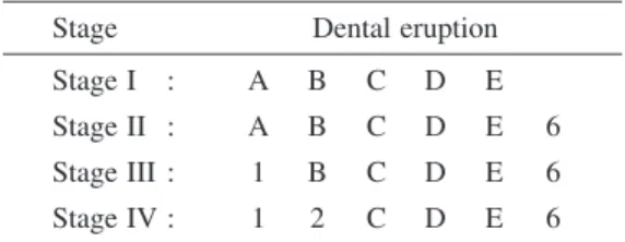
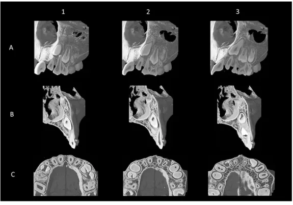
(4) Root resorption of maxillary primary canines. 147. Fig. 1 Reference area A: coronal direction, B: sagittal direction, C: horizontal direction. Fig. 2 Micro-CT image of Stage I This picture was observed coronal direction (A1-3: from labial to lingual), sagittal direction (B1-3: from mesial to distal) and horizontal direction (C1-3: from below to above).. angles of the primary canines (Fig. 1-B). In the horizontal direction, the area which is parallel to the Frankfurt plane, and from the labial alveolar crest of the primary canines to the anterior nasal spine. (Fig. 1-C). Observation was performed from the labial side in the coronal direction, mesial side in the sagittal direction, and lower side in the horizontal direction..
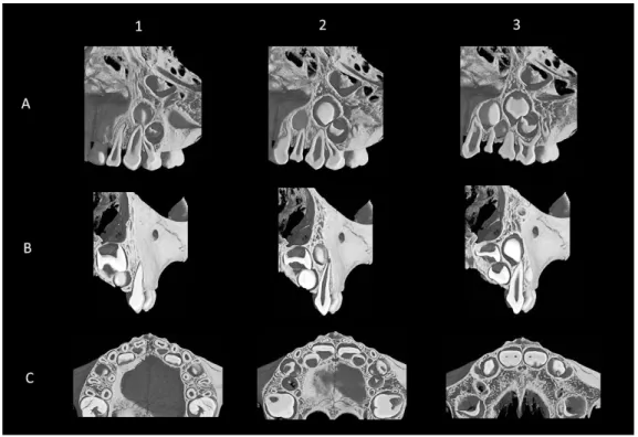
(5) 148. Saka, H., Koyama, T., Tamatsu, Y. et al.. Fig. 3 Micro-CT image of Stage II This picture was observed coronal direction (A1-3: from labial to lingual), sagittal direction (B1-3: from mesial to distal) and horizontal direction (C1-3: from below to above).. 2. Measurement of the distance between the root surface and bony crypt To elucidate the relationship between the roots of maxillary primary canines and the bony crypts of maxillary canine observed on Micro-CT images, the shortest distance between the root surface and bony crypt was measured. Measurement was carried out in the horizontal direction in the range of the root and bony crypt. The mean value and the standard deviation were calculated by the statistical method. Differences in the mean value between the dentition stages were analyzed by Tukey’s t-test (P<0.05), statistical analysis software (Statmate III, Atms) was used.. Results 1. Observation of the positional relationship between the roots of primary canines and successive permanent canines in the dry skulls of children 1) Stage I (Fig. 2). a. Coronal direction The mesio-distal position of the bony crypt of canine. was observed that the inferior margin was almost directly above the root of primary canine and the superior margin was slightly distally positioned (A-2, 3). The supero-inferior position was located superior to the tooth germs of lateral incisor, first premolar, and central incisor (A-3). The shape of bony crypt was observed as quadrangle in the central area (A-2), and triangle on the lingual side (A-3). b. Sagittal direction In the mesial area, although the central and lateral incisors were observed, the canine was not observed (B-1). In the central area, the canine was observed slightly superior to the lingual side of the root apex of primary canine whose root was uncompleted (B-2). In the central and distal areas, in the order of canine, lateral incisor and central incisor were located from the labial to lingual side (B-2, 3). c. Horizontal direction In the inferior area, the canine was not observed, and the mesio-lingual area of the primary canine root was adjacent to the distal area of the bony crypt of lateral incisor (C-1). In the central area, the bony crypt of canine was observed in the lingual side of the root of primary canine (C-2). In the superior area, the central incisor and canine were positioned.
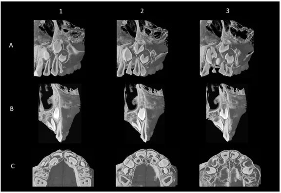
(6) Root resorption of maxillary primary canines. 149. Fig. 4 Micro-CT image of Stage III This picture was observed coronal direction (A1-3: from labial to lingual), sagittal direction (B1-3: from mesial to distal) and horizontal direction (C1-3: from below to above).. side by side (C-3). 2) Stage II (Fig. 3) a. Coronal direction The mesio-distal position of the bony crypt of canine was observed that the inferior margin was almost directly above the root of primary canine and the superior margin was slightly distally positioned similar to Stage I (A-1, 2, 3). Every permanent tooth germ grew supero-inferiorly in comparison with Stage I (A-2, 3). The adjoining bony crypts overlapped and boundaries became unclear in some areas (A-2, 3). b. Sagittal direction The canine and lateral incisor were observed on the lingual side of the primary canine root in all areas (B-1, 2, 3). The canine was located superior to the root apex of primary canine, the lateral incisor was present around the central area on the lingual side of the primary canine root and the canine was adjacent to the nasal cavity (B-1, 2, 3). Regarding the labio-lingual positional relationship, the canine was present on the labial side and the lateral incisor was located on the lingual side (B-1, 2, 3). c. Horizontal direction In the inferior area, the canine was not observed as. in Stage I and the mesio-lingual area of primary canine root was adjacent to the distal area of the bony crypt of lateral incisor (C-1). In the central area, the inferior margin of the bony crypt of canine was located between the primary canine root and the bony crypt of lateral incisor (C-2). Also in the superior area, the bony crypt of canine was located between the primary canine root and bony crypt of lateral incisor. The bony crypts from central incisor to first premolar overlapped labio-lingually (C-3). 3) Stage III (Fig. 4) a. Coronal direction The superior margin of canine was further distally inclined in comparison with Stage II (A-2, 3). In the central area, the bony crypt of canine was observed to be quadrangular (A-2). The bony crypt of canine was connected with the bony crypt of first premolar in lingual area (A-3). Resorption of the root apex of primary canine was observed in the labial area (A-1). b. Sagittal direction The lateral incisor was present around the central area on the lingual side of primary canine root similar to Stage II in every area (B-1, 2, 3). Resorption of the lingual side nearby the root apex of primary.
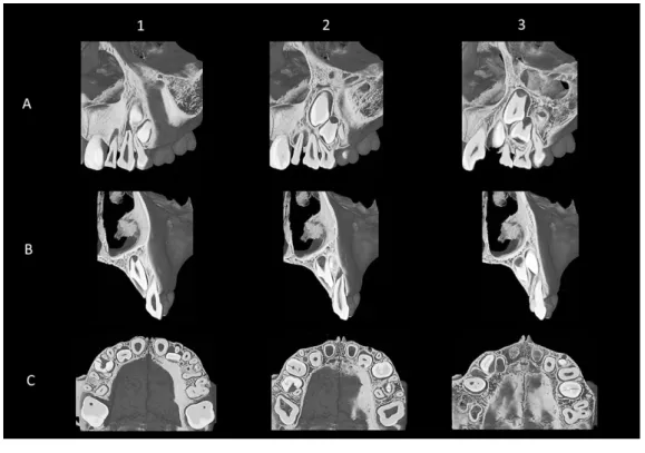
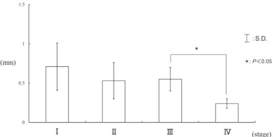
(7) 150. Saka, H., Koyama, T., Tamatsu, Y. et al.. Fig. 5 Micro-CT image of Stage IV This picture was observed coronal direction (A1-3: from labial to lingual), sagittal direction (B1-3: from mesial to distal) and horizontal direction (C1-3: from below to above).. canine was observed in the central area (B-2). c. Horizontal direction In the inferior area, the primary canine root was adjacent to the distal area of the bony crypt of lateral incisor (C-1). The bony crypt of canine was observed on the lingual side in the inferior area (C-1), whereas its central and superior areas were observed on the labial side (C-2, 3). 4) Stage IV (Fig. 5) a. Coronal direction The bony crypt of canine was located slightly mesial to the primary canine root in the labial side (A-1). The superior margin of the bony crypt of canine was distally inclined, and its inferior margin was mesially inclined, being adjacent to the tooth root of the lateral incisor in the area from central to lingual side (A-2, 3). Resorption nearby the root apex of primary canine was observed in the labial side (A-1). b. Sagittal direction The bony crypt of canine was adjacent to the nasal cavity in all areas (B-1, 2, 3). In the central area, it approached the lingual side nearby the root apex of primary canine, showing root resorption (B-2). In the distal area, the first premolar being observed. in the inferior area was adjacent to the primary canine (B-3). c. Horizontal direction The bony crypt of canine was positioned on the labial side in all areas (C-1, 2, 3). In the inferior area, the bony crypt was observed between the lateral incisor and primary canine (C-1). The labial side of lateral incisor’s root was surrounded by the bony crypt of canine and the compact bone on the labial side was resorbed from central to superior area (C-2, 3). 2. Measurement of the distance between the root surface and bony crypt The shortest distance between the root surface of maxillary primary canine and the bony crypt of maxillary canine was measured (Fig. 6). The mean distance in Stage I was 0.71 mm, the maximum 1.26 mm, and the minimum 0.00 mm. The mean distance in Stage II was 0.53 mm, the maximum 0.84 mm, and the minimum 0.00 mm. The mean distance in Stage III was 0.55 mm, the maximum 0.77 mm, and the minimum 0.00 mm. The mean distance in Stage IV was 0.24 mm, the maximum 0.35 mm, and the minimum 0.00 mm..
(8) Root resorption of maxillary primary canines. 151. Fig. 6 The shortest distances between the root surface of maxillary primary canine and the bony crypt of maxillary canine Differences in the mean value between the dentition stages were analyzed by Tukey’s t-test (P<0.05).. Significant differences were observed between Stage III and Stage IV.. Discussion 1. Observation of the positional relationship between the roots of primary canines and successive permanent canines in the dry skulls of children In the jaw of the dry skulls of children, the permanent tooth germs are not fixed. Furthermore, regarding primary root resorption, not only the tooth germs of successive permanent teeth, but also the bony crypts become a major factor. Therefore, we observed the positional relationship between the roots of maxillary primary canines and the bony crypts of successive canines. Previous studies in our department have shown changes in the size, shape, and position of maxillary bony crypts with changes in the tooth eruption phase7,8,12,15–17). Observing the positional changes of the bony crypts of maxillary permanent tooth germs in the primary dentition stage, Kawashima15) reported that the bony crypts of central incisor and canine are located in the labio-superior position, that of lateral incisor is present in the lingo-inferior position and first premolar is in the labio-inferior position. Therefore, central incisor, lateral incisor and first premolar show expansion in the inferior direction with the shift of the tooth eruption stage, whereas only canine is present in the superior position, and descends in. the inferior direction immediately before eruption. Furthermore, Kawashima15) also reported that because all bony crypts are very large, in contrast to the size of the jaw in the primary dentition stage, they show overlap and deviation of their mesio-distal arrangement, however, their arrangement gradually becomes linear after the eruption of lateral incisor. Ihara16) reported that because the shape of the bony crypts develops in the supero-inferior direction, their triangle or quadrangle at the first stage changes to rectangle. Regarding the growth of the superior and inferior margins of the bony crypt containing its permanent tooth germ, Hanai17) reported that the superior margin of the bony crypt of central incisor and first premolar shows no change, whereas their inferior margin descends, the superior margin of lateral incisor ascends, but its inferior margin shows no change and the superior margin of canine ascends, whereas its inferior margin descends. The observation-based results obtained in this study were similar to these previous reports. In particular, the bony crypt of maxillary canine not only showed the growth of the superior and inferior margins, but also presented the finding that the superior margin was distally inclined, and inferior margin was mesially inclined, which were not observed in other areas. Among all teeth, the canine is longest, located in a high position until its eruption, and it needs to avoid the nasal cavity which is present in the supero-internal area of canine..
(9) 152. Saka, H., Koyama, T., Tamatsu, Y. et al.. inden et al.18) observed the internal area of the jaw L of children’s skulls and reported that the position of canine is influenced by the width of the piriform aperture. From these findings, it was considered that mesio-distal inclination is necessary for the growth of canine in the maxilla. The root resorption of maxillary primary canine was initiated from the lingual side nearby the root apex. Linden et al.19) reported that the root resorption of primary canine was morphologically in accordance with the coronal outline of the canine. The position of the bony crypt containing the canine tooth germ was higher than that of other teeth. Furthermore, from superior to central areas of the bony crypt of canine were positioned on the labial side, its inferior area was located on the lingual side. These findings were morphologically almost in accordance with the form of the labial margin of the coronal outline of canine. Therefore, it was considered that the root resorption of primary canine was initiated from the area of the lingual side of root apex. Observing another primary anterior area of maxilla, Hiraide et al.12) reported that the root resorption of primary central incisor is initiated from the lingual surface, and that of the primary lateral incisor is initiated from the mesio-lingual surface being influenced by the growth of central incisor. These findings revealed that not only the successive permanent teeth, but also the adjoining permanent teeth are related in the root resorption of primary anterior teeth, showing differing resorption patterns in each case. Comparing the maxilla and mandible, Lu et al.13) reported that the growth was noted in both the superior and inferior margins of the bony crypt of mandibular canine. This result was similar to the observation-based result in this study. Furthermore, Lu et al.13) stated that since the bony crypt of mandibular canine being located below the primary canine moves to the disto-lingual side with growth, root resorption of mandibular primary canine starts from the disto-lingual side. However, the result for the maxilla in this study showed that since the bony crypt of maxillary canine was located in a high position and mesio-distally inclined, root resorption of primary canine was only partly observed nearby its root apex, even the eruption stage of lateral incisor. As the position of the bony crypt of successive permanent canine in the maxilla was different from that in the mandible, the initiation area of root. resorption also differed. This was considered to be one factor associated with the difference in the eruption order of canine between the maxilla and mandible. 2. Measurement of the distance between the root surface and bony crypt The changes in the positional relationship between the primary teeth and successive permanent teeth with advancement of the eruption stage could be quantitatively analyzed using Micro-CT. Therefore, to elucidate the relationship between the root surface of maxillary primary canines and the bony crypts of maxillary canines observed on images obtained using Micro-CT, the shortest distance between the root surface and bony crypt was measured. The measurements in this study quantitatively showed that the distance between the canine bony crypts and roots reduced with progression of the eruption stage and significant differences were observed between Stage III and Stage IV. Then, comparing the maxilla and mandible, the distances in all eruption stages of maxilla is closer than that of mandible13). It was considered that the reason why canine area in the maxilla is smaller than that of mandible and maxillary canine is longest among all teeth. The minimum distance was 0.00 mm in every stage. This result meant that a part of canine bony crypt was bounded on the lingual side nearby the primary canine’s root apex without resorption even the early dentition stage. Then root resorption was advanced gradually with the shift of the tooth eruption stage.. Conclusion We classified 16 dry skulls of Indian children stored in the Department of Anatomy in Tokyo Dental College into four stages according to the eruption state of primary and permanent teeth, and observed the relationship between the root of maxillary primary canines and successive permanent teeth using Micro-CT. 1. Observation of the positional relationship between the roots of primary canines and successive permanent canines in the dry skulls of children In the primary dentition stage (Stage I), the bony crypts including successive canines was positioned.
(10) Root resorption of maxillary primary canines. almost directly above the root of primary canine and located superior to another bony crypts. After the first molars reached the alveolar crest in addition to the primary dentition stage (Stage II), the bony crypt of canine grew, showing the distal inclination of the superior margin and mesial inclination of the inferior margin. After the central incisors reached the alveolar crest in addition to Stage II (Stage III), root resorption of primary canines was observed on the lingual side nearby the root apex and the bony crypt of canine was adjacent to the nasal cavity. 2. Measurement of the distance between the root surface and bony crypt From central incisors reached the alveolar crest to lateral incisors reached that, it was quantitatively shown that the distance between the roots of primary maxillary canine and the bony crypt of canine reduced. Acknowledgments We would like to thank our staff for all their advice and valuable suggestions.. References 1) Orban, B.: Resorption and repair on the surface of the root. J Am Dent Assoc 15: 1768–1777, 1928. 2) Kronfeld, R.: The resorption of the roots of deciduous teeth. Dent Cosmos 74: 103–120, 1932. 3) Ten Cate, A.R.: Orban’s Oral Histology and Embryology. 10th ed. (Bhaskar, S.N. ed.) The CV Mosby Co., St. Louis, 1986, pp.361–374. 4) Nagatani, S.: A study on the root resorption of human teeth. J Osaka Odontol Soc 51: 859–881, 1988. (in Japanese) 5) Haavikko, K.: Correlation between the root resorption of deciduous teeth and the formation of the corresponding permanent teeth. Proc Finn Dent Soc 69: 191–201, 1973. 6) Matsumoto, H., Matsumoto, T. and Okamoto, Y.: Morphological study of the root resorption of milk teeth. J Osaka Odontol Soc 19: 39–43, 1956. (in Japanese). 153. 7) Saka, H.: A morphological study of the root resorption of the maxillary second deciduous molars. Shikwa Gakuho 94: 911–940, 1994. (in Japanese) 8) Saka, H., Kikuchi, A. and Ide, Y.: A morphological study of root resorption of the maxillary first deciduous molars. Bull Tokyo Dent Coll 37: 137–144, 1996. 9) Kikuchi, A.: A morphological study of root resorption of the mandibular first deciduous molars. Shikwa Gakuho 99: 137–155, 1999. (in Japanese) 10) Kitamura, S., Saka, H. and Ide, Y.: A morphological study of root resorption of the mandibular first deciduous molars. Jpn J Ped Dent 38: 548–561, 2000. (in Japanese) 11) Iida, T., Saka, H. and Ide, Y.: A study of root resorption of the mandibular deciduous incisors and its relationship to the position of successional permanent teeth using Micro-CT. Jpn J Ped Dent 40: 19–31, 2002. (in Japanese) 12) Hiraide, Y., Saka, H., Tamatsu, Y., Akinobu, U., Yanagisawa, N. and Ide, Y.: Root resorption of maxillary primary incisors in relation to position of successive permanent incisors by Micro-CT. Ped Dent J 18: 15–23, 2008. 13) Lu, W.-Y., Saka, H., Tamatsu, Y., Nakahara, K., Agematsu, H. and Ide, Y.: The morphological analysis of root resorption of mandibular primary canines and their relationship with the position of successive permanent teeth using Micro-CT. Ped Dent J 19: 187–195, 2009. 14) Kamioka, H.: Positional relationship between the deciduous molar and the successional permanent teeth — three dimensional observation of the deciduous second molars and second premolars by X-ray CT —. Ohu Univ Dent J 32: 67–78, 2005. (in Japanese) 15) Kawashima, I.: Studies on the changes of the crypt of the maxilla occurring as permanent teeth erupt. Shikwa Gakuho 76: 1041–1098, 1976. (in Japanese) 16) Ihara, Y.: Studies on internal structure of children’s maxilla 2. Form and size of the crypt and the thickness of the compact bone. Shikwa Gakuho 78: 587–641, 1978. (in Japanese) 17) Hanai, S.: Studies on the movement and the growth of the permanent tooth germs in the maxilla of the children with the eruption of the permanent teeth. Shikwa Gakuho 76: 1351–1412, 1976. (in Japanese) 18) Linden, F.P.G.M. and Duterloo, H.S.: Development of the Human Dentition: An Atlas. Harper & Row, Hagerstown, 1976, p.158. 19) Linden, F.P.G.M. and Duterloo, H.S.: Development of the Human Dentition: An Atlas. Harper & Row, Hagerstown, 1976, p.160..
(11)
図
+3
関連したドキュメント
An easy-to-use procedure is presented for improving the ε-constraint method for computing the efficient frontier of the portfolio selection problem endowed with additional cardinality
The inclusion of the cell shedding mechanism leads to modification of the boundary conditions employed in the model of Ward and King (199910) and it will be
(Construction of the strand of in- variants through enlargements (modifications ) of an idealistic filtration, and without using restriction to a hypersurface of maximal contact.) At
It is suggested by our method that most of the quadratic algebras for all St¨ ackel equivalence classes of 3D second order quantum superintegrable systems on conformally flat
Keywords: continuous time random walk, Brownian motion, collision time, skew Young tableaux, tandem queue.. AMS 2000 Subject Classification: Primary:
Answering a question of de la Harpe and Bridson in the Kourovka Notebook, we build the explicit embeddings of the additive group of rational numbers Q in a finitely generated group
Then it follows immediately from a suitable version of “Hensel’s Lemma” [cf., e.g., the argument of [4], Lemma 2.1] that S may be obtained, as the notation suggests, as the m A
In our previous paper [Ban1], we explicitly calculated the p-adic polylogarithm sheaf on the projective line minus three points, and calculated its specializa- tions to the d-th
