Posted at the Institutional Resources for Unique Collection and Academic Archives at Tokyo Dental College, Available from http://ir.tdc.ac.jp/
Title
Localization of palatal area in human somatosensory
cortex
Author(s)
Alternative
Bessho, H; Shibukawa, Y; Shintani, M; Yajima, Y;
Suzuki, T; Shibahara, T
Journal
Journal of Dental Research, 86(3): 265-270
URL
http://hdl.handle.net/10130/446
INTRODUCTION
S
omatosensory information from the orofacial area is received by peripheral sensory neurons (trigeminal neurons) and projected into the thalamus via the trigeminothalamic tract. It is then projected into the primary somatosensory cortex, which is involved in somatotopic functional organization. The cortical representation of orofacial sensation in the human primary somatosensory cortex has been demonstrated by invasive direct cortical stimulation and intracranial recording of somatosensory-evoked potentials (Penfield and Rasmussen, 1950; Picard and Olivier, 1983; Baumgartner et al., 1992; McCarthy et al., 1993). The results of these studies indicated that the orofacial regions are located on the lateral inferior primary somatosensory cortex in humans.Concerning the location of the 'hard palate representing' area in the primary somatosensory cortex, clear individual differences have been reported following stimulation evoking subjective sensation in the palate (intracranial recordings of somatosensory-evoked potentials; McCarthy et
al., 1993). Two spatially separated distributions of the palatal area on the
primary somatosensory cortex were identified in half of all persons examined, while one single focal primary somatosensory cortex palatal area was found in the remaining individuals (McCarthy et al., 1993). In addition, one non-invasive somatosensory-evoked magnetic field recording study with magnetoencephalography suggested that the palatal gingival area has an unexpectedly large distribution in the primary somatosensory cortex, compared with the lip- or tongue-representing areas (Nakahara et al., 2004), although two-point discrimination for the palatal mucosa was found to be poorer than that for the lip or tongue (Ringel and Ewanowski, 1965). The distribution contents of the sensorimotor cortex devoted to a particular structure are directly related to its innervation density (Mountcastle, 1984). Since both of these studies suggested that palatal stimulation evoked a tooth-tap sensation in individuals (McCarthy et al., 1993; Nakahara et al., 2004), pure stimulation was not applied to the palatal mucosa. Periodontal stimulation, however, was included during palatal stimulation in those studies (McCarthy et al., 1993; Nakahara et al., 2004). Therefore, the exact locations and detailed extent of distribution of the pure 'hard palate representing' area in the human primary somatosensory cortex remain to be determined.
The aim of the present study was to determine the feasibility of a precise mapping of the 'hard palate representing' area in the human primary somatosensory cortex. Since the hard palate is innervated by 2 independent sensory nerves, the nasopalatine and the great palatine, we stimulated the right side of the first and third transverse palatine ridges, as well as the right side of the greater palatine foramen. To allow for pure stimulation in these palatal regions, we developed a custom-made stimulation device. Using magnetoencephalography, we recorded somatosensory-evoked magnetic
ABSTRACT
To determine the 'hard palate representing' area in the primary somatosensory cortex, we recorded somatosensory-evoked magnetic fields from the cortex in ten healthy volunteers, using magnetoencephalography. Following electrical stimulation of 3 sites on the hard palate (the first and third transverse palatine ridges, and the greater palatine foramen), magnetic responses showed peak latencies of 15, 65, and 125 ms. Equivalent current dipoles for early magnetic responses were found along the posterior wall of the inferior part of the central sulcus. These dipoles were localized anterior-inferiorly, compared with locations for the hand area in the cortex. However, there were no significant differences in three-dimensional locations among the 3 selected regions for hard palate stimulation. These results demonstrated the precise location of palatal representation in the primary somatosensory cortex, the actual area being small.
KEY WORDS: human, maxillary nerve,
magnetoencephalography, palate, somatosensory-evoked magnetic field.
Received May 1, 2006; Last revision October 6, 2006; Accepted November 7, 2006
Localization of Palatal Area in
Human Somatosensory Cortex
H. Bessho
1*, Y. Shibukawa
2, M. Shintani
3,
Y. Yajima
4, T. Suzuki
2, and T. Shibahara
1 Oral Health Science Center, 1Department of Oral andMaxillofacial Surgery, 2Department of Physiology, 3Laboratory of Brain Research, and 4Department of Oral
and Maxillofacial Implantology, Tokyo Dental College, 1-2-2, Masago, Mihama-ku, Chiba 261-8502, Japan; *corresponding author, [email protected]
J Dent Res 86(3):265-270, 2007 Clinical
266 Bessho et al. J Dent Res 86(3) 2007
fields produced by intracellular currents in the cortical neuron following electrical stimulation, and estimated equivalent current dipoles. Latencies and equivalent current dipole locations generating somatosensory-evoked magnetic fields were analyzed at each stimulation point, so that we could determine the functional organization of the somatotopy of the hard palate primary somatosensory cortex.
MATERIALS & METHODS
ParticipantsRight-handed healthy adults (nine males and one female; mean age, 27 yrs) were studied. All participants gave written informed consent prior to the experiments, and this study was approved by the Ethics Committee of our institute in accordance with the Declaration of Helsinki. None of the participants in our study had either an edentulous maxilla (with recent tooth extraction) or disorders (including acute or chronic pain in the orofacial area) of oral function.
Palatal Stimulation
For electrical stimulation, a constant current of square pulses (SEM-4201, Nihonkohden, Tokyo, Japan) was applied to the first and third transverse palatine ridges, as well as to the greater palatine foramen, with the use of a pair of Ag-AgCl electrodes (Fig. 1A). The electrodes (with their tips about 2.5 mm apart) were embedded on a plastic splint, which was attached to the maxillary dentition so that we could obtain a fixed stimulation position during each session (Figs. 1B, 1C). Stimulus amplitude was set for each person at twice the individual sensory threshold (1-7 mA). Our participants felt neither a tooth-tapping sensation nor any sensation in the upper teeth or gums. In addition, the intensity of these electrical stimulations was confirmed to be non-noxious (producing a non-painful sensation). A 0.05-ms stimulus was applied at intervals of 1-3 sec. To compare cortical responses, we also recorded somatosensory-evoked magnetic fields following stimulation of the median nerve in the right wrist.
Somatosensory-evoked Magnetic Field Recordings
A 306-channel neuromagnetometer (Vectorview, Elekta-Neuromag Co., Helsinki, Finland) was used for recording magnetic fields from 204 positions on the whole scalp. The locations of 3 anatomical landmarks (the nasion and bilateral pre-auricular points) and 2 pairs of head position indicator coils, attached to the forehead and the mastoid process bilaterally, were determined with a three-dimensional digitizer (Isotrak, Polhemus Inc., Colchester, VT, USA). At the start of each recording session, we determined the exact locations and orientations of the sensors, with respect to the head, by measuring the magnetic fields produced by currents applied to the head position indicator coils. In one experiment, we stimulated the 4 sites (3 sites for the palate, and 1 site for the median nerve in the wrist) in each person in a randomized order. Two hundred trials were averaged on magnetic signals in each site. To confirm reproducibility, we obtained a further set of somatosensory-evoked magnetic fields from the 3 palatal sites in a separate experiment on another day. Magnetic resonance images of
Figure 1. Measurement of somatosensory-evoked magnetic fields for the hard palate. (A) Anatomical diagrams showing 3 stimulation sites for the palate. (B,C) The Ag-AgCl electrodes (approximately 2.5 mm between electrodes) were embedded in a custom-made plastic splint attached to the maxillary dentition to obtain a fixed stimulus position.
(D) Typical examples of whole-scalp magnetic responses of somatosensory-evoked magnetic fields following stimulation of the right first transverse palatine ridge. Traces are plotted on the "flattened head", as viewed from above with the person's nose upwards. Each trace began 100 msec prior to and ended 300 msec after stimulus onset. Circles indicate channels showing prominent magnetic signals. (E) Enlarged superimposed traces of the somatosensory-evoked magnetic field evoked by right-side stimulation of the first (upper) and third transverse palatine ridges (middle), and the greater palatine foramen (lower). The 3 successive identifiable peak signal components were designated 1M, 2M, and 3M.
the brain were obtained in each person with a 1.5-T whole-body scanner (Symphony, Siemens, Munich, Germany), so that brain anatomy could be determined.
Data Analysis
Magnetoencephalography signals were digitized at 1 kHz and band-pass-filtered (0.1-330 Hz). Isocontour maps were constructed from the measured data at selected time-points by the minimum norm estimation method (Hämäläinen and Ilmoniemi, 1994). To identify the sources of somatosensory-evoked magnetic fields, we divided the signals into several periods. During each period, one equivalent current dipole was first determined by least-square search for the whole head. Only equivalent current dipoles attaining more than 90% goodness-of-fit were accepted for analysis, in which case the entire time period and all the channels were taken into account for computation of the parameters of a time-varying multidipole model (Hämäläinen et al., 1993). We identified the next equivalent current dipole by removing the effects of the previous sources from the magnetic signal pattern (Uusitalo and Ilmoniemi, 1997), and then searching for additional sources in the responses of the residual waveforms. The three-dimensional location, orientation, and strength of the equivalent current dipole in a spherical conductor model were determined on the three-dimensional coordinate frame detected by head position indicator coils. The equivalent current dipoles were then superimposed on the individual's magnetic resonance images, so that we could determine the source locations with respect to anatomical structures. The differences among the means of the latencies of magnetic fields, as well as the locations of equivalent current dipoles in 3 axes, were statistically tested by the Kruskal-Wallis H-test. Significance was defined as P < 0.05. Post hoc comparisons were performed by the Mann-Whitney U-test with Bonferroni correction.
RESULTS
WaveformsUsing whole-head magnetoencephalography, we obtained magnetic signals produced by neurons in the cortex following stimulation of 3 regions on the hard palate (Figs. 1A-1C). During these stimulations, prominent somatosensory-evoked magnetic field responses were identified in the regions corresponding to subsets of the neuromagnetic sensor array located in the parietotemporal region in the hemispheres both contra- and ipsilateral to the stimulation (circles in Fig. 1D). In each waveform (Fig. 1E) showing somatosensory-evoked magnetic field responses following stimulation of the first and third transverse palatine ridges and the greater palatine foramen, we could consistently identify 3 magnetic components within an analysis period of 300 msec. The early components of peak latency were about 15 msec (1M) in the contralateral hemisphere for all palatal stimulation sites, and in the ipsilateral hemisphere for only the first and third transverse palatine ridge stimulations. The middle (65 msec in latency; 2M) and late components (125 msec in latency; 3M) were also observed in the bilateral hemispheres. No significant interhemispheric differences were found for each of the 3 components (p > 0.05) (Table).
Source Distribution
To make a model with multi-dipoles generating the magnetic signal distribution, we examined the fields by time-varying
multi-dipole analysis (see METHODS). Equivalent current dipoles producing 1M components for all palatal stimulation sites were identified along the posterior wall of the inferior part of the central sulcus (lateral-inferior position of the post-central gyrus corresponding to the orofacial primary somatosensory cortex) (Fig. 2). The equivalent current dipole of 1M for greater palatine foramen stimulation was found in the contralateral hemisphere (Fig. 2C), while those for the first transverse palatine ridge were found bilaterally (Fig. 2A). In the third transverse palatine ridge stimulations, four of ten participants showed bilateral equivalent current dipoles, while the remaining participants showed only contralateral equivalent current dipoles (Fig. 2B, Table) for the 1M magnetic component. The equivalent current dipoles for the 1M and 2M components were located in the same position in the post-central gyrus; however, the equivalent current dipoles for 1M were directed anteriorly, while those for 2M were directed posteriorly (not shown). Furthermore, the equivalent current dipoles of the bilateral 3M components were identified in the upper bank of the Sylvian fissure (corresponding to the secondary somatosensory area) in all participants.
The equivalent current dipole with the initial magnetic component of 20 msec in latency for the median nerve stimulation was localized in the upper area of the post-central gyrus. Following palatal stimulation of the 3 sites, equivalent current dipoles were confirmed to be located in a lower area in the post-central gyrus than those for median nerve stimulation in the three-dimensionally reconstructed magnetic resonance image of the individual brain (Fig. 2D).
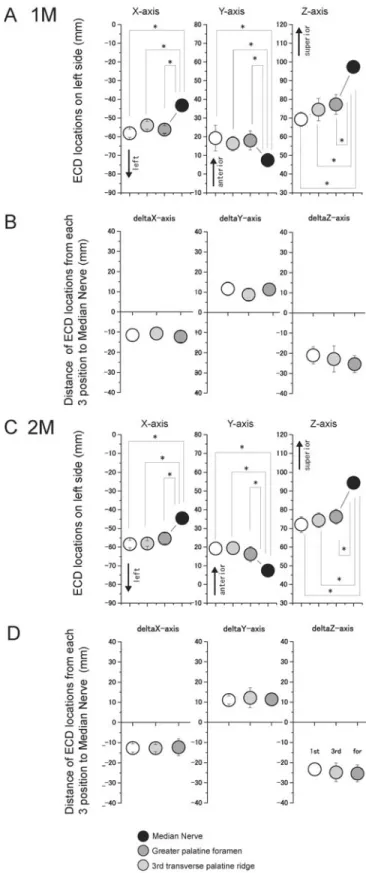
To analyze further the localization of the hard-palate-representing area in the primary somatosensory cortex, we compared the means of the three-dimensional equivalent current dipole locations for contralateral 1M (Figs. 3A, 3B) and 2M (Figs. 3C, 3D) components between each of the 3 palatal and the one median nerve stimulation sites. The equivalent current dipole locations (Figs. 3A, 3C), as well as their relative distances (Figs. 3B, 3D), showed significant anterior and inferior orientation of somatosensory representation of the hard palate area in the primary somatosensory cortex, compared
Table.The Averaged Latencies of 1M to 3M in the Evoked Magnetic Fields following Palatal Stimulation
Peak Latenciesa(msec)
Stimulation Sites 1M 2M 3M
1st transverse palatine ridge
Contralateral 14 ± 2 66 ± 3 135 ± 6 Ipsilateral 16 ± 1* 67 ± 3 116 ± 3 3rd transverse palatine ridge
Contralateral 16 ± 2 68 ± 3 123 ± 8 Ipsilateral 18 ± 3** 65 ± 3 128 ± 6 Greater palatine foramen
Contralateral 17 ± 2 63 ± 2 139 ± 17
Ipsilateral ND 62 ± 3 126 ± 20
a The values are shown as mean ± SE from 10 participants, except
for the latencies of the ipsilateral side of the first (*n = 9) and third transverse palatine ridges (**n = 4).
268 Bessho et al. J Dent Res 86(3) 2007
with that of the wrist area, showing distinct spatial separation between them. However, the 3 regions of the palatal area in the primary somatosensory cortex were localized relatively contiguously. There were no significant differences in distances to the equivalent current dipole locations for each palatal stimulation site from the equivalent current dipioles for median nerve stimulation (Figs. 3B, 3D).
DISCUSSION
The results of this study demonstrated the precise locations and temporal activities of neurons in the hard palate area of the primary somatosensory cortex.
The values of peak latency of 1M for palatal stimulation are in line with results showing that the first responses of somatosensory-evoked magnetic fields by trigeminal nerve stimulation had a peak latency of around 15 msec (Hoshiyama et al., 1996; Yamashita et al., 1999; Nagamatsu et al., 2001; Suzuki et al., 2004). In addition, these values for 1M were consistent with the results from an electroencephalography study showing the latency for the first negative cortical potential following electrical stimulation for the large area of the hard palate (reported to be around 11-13 msec; Maloney et al., 2000). Therefore, our results clearly indicate that the 1M component of the somatosensory-evoked magnetic field reflects the initial cortical response in the palatal representation area in the primary somatosensory cortex.
The equivalent current dipoles producing 1M components were identified on the lateral inferior position of the primary somato-sensory cortex (orofacial primary somatosensory cortex) in the contralateral hemisphere by greater palatine foramen stimulation. However, the equivalent current dipoles for 1M following stimulation of the first transverse palatine ridge (innervated by the nasopalatine nerve) were identified on the orofacial primary somatosensory cortex bilaterally. It has been reported, in humans, that the nasopalatine nerve emerges from the incisive foramen and is distributed
Figure 2. Source locations. (A-C) Magnetic resonance images showing the locations of equivalent current dipoles of the 1M, 2M, and 3M components following electrical stimulation of the first transverse palatine ridge (A), the third transverse palatine ridge (B), and the greater palatine foramen (C). Blue circles show the locations of equivalent current dipoles on frontal planes of magnetic resonance images. The equivalent current dipoles of 1M (left images) and 2M (middle images) are located in the post-central gyrus, and those of 3M (right images) are located in the upper bank of the Sylvian fissure. (D) Locations of contralateral equivalent current dipoles of 1M were superimposed on the three-dimensionally reconstructed magnetic resonance image of the individual's brain. Solid lines indicate the central sulcus, the Sylvian fissure, and the great longitudinal fissure.
over both sides of the anterior part of the palate (Langford, 1989). In contrast, although the greater palatine nerve distributes unilaterally to the posterior part of the palate, the palatal region of the third transverse palatine ridge is anatomically innervated by overlapped distribution via both nasopalatine and greater palatine neurons. In our study, bilateral activation of the primary somatosensory cortex via the 1M component was observed in nine of the ten participants following stimulation of the first transverse palatine ridge (innervated by the nasopalatine nerve), and in four of the ten participants following stimulation of the third transverse palatine ridge. No bilateral activation was observed following stimulation of the greater palatine foramen. Therefore, our results clearly indicate that initial somatosensory information from the anterior part of the palate has a bilateral direct projection to the primary somatosensory cortex, while that from the posterior part has a contralateral projection, supporting evidence for neuronal innervation patterns in hard palate mucosa. In contrast, the equivalent current dipoles of the 2M components from the 'posterior' part were also detected in the bilateral primary somatosensory cortex. These bilateral cortical responses for 2M suggest an ipsilateral response, due to direct projection of uncrossed ascending fibers and/or to projection to both hemispheres from the thalamus (Disbrow et al., 2003).
The equivalent current dipoles of 3M corresponded to activation of the secondary somatosensory cortex in the upper bank of the Sylvian fissure. It is difficult to record a response generated by the secondary somatosensory coretx in somatosensory-evoked potentials recorded from the scalp surface (Hoshiyama et al., 1996), but secondary somatosensory cortex responses can be recorded by magnetoencephalography. Neurons in the secondary somatosensory cortex not only receive somatosensory information from the primary somatosensory cortex, but also respond to ascending signals from both sides of the body (bilateral function) (Hari et al. 1993; Hoshiyama et al., 1996). Therefore, the secondary somatosensory cortex may have an important role in sensory information-processing for the hard palate.
During the somatosensory-evoked magnetic field recordings, none of the volunteers felt either a tooth-tapping sensation or any sensation of the upper teeth or gums. Therefore, our results indicate the exact locations and distribution of the pure 'hard palate representing' area in the human primary somatosensory cortex, revealing anterior and inferior orientations compared with representation of the hand area. In addition, this localization of the palatal primary somatosensory cortex is consistent with the well-known somatotopic organization of the primary somatosensory cortex. In the three-dimensional comparisons of equivalent current dipole locations, each of the 3 sites of the palatal area in the primary somatosensory cortex was localized contiguously, indicating that the hard palate mucosa is
represented by a small region in the primary somatosensory cortex, as expected from the low innervation densities to the hard palate (Ringel and Ewanowski, 1965; McCarthy et al., 1993). The hard palate plays an important role in the precise manipulation necessary for orofacial functions such as articulation and oral object exploration. Therefore, sensory
Figure 3.Averaged equivalent current dipole positions. (A,C) Comparisons of the three-dimensional equivalent current dipole locations on each axis between 3 regions of the hard palate and median nerve stimulations are shown for 1M (A) and 2M (C) components. Somatosensory representation of palatal mucosa in the primary somatosensory cortex shows anterior and inferior orientations in the primary somatosensory cortex, compared with representation of the hand area. (B,D) Distances of equivalent current dipoles from each site on the palate to the median nerve are shown for 1M (B) and 2M (D). Each site for the palatal area in the primary somatosensory cortex was localized contiguously. Statistically significant differences are labeled with asterisks (P < 0.05).
270 Bessho et al. J Dent Res 86(3) 2007
information processing in the palatal area of the primary somatosensory cortex may be integrated with that for lip/tongue sensation, to produce precise sensorimotor functions.
In summary, we clarified the precise location of the 'hard palate representing' area in the primary somatosensory cortex, with the actual area of cortical representation being small, as predicted. In addition, these results provide a detailed organizational map of the orofacial area in the primary somatosensory cortex, by adding the palatal area to the classic "sensory homunculus".
ACKNOWLEDGMENTS
This study was supported by grants for High-Tech Research Center Projects to Y.Y. (HRC3A02 and 6A07) and to Y.S. (6A03) from the MEXT of Japan. I sincerely thank Drs. K. Kubo, E. Takeda, and M. Tazaki for their continuous support of this work. I also thank Professor Jeremy Williams, Tokyo Dental College, for his assistance with the English of this manuscript.
REFERENCES
Baumgartner C, Barth DS, Levesque MF, Sutherling WW (1992). Human hand and lip sensorimotor cortex as studied on electrocorticography. Electroencephalogr Clin Neurophysiol 84:115-126.
Disbrow EA, Hinkley LB, Roberts TP (2003). Ipsilateral representation of oral structures in human anterior parietal somatosensory cortex and integration of inputs across the midline. J Comp Neurol 467:487-495.
Hämäläinen MS, Ilmoniemi RJ (1994). Interpreting magnetic fields of the brain: minimum norm estimates. Med Biol Eng Comput 32:35-42.
Hämäläinen R, Hari R, Ilmoniemi RJ, Knuutila J, Lounasmaa OV (1993). Magnetoencephalography—theory, instrumentation, and applications to noninvasive studies of the working human brain. Rev Mod Phys 65:413-497.
Hari R, Karhu J, Hamalainen M, Knuutila J, Salonen O, Sams M, et al. (1993). Functional organization of the human first and second somatosensory cortices: a neuromagnetic study. Eur J Neurosci 5:724-734.
Hoshiyama M, Kakigi R, Koyama S, Kitamura Y, Shimojo M, Watanabe S (1996). Somatosensory evoked magnetic fields following stimulation of the lip in humans. Electroencephalogr Clin Neurophysiol 100:96-104.
Langford RJ (1989). The contribution of nasopalatine nerve to sensation of the hard palate. Br J of Oral Maxillofac Surg 27:379-386.
Maloney SR, Bell WL, Shoaf SC, Blair D, Bastings EP, Good DC, et al. (2000) Measurement of lingual and palatine somatosensory evoked potentials. Clin Neurophysiol 111:291-296.
McCarthy G, Allison T, Spencer DD (1993). Localization of the face area of human sensorimotor cortex by intracranial recording of somatosensory evoked potentials. J Neurosurg 79:874-884. Mountcastle VB (1984). Central nervous mechanisms in
mechanoreceptive sensibility. In: Handbook of physiology-the nervous system. Darian-Smith I, editor. Bethesda, MD: American Physiological Society, pp. 789-876.
Nagamatsu K, Nakasato N, Hatanaka K, Kanno A, Iwasaki M, Yoshimoto T (2001). Neuromagnetic localization of N15, the initial cortical response to lip stimulus. Neuroreport 12:1-5. Nakahara H, Nakasato N, Kanno A, Murayama S, Hatanaka K, Itoh H,
et al. (2004) Somatosensory-evoked fields for gingiva, lip, and tongue. J Dent Res 83:307-311.
Penfield W, Rasmussen T (1950). A clinical study of localization of function. In: The cerebral cortex of man. New York: Macmillan. Picard C, Olivier A (1983). Sensory cortical tongue representation in
man. J Neurosurg 59:781-789.
Ringel RL, Ewanowski SJ (1965). Oral perception: 1. Two-point discrimination. J Speech Hear Res 8:389-398.
Suzuki T, Shibukawa Y, Kumai T, Shintani M (2004). Face area representation of primary somatosensory cortex in humans identified by whole-head magnetoencephalography. Jpn J Physiol 54:161-169.
Uusitalo MA, Ilmoniemi RJ (1997). Signal-space projection method for separating MEG or EEG into components. Med Biol Eng Comput 35:135-140.
Yamashita H, Kumamoto Y, Nakashima T, Yamamoto T, Inokuchi A, Komiyama S (1999). Magnetic sensory cortical responses evoked by tactile stimulations of the human face, oral cavity and flap reconstructions of the tongue. Eur Arch Otorhinolaryngol 256(Suppl 1):S42-S46.