INTRODUCTION
Laparoscopic colectomy is increasingly performed worldwide, and has been clinically accepted as a key solution for advanced cancer of the colon and rectum. The 3 - year overall survival and disease - free survival in patients who had laparoscopic surgery did not differ from patients who underwent open colectomy (1, 2). Laparoscopic- assisted colorectal surgery has become an accepted surgical modality for advanced cancer of the colon and rectum. Lymph nodes dissection is necessary for advanced colorectal can-cer in both laparoscopic and open method.
Feasibility and safety of D3 lymph node dissection for sigmoid colon and rectal cancer was reported (3, 4), and feasibility of D3 dissection with preservation of the left colic artery (LCA) was also reported (5). In this report, Kobayashi etc. explained that the mean lengths of the sigmoid colon of Japanese is believed to be longer and this anatomic feature has made it possible to preserve the proxi-mal sigmoid colon during the surgery of rectal and lower sigmoid colon cancer. In some cases, we have preserved LCA in both D2 and D3 dissections to maintain the blood supply to the preserved long proximal sigmoid colon. However, lymph nodes dissection around inferior mesenteric artery (IMA) with LCA preservation using basic five ports placement is difficult for anatomical feature of IMA. There are few reports regarding ports placement for lymph nodes dissection. So, we report about the new ports placement where a port is inserted from a suprapubic region. The aim of this study is to evaluate the new ports placement in laparoscopic cen-tral lymph nodes dissection with LCA preservation for sigmoid
colon and rectal cancer.
PATIENTS AND METHODS
PatientsThis study included 22 consecutive patients (new ports place-ment group (new group, n = 15) and basic ports placeplace-ment group (basic group, n = 7)) who performed laparoscopic central lymph nodes dissection with LCA preservation for sigmoid colon and rectal cancer (Figure 1). Patient’s characterlistics and harvested lymphnodes, operation time, intraoperative blood loss were com-pared.
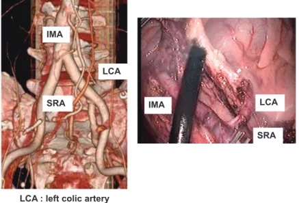
All patients first underwent colonoscopy with biopsy to confirm the diagnosis, multislice spiral computed tomographic (CT) scans of the abdomen, pelvis, and chest to fully stage the extent of tumor before surgery. In addition, 3D- CT colonography and angiography was performed in all cases (Figure 2). Pre and intra- operative simu-lation was performed using these images. These images showed clearly the vascularity, and we could detect the tumor location, IMA, LCA sigmoid colon artery (SA), and superior rectal artery (SRA). And we could preserve LCA cutting the SA or SRA at a suit-able point.
Lymphnodes dissection procedure
The root of the IMA is dissected with an ultrasonic cutting and coagulating surgical device (LCS) to avoid bleeding. The lymph nodes around the IMA are dissected to expose the IMA wall. The bifurcation of the LCA and SRA is exposed. The LCA is exposed until the IMV can be recognized. The IMV is clipped and cut. Adi-pose tissue surrounded by the IMA, LCA, and IMV is dissected with LCS (Figure 3). The mesosigmoid is mobilized posteriorly in a medial - to - lateral fashion to expose the left ureter and gonadal vessels.
ORIGINAL
New ports placement in laparoscopic central lymph nodes
dissection with left colic artery preservation for sigmoid
colon and rectal cancer
Jun Higashijima, Mitsuo Shimada, Takashi Iwata, Kozo Yoshikawa, Toshihiro Nakao, Masaaki Nishi, Hideya Kashihara, and Chie Takasu
Department of Digestive and Pediatric Surgery Tokushima University Hospital, Kuramoto 3-18-15 Tokushima 770-8503, Japan. Abstract : Background : Lymph nodes (LNs) dissection around inferior mesenteric artery (IMA) with left colic artery (LCA) preservation is difficult due to the anatomical feature of IMA. The aim of this study is to evaluate the usefulness of new ports placement inserted from a suprapubic region in laparoscopic LNs dissection around IMA with LCA preservation for sigmoid colon and ractal cancer. Methods : Twenty -two patients who underwent laparoscopic colectomy for sigmoid colon and recal cancer were included. The new ports placement group (n = 15, new group) was compared with the basic ports placement group (n = 7, basic group). Average number of har-vested LNs, total operation time, central LNs dissection time, intraoperative blood loss were compared. Results : There was no significant difference in the average number of harvested LNs. The mean of intraoperative blood loss of new group was significantly lower than that of the basic group (40.0 39.8 ml vs 95.7 81.0 ml, p 0.05). In addition, the mean of operation time of the new group was significantly shorter than that of the basic group (250.0 55.7 min vs 353.4 80.2 min, p 0.05). Conclusions : The new ports placement is useful in laparoscopic LNs dissection around IMA with LCA preservation for sigmoid colon and rectal cancer. J. Med. Invest. 62 : 223-227, August, 2015
Keywords :port placement, laparoscopic colectomy, lymph nodes dissection, inferior mesenteric artery
Received for publication April 23, 2015 ; accepted July 6, 2015. Address correspondence and reprint requests to Jun Higashijima, De-partment of Digestive and Pediatric Surgery, Tokushima University Hospital, Kuramoto 3 18 15 Tokushima 7708503, Japan and Fax : 088 631 -9698.
New ports placement
Basic ports placement
: the port that LCS is inserted at exposing the IMA wall : the working and camera ports
CT colonography ( colonography and angiography )Fusion 3D-CT image Volume rendering image
Tumor
Tumor IMA
SRA S1
IMA : inferior mesenteric artery SRA : superior rectal artery S1 : first sigmoid colic artery
Lymph node
LCA : left colic artery IMA
SRA
LCA
IMA LCA
SRA
Figure 1. Basic ports placement and new ports placement. LCS inserted from a suprapubic region port is parallel to IMA in new ports placement. At exposing the IMA wall, we can effectively use braid length of LCS in new ports placement where a port is inserted from a suprapubic region. On the other hand, we can not use braid full length of LCS in basic ports placement where a port is not inserted from the suprapubic region.
Figure 2. Pre and intra - operative simulation using three - dimensional 3D - CT. CT colonography clearly shows tumor locations, and angiography shows accurate vascularity. Fusion 3D - CT images and volume rendering images shows clearly the tumor location and the feeding artery.
Figure 3. Surgical technique of central lymph nodes dissection. The root of the IMA is dissected with an ultrasonic cutting and coagulating sur-gical device (LCS) to avoid bleeding. The lymph nodes around the IMA are dissected to expose the IMA wall. The bifurcation of the LCA and SRA is exposed. The LCA is exposed until the IMV can be recognized. The IMV is clipped and cut. Adipose tissue surrounded by the IMA, LCA, and IMV is dissected with LCS.
0 2 4 6 8 10 12 14 16 (n)
Lymph nodes
Basic group New group N.S. 0 20 40 60 80 100 120 140
Blood loss
(ml)Basic group New group
p<0.05 0 50 100 150 200 250 300 350 400
Operation time
p<0.05Basic group New group (min)
LCS inserted from a suprapubic region port is parallel to IMA in new ports placement. At exposing the IMA wall, we can effectively use braid length of LCS in new ports placement where a port is in-serted from a suprapubic region. On the other hand, we can not use braid full length of LCS or vessel sealing system in basic ports placement where a port is not inserted from a suprapubic region (Figure 1).
Statistical analysis
In comparison of the two groups, the Student’s t-test for inde-pendent samples and chi - squared test were applied for categorical data. The results are presented as mean!SD. All statistical analy-sis was performed using the JMP 10 statistical software package (SAS Institute Inc, Tokyo, Japan). A p - value less than 0.05 was con-sidered statistically significant.
RESULTS
The baseline data of the patients are shown in Table 1. There were four upper rectal cancer cases in new group. Clinical and pathological TNM stage were II or III in all patients. There were no relevant differences between the two groups in terms of age, sex, BMI, size of tumor, tumor location, and tumor stage (Table 1). There was no significant difference in the average number of harvested lymph nodes of both groups (new group : 12.9!6.3, ba-sic group : 13.5!4.7). The mean of intraoperative blood loss of new group was significantly lower than that of basic group (new group : 40.0!39.8 ml, basic group : 95.7!81.0 ml, p!0.05). And the mean of operation time of new group was significantly shorter than that of basic group (new group : 250.0!55.7 min, basic group : 353.4!80.2 min, p!0.05) (Figure 4).
Lymph nodes dissection around the IMA was performed for all patients by laparoscopic procedure. The laparoscopic D3 lymph node dissection was considered to be a safe method. There were no conversion cases to laparotomy. There was no case of intraop-erative or postopintraop-erative morbidity related to lymph nodes dissec-tion technique, including bleeding due to vessel injury. One pa-tient in basic port placement group, and another papa-tient in new port placement group had anastomotic failure, which improved with conservative treatment. And one patient had chylorrhea. This case also improved with conservative treatment.
Table 1. Patient’s characterlistics Basic group
(n = 7)
New group
(n = 15) p value Age (years, mean!SD) 69.3!10.1 66.2!8.2 n.s. Sex
Men 4 10
Women 3 5 n.s.
BMI (kg/m2, mean!SD) 23.1!1.9 23.2!2.8 n.s.
Size of tumor (cm, mean!SD) 4.3!0.9 4.6!2.0 n.s. Tumor location S 7 11 Rs 0 4 n.s. stage !! 4 8 !!! 3 7 n.s. SD : standard deviation
Figure 4. Average number of harvested lymph nodes, the mean of operation time, the mean of intraoperative blood loss. The mean of intraop-erative blood loss of new group was significantly lower than that of basic group (new group : 40.0!39.8 ml, basic group : 95.7!81.0 ml, p!0.05). And the mean of operation time of new group was significantly shorter than that of basic group (new group : 250.0!55.7 min, basic group : 353.4! 80.2 min, p!0.05).
DISCUSSION
In colorectal cancer, anastomotic leakage is a severe complica-tion, and significant morbidity and mortality are reported and nu-merous risk factors have been identified (6, 7). One of the causes of anastomotic leakage is represented by too much tension on the anastomosis due to a short proximal colon limb leading to decreased perfusion. Many surgeons prefer to perform high- tie ligation (liga-tion of inferior mesenteric artery) because of oncological reasons (8, 9). As a result, the left colic artery must be sacrificed making the blood supply to the anastomosis completely dependent on an intact marginal artery of Drummond (10). However, the marginal artery of Drummond is occasionally tenuous at the splenic flexure, and is absent in 5% of patients, which is called the Griffiths point (11).
There are some reports about the advantage of low - tie ligation (resection of superior rectal artery) in colorectal cancer. Mark Buunen et al. reported that in the majority of cases, it was not nec-essary to perform high- tie ligation in order to create a tension - free anastomosis, and low - tie ligation was applicable in 80% of cases and might prevent anastomotic leakage due to insufficient blood supply of the proximal colon limb (12). Marilyne M et al. reviewed publications concerning the level of ligation in rectal cancer sur-gery (13). In these articles, there is insufficient evidence to sup-port high tie as the first choice, and furthermore, high tie has been proven to decrease perfusion and innervation of the proximal limb. So in some cases, low tie strategy preserving left colic artery may be the preferred method.
The present study demonstrated our laparoscopic central lymph nodes dissection technique for sigmoid colon and rectal cancer in which we strive to preserve the left colic artery to maintain the blood supply to the preserved long proximal sigmoid colon. The blood supply and lymphatic system of the colon are relatively sim-ple compared to other organs, and we could cut the vessels at their root for lymph nodes dissection.
The LCA is absent in 12% of individuals, in whom the colosig-moid artery performs this function (14, 15). There can be a large distance between the root of the IMA and that of the LCA in some cases, which can make LN dissection around the IMA difficult until the LCA is recognized. In these difficult cases, 3D- CT angiography imaging makes it easier to perform central lymph node dissection because the mesenteric vessels, such as the superior mesenteric artery and vein, IMA, and IMV are clearly recognized (16).
In this study, we reported for the first time about the usefulness of the new ports design where a port is inserted from a suprapubic region. This new ports design is our original strategy to perform central lymph node dissection easier and more safely. The intraop-erative blood loss of new ports placement group was significantly lower than that of basic ports placement group, and the mean of operation time was significantly shorter. This is because in new port placement group, LCS could be inserted parallel to IMA from a suprapubic region port. And at exposing the IMA wall, we can effectively use braid length of LCS in new ports placement where a port is inserted from a suprapubic region. On the other hand, we can not use braid full length of LCS or vessel sealing system in ba-sic ports placement where a port is not inserted from a suprapubic region. We adopt this new port placement in sigmoid colon cancer, and rectal cancer, even if in lower rectal cancer. In lower rectal cancer operation, this new port placement is also efficient. When mesorectum of rectum needs to be dissected, we can insert LCS vertically to rectum, and it becomes easy to dissect mesorectum clearly, and anastomosis can be performed safely.
Concerning the complication in this study, anastomotic leakage was occurred in both groups (one case respectively), and chylor-rhea was occurred in basic groups in this study. However, there was no siginificant difference in the complication rate. This study
included only small number of patients, so further examination is needed to prove the usefulness of our method.
Only one port placement change makes it easy to perform lymph node dissection, and operative procedure, so this new port place-ment is very useful method for sigmoid colon and rectal cancer operation.
CONCLUSION
The new ports design where a port is inserted from a suprapu-bic region reduces operation time and is effective in laparoscopic central lymph nodes dissection with left colic artery preservation for sigmoid colon and rectal cancer.
CONFLICT OF INTEREST
I have no financial relationship to disclose.
REFERENCE
1. The Colon Cancer Laparoscopic or Open Resection Study Group : Survival after laparoscopic surgery versus open sur-gery for colon cancer : long - term outcome of a randomized clinical trial. Lancet Oncol 10 : 44 - 52, 2009
2. Jayne DG, Guillou PJ, Thorpe H, Quirke P, Copeland J, Smith AM, Heath RM, Brown JM : Randomized Trial of Laparoscopic-Assisted Resection of Colorectal Carcinoma : 3 - Year Results of the UK MRC CLASICC Trial Group. J Clin Oncol 25(21) : 3061 - 3068, 2007
3. Liang JT, Huang KC, Lai HS, Lee PH, Sun CT : Feasibility of laparoscopic D3 lymphadenectomy for male rectosigmoid can-cer with clinically positive lymph nodes. Surg Endosc 22 : 2514 - 2517, 2008
4. Liang JT, Huang KC, Lai HS, Lee PH, Sun CT : Oncologic Re-sults of Laparoscopic D3 Lymphadenectomy for Male Sigmoid and Upper Rectal Cancer with Clinically Positive Lymph Nodes. Ann Surg Oncol 14(7) : 1980 - 1990, 2007
5. Kobayashi M, Okamoto K, Namikawa T, Okabayashi T, Araki K : Laparoscopic lymph node dissection around the inferior mesenteric artery for cancer in the lower sigmoid colon and rectum Is D3 lymph node dissection with preservation of the left colic artery feasible? Surg Endosc 20 : 563 - 569, 2006 6. Soeters PB, de Zoete JP, Dejong CH, Williams NS, Baeten
CG : Colorectal surgery and anastomotic leakage. Dig Surg 19(2) : 150 - 155, 2002
7. Peeters KC, Tollenaar RA, Marijnen CA, Klein Kranenbarg E, Steup WH, Wiggers T, Rutten HJ, van de Velde CJ : Risk fac-tors for anastomotic failure after total mesorectal excision of rectal cancer. Br J Surg 92(2) : 211 - 216, 2005
8. Pezim ME, Nicholls RJ : Survival after high or low ligation of the inferior mesenteric artery during curative surgery for rec-tal cancer. Ann Surg 200(6) : 729 - 733, 1984
9. Surtees P, Ritchie JK, Phillips RK : High versus low ligation of the inferior mesenteric artery in rectal cancer. Br J Surg 77 (6) : 618 - 621, 1990
10. Nano M, Dal Corso H, Ferronato M, Solej M, Hornung JP, Dei Poli M : Ligation of the inferior mesenteric artery in the surgery of rectal cancer : anatomical considerations. Dig Surg 21(2) : 123 - 126 discussion 6 - 7, 2004
11. Griffiths JD : Surgical anatomy of the blood supply of the dis-tal colon. Ann R Coll Surg Engl 19 : 241 - 256, 1956
12. Mark Buunen & Marilyne M. Lange & Max Ditzel & Geert- Jan Kleinrensink & Cees J. H. van de Velde & Johan F. Lange
Level of arterial ligation in total mesorectal excision (TME) : an anatomical study. Int J Colorectal Dis 24 : 1317 - 1320, 2009 13. Marilyne M. Lange, M.Sc.Mark Buunen, M.D.Cornelis J. H. van de Velde, M.D.Johan F. Lange, M.D. Level of Arterial Ligation in Rectal Cancer Surgery : Low Tie Preferred over High Tie. A Review. Dis Colon Rectum (51) : 1139 - 1145, 2008 14. Kornblith PL, Boley SJ, Whitehouse BS : Anatomy of the splanchnic circulation. Surg Clin North Am 72 : 1 - 30, 1992
15. Rosenblum JD, Boyle CM, Schwartz LB : The mesenteric cir-culation : anatomy and physiology. Surg Clin North Am 77 : 289 - 306, 1997
16. Horton KM, Fishman EK : 3D CT angiography of the celiac and superior mesenteric arteries with multidetector CT data sets : preliminary observations. Abdom Imaging 25 : 523 - 525, 2000