INTRODUCTION
Pleural effusion is associated with both benign and malignant diseases. It is divided into exudative ef-fusion and transudative efef-fusion (1). While transu-dative effusion is mainly seen in patients with heart
failure, renal failure, and liver cirrhosis, exudative effusion is seen in patients with various diseases, including malignant diseases, pneumonia, pulmonary tuberculosis, and collagen diseases (1-4). In particu-lar, malignant effusion is seen frequently in patients with lung cancer and is known as a marker of poor prognosis for the patients (5). Therefore, the man-agement of pleural effusion is clinically important.
Pleural effusion formation is multifactorial. At least, pleural effusion formation is associated with 1) im-paired drainage of the pleural space due to obstruc-tion of vessels and lymphatics of the lungs and
ORIGINAL
Levels of soluble vascular endothelial growth factor
re-ceptor 1 are elevated in the exudative pleural effusions
Hideki Tomimoto, Seiji Yano, Hiroaki Muguruma, Soji Kakiuchi, and Saburo Sone
Department of Internal Medicine and Molecular Therapeutics, Institute of Health Biosciences, The University of Tokushima Graduate School, Tokushima, JapanAbstract :
Purpose : Vascular endothelial growth factor (VEGF) plays a critical role in the produc-tion of malignant pleural effusions. In the present study, we examined the levels of soluble VEGF receptor-1 (sVEGFR-1) and angiopoietin-2 (Ang-2), as possible regulators of VEGF activity, in transudative and exudative pleural effusions.
Methods : Forty-two patients were included in this study : 4 with transudative pleural effu-sions due to heart failure (HF), 38 with exudative pleural effueffu-sions (lung cancer [LC], 22 ; other malignant diseases [MD], 10 ; tuberculosis [TB], 6) . The levels of VEGF, Ang-2, and sVEGFR-1 in the pleural effusions were measured by an enzyme-linked immunosorbent assay.
Results : The levels of VEGF, Ang-2, and sVEGFR-1 in exudative effusions were higher than those in transudative effusions. Interestingly, the levels of VEGF and Ang-2 in bloody ef-fusions were significantly higher than those in non-bloody efef-fusions (p < 0.05), but the level of sVEGFR-1 in bloody effusions was lower than that in non-bloody effusions. The levels of VEGF and Ang-2 were significantly higher in the malignant effusions, compared with fusion from HF and TB (p < 0.05). In addition, sVEGFR-1 was significantly higher in the ef-fusion from LC, MD, and TB compared with efef-fusion from HF (p < 0.05). In the malignant effusions, direct correlations were observed among VEGF, sVEGFR-1, and Ang-2.
Conclusions : The sVEGFR-1 levels were elevated in exudative pleural effusions, and were lower in bloody effusions than in non-bloody effusions, thus suggesting the regulatory role of sVEGFR-1 in the exudative pleural effusions. J. Med. Invest. 54 : 146-153, February, 2007 Keywords : bloody effusions, VEGF, angiopoietin-2
Received for publication November 30, 2006 ; accepted January 9, 2007.
Address correspondence and reprint requests to Dr. Saburo Sone, Department of Internal Medicine and Molecular Therapeutics, Institute of Health Biosciences, The University of Tokushima Graduate School, Kuramoto-cho, Tokushima 770-8503, Japan and Fax : +81-88-633-2134
The Journal of Medical Investigation Vol. 54 2007
pleura, and 2) increased pleural fluid formation (6-8). Considering the latter mechanism, vascular endo-thelial growth factor (VEGF), a 34 to 42-kDa dimeric protein, is a potent mediator (9, 10). Initially discov-ered because of its ability to increase vascular meability, the molecule was first called vascular per-meability factor (VPF) (11). We previously reported that exudative pleural effusions associated with lung cancer contained significantly higher amounts of VEGF than transudative pleural effusions (8). In ad-dition, tumor-cell derived VEGF facilitated the pro-duction of bloody pleural effusions by inducing hy-perpermeability in the thoracic cavity (12), indicat-ing the critical role of VEGF in the production of bloody malignant pleural effusions. However, the presence of other cofactors or inhibitors with VEGF/ VPF might regulate pleural fluid formation.
In addition, the biological activity of VEGF has been previously been documented to be modified or regulated by several VEGF-related molecules. For example, VEGF binds to specific receptors, such as VEGFR-1 (Flt-1) and VEGFR-2 (Flk-1/KDR), and this binding is augmented by the presence of co-receptors (neuropilin-1 and -2) (9, 13, 14). VEGFR-2-mediated signaling is thought to be necessary for fully expressing biological function of VEGF. VEGFR-1 acts as a negative regulator of VEGF activity, as VEGF-mediated stimulation of VEGFR-1 autophos-phorylation is weak in endothelial cells, while 1 has 10 times higher affinity to VEGF than VEGFR-2 (15). Much attention has been paid to a naturally occurring, alternatively spliced soluble form of VEGFR-1 (sVEGFR-1). sVEGFR-1 binds to VEGF with high affinity and neutralizes VEGF activity (16, 17). sVEGFR-1 is reported to present in the serum and amniotic fluid of pregnant women, but it is un-known whether sVEGFR-1 exists in pleural effusion (18-20).
Angiopoietin (Ang)-1 and -2 have been identified as ligands for Tie-2, which is a receptor tyrosine kinase specifically expressed on endothelial cells, and Angs play critical roles in angiogenesis in con-cert with VEGF (21-23). Ang-1 binds to Tie-2 and maintains and stabilizes mature vessels by promot-ing the interaction between endothelial cells and surrounding extracellular matrix (23). Ang-2 com-petitively binds to Tie-2, and antagonizes the stabi-lizing action of Ang-1, which results in destabiliza-tion of vessels (22, 23). Recent studies reported that Ang-2 induced peritoneal bleeding in hepatic tumor models. In addition, elevated levels of Ang-2, but not Ang-1, have recently been reported to exist in
exu-dative pleural effusion (24). However, the role of Ang-2 or relationship of Ang-Ang-2 with sVEGFR-1 in pleural effusion is not fully understood.
In the present study, we examined the levels of sVEGFR-1 and Ang-2 as possible regulators of VEGF activity in transudative and exudative pleural effu-sions. The significance of these VEGF-associated molecules in pleural effusion is herein discussed.
MATERIALS AND METHODS
Reagents.Recombinant VEGF165 was obtained from R&D Systems (Inc., Minneapolis, MN), and Recombinant sVEGFR-1 from Fitsgerald Industries International (Inc.,Concord,MA).
Patient Characteristics.
A total of 42 pleural fluid samples, obtained from patients (34 males and 8 females) who underwent thoracentesis in Tokushima University Hospital be-tween April 2001 and February 2005, were examined after written informed consent had been obtained (Table 1). The mean age of the patients was 63.6±
14 years. A pleural effusion was categorized as ma-lignant if the results of pleural fluid cytology test-ing or pleural biopsy were positive for malignancy or if the patient had a known metastatic malignancy without other cause for the effusion. The 32 patients in the malignant effusion group were diagnosed as follows : Primary lung carcinomas - 22 (4 small cell, 12 adenocarcinoma, 4 squamous cell, 2 large cell), Malignant mesothelioma - 2, Malignant lymphoma - 2, Adult T-cell leukemia - 1, Renal cancer - 1, Epipha-ryngial cancer - 1, Acral myxoinflammatory fibro-blastic sarcoma - 1, Skin cancer (squamous cell car-cinoma) - 1, Adenocarcinoma of unknown origin - 1. Tuberculous pleuritis (n=6) was categorized as one that was either microbiologically positive or a tuber-culous lesion in pleural biopsy or purulent fluid with pulmonary tuberculosis which had no features of bacterial pneumonia and malignant. Pleural effusion with cardiac insufficiency (n=4) was categorized as a type of transudative effusion in a patient with symp-toms and signs of CHF who responded to appropri-ate therapy.
Measurements of VEGF, Ang-2, and sVEGFR-1. After thoracentesis, pleural fluid was collected and submitted to routine laboratory examinations, includ-ing the appearance (bloody or non-bloody), a protein
analysis, and a lactate dehydrogenase analysis. For the measurement of VEGF, sVEGFR-1, and Ang-2, the pleural fluids were immediately centri-fuged at 1200 rotations/min for 7 min at 4℃, and the supernatant was stored at -80℃ until analysis. The levels of VEGF, sVEGFR-1 and Ang-2 were deter-mined using sandwich ELISA kits (R&D Systems, Inc., Minneapolis, MN) according to the manufac-turers instructions.
Statistical Analysis.
Mann-Whitney tests were used throughout the study to compare individual groups. The correlation was analyzed with the Speaman correlation test. The criterion of statistical significance was p < 0.05. Sta-tistical significance was analyzed using GraphPad Prism software program Ver.4.01.
RESULTS
Measurement of sVEGFR-1 bound to VEGF by ELISA.
In the first set of experiments, we determined whether VEGF-sVEGFR-1 complex was measured by ELISA for VEGF and sVEGFR-1. Recombinant VEGF165 and recombinant sVEGFR-1 were incu-bated in medium with 10% FBS for 1 hour at 37℃, and the resultant solutions were measured by ELISA for VEGF and sVEGFR-1, respectively. As shown in Fig. 1A, B, the addition of VEGF did not affect the
level of 1, thus suggesting that sVEGFR-1 values determined by this ELISA contain free sVEGFR-1 and sVEGFR-1 bound to VEGF. On the
Fig. 1. Measurement of sVEGFR-1 bound to VEGF by ELISA. Recombinant VEGF 165 and recombinant sVEGFR-1 were in-cubated in medium with 10% FBS for 1 hour at 37℃, and the resultant solutions were measured by ELISA for VEGF and sVEGFR-1, respectively.
Table 1 Characteristics and Laboratory Findings of the Subjects
LC (n=22) Ma (n=10) Tb (n=6) HF (n=4) Gender Male 18 8 4 4 Female 4 2 2 0 Age average 65 59.6 64.6 64.3 range 42-92 19-89 31-86 56-71 LDH (IU/L) average 631 416 225 180 range 108-1852 95-1775 137-365 111-209 LDH ratio average 2.98 1.5 2.2 0.45 range 0.5-8.87 0.74-3.29 0.74-3.29 0.31-0.56 Protein (g/dL) average 4.02 3.13 3.45 1.48 range 1.88-5.56 1.41-4.77 1.61-5.01 0.14-2.96 Appearance bloody 14 6 1 1 non-bloody 8 4 5 3 VEGF (ng/ml) average 3.12 0.8 0.47 0.087 range 0.016-22.5 0.025-2.66 0.005-2.73 0-0.34 Ang-2 (ng/ml) average 39.9 41.9 19.7 10.5 range 5.65-82.5 15.1-93.9 13.2-30.6 8.38-13.7 sVEGFR-1 (ng/ml) average 0.722 0.967 6.76 0 range 0-4.57 0-5.41 0.007-35.9 0
H. Tomimoto, et al. sVEGFR-1 in pleural effusions
other hand, the addition of sVEGFR-1 resulted in a decrease in the level of VEGF, thus suggesting that VEGF values determined by this ELISA indicate free VEGF.
Levels of VEGF, sVEGFR-1, and Ang-2 in exudative and transudative effusion.
We next measured the levels of VEGF, sVEGFR-1, and Ang-2 in the pleural effusions from 42 patients, and compared the levels in exudative effusions and transudative effusions. Since the ratio of LDH in the pleural effusion/LDH in the serum is commonly used as a marker to distinguish exudative effusion from transudative effusion, we considered such ef-fusion to be exudative when a ratio of more than 0.6 was observed. According to the results of previous reports, the levels of VEGF and Ang-2 in exudative effusion were higher than those in transudative ef-fusion (Fig. 2 A, B). Moreover, the levels of sVEGFR-1 were also higher in the exudative effusions com-pared with transudative effusions (Fig. 2 C).
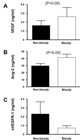
Bloody effusion is frequently associated with ma-lignant diseases. Interestingly, the levels of VEGF and Ang-2 in bloody effusion were significantly higher than those in non-bloody effusion (p < 0.05) (Fig. 3 A, B). In contrast, the level of sVEGFR-1 in bloody effusion was lower than that in non-bloody effusion (Fig. 3C).
Comparison of the levels of VEGF, Ang-2, and sVEGFR-1 in the pleural effusions between the dis-ease groups.
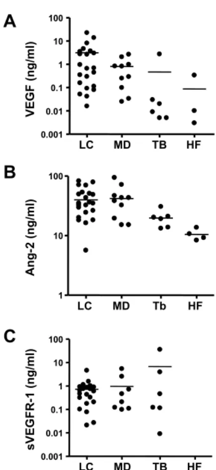
We next compared the levels of VEGF, Ang-2, and sVEGFR-1 in the pleural effusion among various diseases. As shown in Fig. 4A∼C and Table 1, the levels of VEGF and Ang-2 were significantly higher in the effusion from LC and MD, compared with effusion from HF and TB (p < 0.05). In addition, sVEGFR-1 was significantly higher in the effusion from LC, MD, and TB compared with effusion from HF (p < 0.05).
Correlation of the levels of VEGF, Ang-2, and/or sVEGFR-1 in malignant pleural effusions.
Since the high levels of VEGF, Ang-2, and sVEGFR-1 were detected in malignant effusions, we further evaluated the correlation of these three factors in malignant effusions by the Speaman cor-relation test analyses. There are direct corcor-relations between VEGF and Ang-2, VEGF and sVEGFR-1, and sVEGFR-1 and Ang-2 (Fig. 5A∼C).
Fig. 2. Levels of VEGF, sVEGFR-1, and Ang-2 in exudative and transudative effusions. Horizontal lines represent the median values.
Fig. 3. Levels of VEGF, sVEGFR-1, and Ang-2 in bloody- and non-bloody effusions. Horizontal lines represent the median values.
DISCUSSION
We recently reported that VEGF presented within exudative pleural effusion, and it facilitated the pro-duction of bloody pleural effusion by inducing vas-cular hyperpermeability in the thoracic cavity (12). In addition, Ang-2 was shown to present in the exu-dative effusion (24). In the present study, we con-firmed that higher levels of VEGF and Ang-2 existed in the exudative effusion compared with transuda-tive effusion, and further demonstrated that an en-dogenous inhibitor of VEGF, sVEGFR-1, also existed in the exudative effusion. Moreover, higher levels of VEGF and Ang-2 were detected in the bloody ef-fusion than in the non-bloody efef-fusion. In contrast, sVEGFR-1 levels in bloody effusion were lower than that in non-bloody effusion, suggesting the regula-tory role of sVEGFR-1 in the exudative pleural ef-fusion.
sVEGFR-1, found in trophoblasts and endothelial cells of normal placental tissue, was abnormally de-tected in the serum of preeclamptic patients (25). Clinical symptoms in preeclamptic patients are simi-lar with the side effects of VEGF neutralizing anti-body in cancer patients (26, 27), thus suggesting that the preeclamptic symptoms on the maternal side are due to an abnormal suppression of endogenous VEGF by sVEGFR-1. Therefore, sVEGFR-1 is thought to regulate or modulate VEGF activity as an endoge-nous inhibitor. Recent clinical trials showed thera-peutic potential of VEGF/VEGFR inhibitors, in-cluding anti-VEGF antibody (bevacizumab), against various solid tumors, including renal cell carcinoma and non-small cell lung cancer (17, 28, 29).
It is thought that bloody pleural effusions result from vascular wall rupture or leakage of blood cells. The bloody pleural effusions are observed frequently and associated with both of benign and malignant disease. We found that the level of VEGF in the bloody effusions was higher than that in the non-bloody effusions. On the other hand, the level of sVEGFR-1 in the bloody effusions was lower than that in the non-bloody effusions. Since sVEGFR-1 is an endogenous inhibitor of VEGF (16, 17), sVEGFR-1 might be produced by host cells in response to suppression of VEGF activity. If the production level of sVEGFR-1 is insufficient, then bloody effusion might be produced because of the high VEGF ac-tivity. Further studies are warranted to examine this hypothesis.
Ang-2 has been shown to be involved in the patho-genesis of a variety of human diseases including
can-Fig. 4. The levels of VEGF, Ang-2, and sVEGFR-1 in the pleural effusions due to lung cancer (LC), other malignant dis-eases (MD), tuberculosis (TB), and heart failure (HF). Hori-zontal lines represent the median values.
Fig. 5. Correlation of the levels of VEGF, Ang-2, and/or sVEGFR-1 in malignant pleural effusions.
H. Tomimoto, et al. sVEGFR-1 in pleural effusions
cer, diabetic retinopathy, pulmonary hypertension, and coronary artery disease (30-33). Concerning ma-lignant diseases, Ang-2 induces the instability of the blood vessels and facilitates the angiogenesis in the presence of VEGF, and it is one of the poor prognos-tic factors of non-small cell lung cancer (34). More-over, a recent report showed that Ang-2 was sup-posed to be produced locally in the pleural space, and that elevated levels of Ang-2, as well as VEGF, in exudative pleural effusions were detected. How-ever, the functional significance of Ang-2 in pleural disease is poorly understood. We confirmed that a high level of VEGF and Ang-2 coexisted in the exu-dative effusion, especially bloody effusion. Moreover, there was a direct correlation between the levels of VEGF and Ang-2. Since Ang-2 overexpression in he-patocellular carcinoma caused the abdominal bleed-ing in orthotopic animal models (35), VEGF and Ang-2 might thus synergistically induce not only angio-genesis, but also vascular hyperpermeability.
In the present study, the cellular origins of the VEGF, Ang-2, and sVEGFR-1 were not determined because of the lack of the corresponding tissue sam-ples and techniques to detect these factors appro-priately. However, several lines of evidence show that VEGF is produced by various types of cells, in-cluding tumor cells, muscle cells, pericytes, glia cell, and fibroblasts (9). Ang-2 is mainly produced by endothelial cells and cancer cells (22). sVEGFR-1 is mainly produced by trophoblasts (18-20), but it might be produced by endothelial cells and macro-phages because these cells express sVEGFR-1 (36). Lung cancer is the leading cause of malignant pleu-ral effusion (37). At least 25% of all patients with lung cancer tend to develop pleural effusion at some time during the course of the disease. The standard treat-ment for malignant pleural effusion is drainage fol-lowed by the instillation of sclerosing agents, but the clinical efficacy of this treatment varies (38). VEGF/ VPF is reported to be responsible for malignant pleu-ral effusion formation and targeting the production of VEGF/VPF and/or blocking the VEGF/VPF re-ceptor may be a way to control malignant pleural ef-fusion. In addition to the anti-VEGF antibody (bevaci-zumab) which has been shown to have a therapeu-tic potential against non-small cell lung cancer, an endogenous VEGF inhibitor, sVEGFR-1, may be useful for controlling the malignant pleural effusion associated with VEGF-induced vascular hyperper-meability.
REFERENCES
1. Light RW, Macgregor MI, Luchsinger PC, Ball WC Jr : Pleural effusions : The diagnostic sepa-ration of transudates and exudates. Ann Intern Med 77 : 507-13, 1972
2. Porcel JM, Light RW : Diagnostic approach to pleural effusion in adults. Am Fam Physician 73 : 1211-20, 2006
3. Vives M, Porcel JM, Vicente de Vera M, Ribelles E, Rubio M : A study of Light’s criteria and possible modifications for distinguishing exudative from transudative pleural effusions. Chest 109 : 1503-7, 1996
4. Light RW : Clinical practice : Pleural effusion. N England J Med 346 : 1971-7, 2002
5. Fontanini G, Vignati S, Boldrini L, Chine S, Silvestri V, Lucchi M, Mussi A, Angeletti CA, Bevilacqua G : Vascular endothelial growth fac-tor is associated with neovascularization and in-fluences progression of non-small cell lung car-cinoma. Clin Cancer Res 3 : 861-5, 1997 6. Kinasewitz GT : Transudative effusions. Eur
Respir J 10 : 714-8, 1997
7. Light RW : Pleural diseases. Dis Mon 38 : 261-331, 1992
8. Yanagawa H, Takeuchi E, Suzuki Y, Ohmoto Y, Bando H, Sone S : Vascular endothelial growth factor in malignant pleural effusion associated with lung cancer. Cancer Immunol Immunother 48 : 396-400, 1999
9. Ferrara N, Davis-Smyth T : The biology of vas-cular endothelial growth factor. Endocr Rev 18 : 4-25, 1997
10. Yuan A, Yu CJ, Kuo SH, Chen W, Lin FY, Luh KT, Yang PC, Lee YC : Vascular endothelial growth factor 189 mRNA isoform expression specifically correlates with tumor angiogenesis, patient survival, and postoperative relapse in non-small-cell lung cancer. J Clin Oncol 19 : 432-41, 2001
11. Senger DR, Galli SJ, Feder J, Dvorak HF : Tu-mor cells secrete a vascular permeability factor that promotes accumulation of ascites fluid. Sci-ence 219 : 983-5, 1983
12. Yano S, Shinohara H, Herbst RS, Kuniyasu H, Bucana CD, Ellis LM, Fidler IJ : Production of experimental malignant pleural effusions is de-pendent on invasion of the pleura and expres-sion of vascular endothelial growth factor/vas-cular permeability factor by human lung cancer cells. Am J Pathol 157 : 1893-903, 2000
13. Shibuya M : Structure and dual function of vas-cular endothelial growth factor receptor-1 (Flt-1). Int J Biochem Cell Biol 33 : 409-20, 2001 14. Soker S, Takashima S, Miao HQ, Neufeld G,
Klagsbrun M : Neuropilin-1 is expressed by en-dothelial and tumor cells as an isoform-specific receptor for vascular endothelial growth factor. Cell 92 : 735-45, 1998
15. Takahashi H, Shibuya M : The vascular endo-thelial growth factor (VEGF)/VEGF receptor system and its role under physiological and pathological conditions. Clin Sci (Lond) 109 : 227-41, 2005
16. Shibuya M : Structure and dual function of vas-cular endothelial growth factor receptor-1 (Flt-1). Int J Biochem Cell Biol 33 : 409-20, 2001 17. Malecki M, Trembacz H, Szaniawska B,
Przy-byszewska M, Janik P : Vascular endothelial growth factor and soluble FLT-1 receptor in-teractions and biological implications. Oncol Rep 14 : 1565-9, 2005
18. Banks RE, Forbes MA, Searles J, Pappin D, Canas B, Rahman D, Kaufmann S, Walters CE, Jackson A, Eves P, Linton G, Keen J, Walker JJ, Selby PJ : Evidence for the existence of a novel pregnancy-associated soluble variant of the vascular endothelial growth factor receptor, Flt-1. Molecular Human Reproduction 4 : 377-386, 1998
19. Clark DE, Smith SK, He Y, Day KA, Licence DR, Corps AN, Lammoglia R, Charnock-Jones DS : A vascular endothelial growth factor an-tagonist is produced by the human placenta and released into the maternal circulation. Biol Re-prod 59 : 1540-8, 1998
20. He Y, Smith SK, Day KA, Clark DE, Licence DR, Charnock-Jones DS : Alternative splicing of vascular endothelial growth factor (VEGF)-R1 (FLT-1) pre-mRNA is important for the regu-lation of VEGF activity. Mol Endocrinol 13 : 537-45, 1999
21. Polverini PJ : Angiogenesis in health and dis-ease : Insights into basic mechanisms and thera-peutic opportunities. J Dent Educ 66 : 962-75, 2002
22. Maisonpierre PC, Suri C, Jones PF, Bartunkova S, Wiegand SJ, Radziejewski C, Compton D, McClain J, Aldrich TH, Papadopoulos N, Daly TJ, Davis S, Sato TN, Yancopoulos GD : An-giopoietin-2, a natural antagonist for Tie 2 that disrupts in vivo angiogenesis. Science 277 : 55-60, 1997
23. Hanahan D : Signaling vascular morphogenesis and maintenance. Science 277 : 55-60, 1997 24. Kalomenidis I, Kollintza A, Sigala I, Papapetropoulos
A, Papiris S, Light RW, Roussos C : Angio-poietin-2 levels are elevated in exudative pleu-ral effusions. Chest 129 : 1259-66, 2006
25. Koga K, Osuga Y, Yoshino O, Hirota Y, Ruimeng X, Hirata T, Takeda S, Yano T, Tsutsumi O, Taketani Y : Elevated serum soluble vascular endothelial growth factor receptor 1 (sVEGFR-1) levels in women with preeclampsia. J Clin Endocrinol Metab 88 : 2348-51, 2003
26. Bdolah Y, Karumanchi SA, Sachs BP : Recent advances in understanding of preeclampsia. Croat Med J 46 : 728-36, 2005
27. Maione P, Gridelli C, Troiani T, Ciardiello F : Combining targeted therapies and drugs with multiple targets in the treatment of NSCLC. Oncologist 11 : 274-84, 2006
28. Yang JC : Bevacizumab for patients with me-tastatic renal cancer : An update. Clin Cancer Res 10 : 6367S-70S, 2004
29. Midgley R, Kerr D : Bevacizumab - current status and future directions. Ann Oncol 16 : 999-1004, 2005
30. Tanaka F, Ishikawa S, Yanagihara K, Miyahara R, Kawano Y, Li M, Otake Y, Wada H : Expres-sion of angiopoietins and its clinical significance in non-small cell lung cancer. Cancer Res 62 : 7124-9, 2002
31. Takagi H, Koyama S, Seike H, Oh H, Otani A, Matsumura M, Honda Y : Potential role of the angiopoietin/tie2 system in ischemia-induced retinal neovascularization. Invest Ophthalmol Vis Sci 44 : 393-402, 2003
32. Zhao YD, Campbell AI, Robb M, Ng D, Stewart DJ : Protective role of angiopoietin-1 in experi-mental pulmonary hypertension. Circ Res 92 : 984-91, 2003
33. Calvi C, Dentelli P, Pagano M, Rosso A, Pegoraro M, Giunti S, Garbarino G, Camussi G, Pegoraro L, Brizzi MF : Angiopoietin 2 induces cell cycle arrest in endothelial cells : a possible mecha-nism involved in advanced plaque neovasculari-zation. Arterioscler Thromb Vasc Biol 24 : 511-8, 2004
34. Tanaka F, Yanagihara K, Otake Y, Kawano Y, Miyahara R, Takenaka K, Katakura H, Ishikawa S, Ito H, Wada H : Prognostic factors in resected pathologic (p-) stage IIIA-N2, non-small-cell lung cancer. Ann Surg Oncol 11 : 612-8, 2004 35. Tanaka S, Mori M, Sakamoto Y, Makuuchi M, H. Tomimoto, et al. sVEGFR-1 in pleural effusions
Sugimachi K, Wands JR. Biologic significance of angiopoietin-2 expression in human hepato-cellular carcinoma. J Clin Invest 103 : 341-5, 1999
36. Barleon B, Sozzani S, Zhou D, Weich HA, Mantovani A, Marme D : Migration of human monocytes in response to vascular endothelial
growth factor (VEGF) is mediated via the VEGF receptor flt-1. Blood 87 : 3336-43, 1996
37. Kvale PA, Simoff M, Prakash UB, American College of Chest Physicians : Lung cancer : Pal-liative care. Chest 123 : 284S-311S, 2003 38. Antunes G,Neville E : Management of
malig-nant pleural effusions. Thorax 55 : 981-3, 2000