Case Report
J. St. Marianna Univ.Vol. 11, pp. 165–171, 20201 Division of Gastrointestinal and General Surgery, St. Marianna University School of Medicine 2 Department of Pathology, St. Marianna University School of Medicine
A Case of Inflammatory Myofibroblastic Tumor Originating in the Lesser
Omentum Treated by Laparoscopic Resection
Masaki Hiwatari
1, Shinya Mikami
1, Taichi Mafune
1, Sota Usui
1, Yuki Amano
1,
Asako Fukuoka
1, Takeharu Enomoto
1, Junichi Tsuchiya
2, and Takehito Otsubo
1(Received for Publication: August 20, 2020)
Abstract
A 51-year-old woman underwent a CT scan performed that revealed a neoplastic lesion in the upper abdo‐ men, and she was referred to our surgical department. Abdominal CT showed a tumor of about 50 mm lying between the posterior wall of the gastric minor curvature and the pancreas. Continuity with the surrounding tissue was not clear. Ultrasonography showed that the tumor was mobile on postural change, and it was consid‐ ered to originate from the mesentery. In addition, PET scanning showed mild accumulation, and MRI showed limited diffusion, suggesting the possibility of malignancy. Laparoscopic tumor resection was performed as a diagnostic treatment. Intraoperative findings showed tumor continuity only with the lesser omentum, and the patient was judged to have a primary tumor of the lesser omentum. Histopathological findings showed prolifera‐ tion of myofibroblasts and lymphocyte infiltration, and the patient was thus diagnosed as having an inflamma‐ tory myofibroblastic tumor. Inflammatory myofibroblastic tumor as a primary tumor of the lesser omentum is rare and is reported here along with a literature review.
Key words
Inflammatory myofibroblastic tumor, laparoscopic surgery, lesser omentum
Introduction
Inflammatory myofibroblastic tumors (IMTs) are neoplastic lesions that are characterized by the proliferation of myofibroblasts and significant inflam‐ matory cell infiltrate. This report documents a case of IMT originating in the lesser omentum that we en‐ countered and resected laparoscopically, along with a review of the relevant literature.
Case Presentation
The patient was a 51-year-old woman with no chief complaints. She had a medical history of hyper‐ lipidemia and cholecystolithiasis but no family his‐ tory of note. The patient noticed a tumor in her right breast and was examined by our hospital, where she was diagnosed as having right breast cancer. A com‐
puted tomography (CT) scan performed for detailed investigation revealed a neoplastic lesion in the epi‐ gastric region, so she was referred to our surgical de‐ partment.
At the time of admission, her vital signs were height, 152 cm; weight, 53 kg; blood pressure, 119/80 mmHg; pulse, 70 bpm; body temperature, 36.2°C. Her palpebral and scleral conjunctiva showed no signs of anemia or jaundice. There were no palpa‐ ble superficial lymph nodes. During examination of the abdomen, we did not observe any findings sug‐ gesting peritoneal irritation, such as tenderness or re‐ bound tenderness. There was no obvious, palpable mass, and the patient did not exhibit hepatosplenome‐ galy.
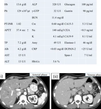
Blood test findings did not show any specific ab‐ normal findings (Table 1). An abdominal ultrasound
Table 1. Preoperative Test Results
Fig. 1. On CT imaging, a smooth tumor with calcification measuring 45 × 38 mm
was observed close to the stomach. The tumor was enhanced strongly in the arterial phase. Because the border between the tumor and stomach was ob‐ scure, the tumor was thought to have invaded the stomach. (i) Arterial phase. (ii) Venous phase. (iii) Coronal plane. (iv) Sagittal plane.
showed a richly perfused tumor measuring 58 × 46 mm in size between the stomach and tail of the pan‐ creas. The tumor position changed when the patient was moved into an upright position, so we believed that the tumor was of mesenteric origin. A contrast-enhanced CT scan showed a partially calcified tumor with distinct margins measuring 42 × 38 mm near the
posterior gastric wall along the lesser curvature. There was intense contrast enhancement during the early phase. The degree of continuity with the sur‐ rounding organs was unclear, but the findings sug‐ gested that the tumor could be of gastric origin (Fig. 1).
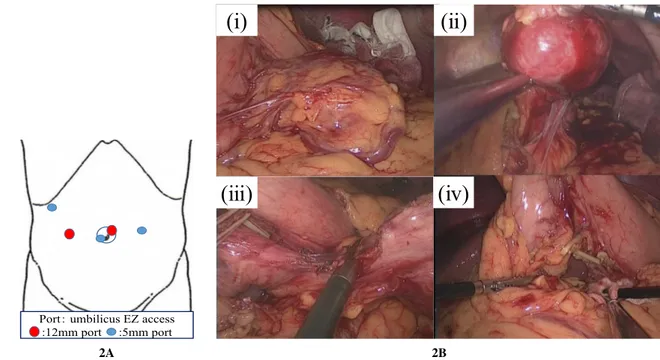
2A 2B
Fig. 2. Port placement and intraoperative images. A Port placement. B- (i) The tumor was covered with greater omentum.
(ii) On opening the omental bursa, we observed a smooth red tumor. Because it continued not with the stomach and pancreas but with the lesser omentum, we considered that the tumor had arisen from the lesser omentum. (iii) We separated the tumor from lesser omentum. (iv) We resected the tumor.
fundus gland polyp, but no other elevated lesions were observed. Magnetic resonance imaging (MRI) findings revealed a tumor appearing hypointense on T1 images and mildly hyperintense on T2 images that was not continuous with either the pancreas or stom‐ ach. Diffusion-weighted images suggested the possi‐ bility of malignancy due to restriction of diffusion. Positron emission tomography-CT (PET-CT) scan‐ ning showed that the tumor exhibited mildly in‐ creased uptake with a maximum standardized uptake value (SUVmax) of 3.5.
Although the above-mentioned findings did not allow us to reach a diagnosis, we believed that this tumor was of mesenteric origin and that the potential for malignancy could not be ruled out. Leiomyosar‐ coma, schwannoma, and GIST were considered as differential diagnoses. We thus performed laparo‐ scopic tumor resection for the purpose of diagnosis and treatment and planned to perform surgery for the right breast cancer thereafter.
Surgical findings: The surgery was performed lapa‐
roscopically under general anesthesia with the patient in the supine position. We inserted a total of three ports: a 10-mm umbilical camera port, and two 5-mm ports in the left flank and in the left hypogastrium be‐
low the midline. When we opened the omental bursa, we observed a red tumor with a smooth surface. The tumor was not continuous with either the pancreas or stomach and only exhibited continuity with the lesser omentum, so we determined that it was of omental origin (Fig. 2 A, B). We then resected the tumor. Sur‐ gery lasted 160 min, and the volume of blood loss was 70 ml.
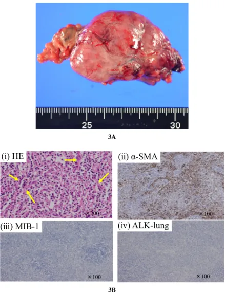
Resected specimen findings: Macroscopic examina‐
tion of the resected specimen revealed a partially cal‐ cified solid tumor measuring 55 × 45 × 25 mm. The histological examination revealed atypical, fusiform, oval-shaped cells, and immunological staining re‐ vealed increased proliferation of α-smooth muscle ac‐ tin (αSMA)-positive myofibroblasts and extensive in‐ terstitial infiltration by non-atypical lymphocytes. We therefore diagnosed the patient as having IMT. The MIB-1 index was <5%. In addition, the patient tested negative for anaplastic lymphoma kinase-1 (ALK), which is thought to be a highly specific test for IMT (Fig. 3 A, B).
Postoperative clinical course: The patient’s postop‐
erative course was favorable, and she was discharged on postoperative day 5. To date, at 4 years and 5 months after the surgery, no recurrence of findings
3A
3B
Fig. 3. Macroscopic and microscopic findings of the tumor. A. Macroscopically, the
tumor was solitary and measured 55 × 45 × 25 mm. B. (i) Pathology find‐ ings showed the tumor cells to be in a deformed spindle or oval shape, and many lymphocytes had invaded the tumor. The yellow arrows indicate the spindle tumor cells. (ii) The tumor was positive for αSMA. (iii) The rumor was negative for ALK. (iv) The MIB-1 index was <5%.
has been observed.
Discussion
IMT develops as a result of myofibroblast prolif‐ eration, and these tumors are characterized by the presence of myofibroblasts and extensive lympho‐ cytic infiltrates1). Although numerous causes have
been proposed, such as infection or inflammation as a result of trauma, or surgery or malignant tumors, their origin remains unknown. According to reports, the lungs are the most common site of onset, al‐ though IMTs may arise throughout the body, includ‐
ing the peritoneum, mesentery, and bowel. Onset has also been reported in the bladder, endocrine organs such as the prostate, and in the breast, spleen, pan‐ creas, parenchymal intra-abdominal organs such as the liver, and even in the soft tissues2,3). The condition
tends to occur from childhood to young adulthood4).
Symptoms vary based on the site of onset and tumor diameter. However, as in our case, a few reports state that the tumor may be asymptomatic, but the patient may present with pyrexia or abdominal pain. Imaging is not adequately specific for diagnosis, so the diag‐ nosis is based on histopathological tests4). Differenti‐
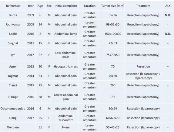
Table 2. Review of 12 Cases of IMT Originating in the Omentum ating diseases of primary IMT in the abdominal cav‐
ity include schwannoma, GIST, and leiomyosarcoma. It is important to identify the primary organ for dif‐ ferentiation, and this can be judged comprehensively by combining CT, MRI, and endoscopy. In addition, PET may be used to judge benign/malignant tumors, but IMT may show faint accumulation on PET, as in our case, which is a reference-level finding. None of these diseases have specific diagnostic imaging char‐ acteristics, and it is considered difficult to distinguish them. IMT may present with the following three his‐ tological patterns5), 1) a myxoid/vascular pattern,
with scant distribution of spindle-shaped cells accom‐ panied by an inflammatory cell infiltrate, 2) a com‐ pact spindle cell pattern, with a dense arrangement of hyperproliferative spindle-shaped cells, and 3) a hy‐ pocellular fibrous pattern, with scant spindle-shaped cells in a collagen fiber matrix resembling scar tissue, accompanied by an inflammatory cell infiltrate, and occasionally associated with calcification or ossifica‐ tion. We believe that we observed pattern 1 in the present case.
Immunologically, myofibroblast cell lines tend
to test positive for αSMA, vimentin, and desmin at higher rates than normal2,4,6). Characteristically, these
cell lines are also said to test positive for chromoso‐ mal anaplastic lymphoma kinase (ALK) at a higher rate. Our patient tested positive for αSMA but nega‐ tive for ALK. The ALK inhibitor crizotinib has been shown to be effective for ALK-positive IMT, whereas testing negative for ALK has also been reported to be a factor indicating a poor prognosis for this condi‐ tion7).
When we searched PubMed using the keywords “omentum” and “inflammatory myofibroblastic tu‐ mor”, we found 12 reported cases of onset in the greater or lesser omentum, including the present case (we excluded cases in which we were unable to ob‐ tain the full report, or in which information was in‐ sufficient). Table 2 summarizes our case and the 11 other reported cases8–18). The median age of onset was
20 years, and the initial symptom was abdominal pain in seven cases, a feeling of abdominal discom‐ fort in one case, awareness of an abdominal mass in three cases, and one case was asymptomatic. There were various imaging findings, although findings sug‐
gestive of IMT were observed on PET in certain cases, and these findings were difficult to differenti‐ ate from malignancy in some cases. Surgical resec‐ tion was performed in all cases, although including our case, laparoscopic resection was performed in only three cases. We believe that the facts that the tu‐ mors were comparatively large at the time of detec‐ tion and that there were numerous young patients may have contributed to this. The results for immu‐ nostaining showed that six patients tested positive for the aforementioned ALK, whereas four tested nega‐ tive, and the outcome in two patients was unknown. Therefore, approximately half of all cases tested posi‐ tive for ALK. Reports indicate that between 5 and 60% of patients test positive for ALK, and the rate of positive tests reportedly varies based on the site of onset2,6). The results in the present tabulation resem‐
ble those in previous reports. It would be desirable to accumulate further cases going forward.
According to the World Health Organization (WHO) classification, IMT is an intermediate (rarely metastasizing) tumor19). Originally, the observation of
a mixture of proliferating fibroblasts, myofibroblasts, and inflammatory cells was associated with inflam‐ matory changes, such as granulation tissue, so previ‐ ously, these lesions were understood to be reactive le‐ sions and were referred to in general terms, such as plasma cell granulomas and inflammatory pseudotu‐ mors. However, in the interim, there have been cases in which these tumors exhibited local recurrence, dis‐ tant metastasis, and infiltration, so they are now de‐ fined as intermediate tumors20). Cases of local recur‐
rence usually occur as a result of inadequate resection or careless biopsy, so generally, complete resection is considered to be the appropriate form of treatment. However, when we consider the fact that IMT onset may be caused by invasive surgical procedures1), we
believe that it is desirable to perform minimally inva‐ sive surgery. If elective surgery is permissible, then we believe that laparoscopic surgery is a useful tech‐ nique that will allow both examination and diagnosis and minimally invasive treatment in comparison with open resection.
Conclusion
We experienced a case of IMT originating from the lesser omentum. Although IMT originating from the lesser omentum is extremely rare, it should be kept in mind as a differential disease when evaluating nonspecific intra-abdominal tumors.
Conflicts of Interest
The authors have nothing to disclose.
References
1) Pettinato G, Manivel JC, De Rosa N, et al. In‐ flammatory myofibroblastic tumor (plasma cell granuloma). Clinicopathologic study of 20 cases with immunohistochemical and ultrastructural observations. Am J Clin Pathol 1990; 94: 538– 546.
2) Coffin CM, Watterson J, Priest JR, et al. Extrap‐ ulmonary inflammatory myofibroblastic tumor (inflammatory pseudotumor): A clinicopatho‐ logic and immunohistochemical study of 84 cases. Am J Surg Pathol 1995; 19: 859–872. 3) Kovach SJ, Fischer AC, Katzman PJ, et al. In‐
flammatory myofibroblastic tumors. J Surg On‐ col 2006; 94: 385–391.
4) Coffin CM, Humphery PA, Dehner LP. Extrap‐ ulmonary inflammatory myofibroblastic tumor: a clinical and pathological survey. Semin Diagn Pathol 1998; 15: 85–101.
5) Hisaoka M, Hashimoto H. Inflammatory myofi‐ broblastic tumor. Pathol Clin Med 2003; 21: 413–418.
6) Gleason BC, Hornick JL. Inflammatory myofi‐ broblastic tumours: where are we now? J Clin Pathol 2008; 61: 428–437.
7) Butrynski JE, D’Adamo DR, Hornick JL, et al. Crizotinib in ALK-rearranged inflammatory my‐ ofibroblastic tumor. N Engl J Med 2010; 363: 1727–1733.
8) Uchiyama Y, Matsuyama R, Suzuki K, et al. A case of inflammatory myofibroblastic tumor of the lesser omentum with intratumoral hemor‐ rhage. J Gastroenterol Nihon Shokakibyo Gak‐ kai Zasshi [in Japanese] 2009; 106: 1321–1326. 9) Liang W, Lin S, Chen Z. Imaging findings of in‐
flammatory myofibroblastic tumor from the greater omentum: one case report. Medicine (Baltimore) 2017; 96: e8297. doi:10.1097/MD. 0000000000008297.
10) Oeconomopoulou A, Verney Y, Kanavaki K, et al. Inflammatory myofibroblastic tumor of the small intestine mimicking acute appendicitis: a case report and review of the literature. J Med Case Rep 2016; 10: 100. doi:10.1186/ s13256-016-0880-0.
11) El Hage Chehade HH, Zbibo RH, Abou Hussein BM, et al. Highly vascularized primarily inflam‐
matory pseudotumor of the omentum in an adult male: a case report. Am J Case Rep 2016; 17: 79–83.
12) Cianci P, Ambrosi A, Fersini A, et al. Volumi‐ nous omental inflammatory myofibroblastic tu‐ mor in an elderly man: a case report and litera‐ ture review. Case Rep Surg 2015; 2015: 1–5. 13) Yagmur Y, Akbulut S, Gumus S. Mesenteric in‐
flammatory pseudotumor: a case report and comprehensive literature review. J Gastrointest Cancer 2014; 45: 414–420.
14) Aptel S, Gervaise A, Fairise A, et al. Abdominal inflammatory myofibroblastic tumor. Diagn In‐ terv Imaging 2012; 93: 410–412.
15) Kye BH, Kim HJ, Kang SG, et al. A case of in‐ flammatory myofibroblastic tumor originated from the greater omentum in young adult. J Ko‐ rean Surg Soc 2012; 82: 380–384.
16) Singhal M, Ramanathan S, Das A, et al. Omen‐ tal inflammatory myofibroblastic tumor mimick‐ ing peritoneal carcinomatosis. Cancer Imaging 2011; 11: 19–22.
17) Sodhi KS, Virmani V, Bal A, et al. Inflamma‐ tory pseudotumor of the omentum. Indian J Pe‐ diatr 2010; 77: 687–688.
18) Gupta CR, Mohta A, Khurana N, et al. Inflam‐ matory pseudotumor of the omentum: an un‐ common pediatric tumor. Indian J Pathol Micro‐ biol 2009; 52: 219–221.
19) Weiss SW. Histological typing of soft tissue tu‐ mors, Second edition, WHO, Springer-Verlag, Berlin, 1994: 48.
20) Yamazawa H. Why is the inflammatory myofi‐ broblastic tumor of the heart noted? Nihon Shoni Junkanki Gakkai Zasshi [in Japanese] 2016; 32: 321–322.