INTRODUCTION
Homeostasis is one of the most important con-cepts in our understanding of the biological mecha-nisms of somatic, autonomic and endocrine controls. The constant internal environment which consists of the circulatory organic fluid which surrounds and bathes all body tissue is the result of a control mechanism that limits the variability of body state. It is well accepted that arterial blood pressure (AP) is maintained at a constant level via the neural mechanisms of baroreflex, chemoreflex and/or circulating hormonal control, which include free radicals, such as NO. The key neuronal and hor-monal mechanisms related to maintaining homeo-stasis are located in the hypothalamus. Although, during the waking period, the emotional reactions or exercise affect AP, it is reasonable to assume that AP is controlled at a constant level during sleep, since this activity does not involve voluntary movement and/or emotional behavior.
Two stages of sleep exist ; one is rapid-eye-movement (REM) sleep and the other is non-REM
sleep. REM sleep is characterized by arousal elec-troencephalogram (EEG), rapid eye movements (REMs) and muscle atonia, while non-REM sleep is characterized by a dominant slow wave activity in EEG. REM sleep is also called dreaming sleep, and dreams which contain vivid imagery are often experienced during this state.
During non-REM sleep, autonomic functions such as AP, heart rate (HR) and respiration are con-trolled and quite stable, showing constant values throughout this stage. However, during REM sleep, significant fluctuations in autonomic functions can be observed (Fig. 1 for example in a rat). AP in-creases in the transition from non-REM to REM sleep, exhibiting large phasic surges during REM sleep, although the skeletal muscles lose their activity completely. Great autonomic fluctuations during REM sleep can be seen in various mam-mals, including humans. During REM sleep, our bodies no longer appear to be under the homeostatic control (1).
Why does AP rise actively during REM sleep? Although, at present, this issue remains to be re-solved, we report here, in the form of a mini review, A) the neural mechanisms for REM sleep genera-tion, B) the characteristics of REM sleep-associated AP changes, and C) possible mechanisms for AP control during REM sleep.
Why does arterial blood pressure rise actively during REM
sleep?
Hiroyoshi Sei, and Yusuke Morita
Department of Physiology, The University of Tokushima School of Medicine, Tokushima, Japan Abstract : A large fluctuation in autonomic function is one of the most important charac-teristics of REM sleep. Arterial blood pressure (AP) increases during the transition from non-REM to REM sleep, showing phasic surges during REM sleep. REM-associated AP changes involve 1) a long-term recovery process after surgery, 2) circadian rhythm, 3) relationships with ambient temperature. REM-associated AP changes are mediated by sympathetic nerves, buffered by baroreflex, abolished in decerebrated cats, and related to hippocampal theta activity in rats. Furthermore, the midbrain dopaminergic system has been recently found to be involved in increases in REM-associated AP. J. Med. Invest. 46 : 11-17, 1999
Key words : REM sleep, blood pressure, dopamine
Received for publication December 1, 1998 ; accepted January 7, 1999.
Address correspondence and reprint requests to Hiroyoshi Sei, Department of Physiology, The University of Tokushima School of Medicine, Tokushima 770-8503, Japan and Fax : +81-88-633-9251.
The Journal of Medical Investigation Vol.46 1999 11 11
A. Neural mechanisms for REM sleep generation
In the decerebrated cat whose brain stem is transected at a pre-pontine level, the REM sleep-like stage is characterized by the occurrence of muscle atonia, pontine ponto-geniculo-occipital (PGO) waves and REMs (2, 3). This strongly indicates that the center for generation of REM sleep exists not in the forebrain but in the brain stem.
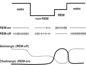
Sleep-neurophysiolosists have considered that interaction between REM-on and REM-off neurons in the brain stem play a critical role in the genera-tion of REM sleep (4-6, Fig. 2). The REM-on neuron is completely silent or functions at a considerably lower level of discharge during waking and non-REM sleep, but their discharge rate is augmented prior to the onset of REM sleep, and then stops firing completely at the end of REM sleep. The REM-off neuron shows a reverse discharge pattern to REM-on neurons. To date, there is abundant evidence that REM-on neurons are largely cholinergic neurons in the pontine tegmentum area, while REM-off neurons include serotonergic neurons in the raphe nucleus, noradrenergic neurons in the locus coeruleus and histaminergic neurons in the posterior hypo-thalamus. The cholinergic REM-on neurons and aminergic REM-off neurons show the opposite firing pattern with a vigilance change. The increase of firing rate in REM-on and the decrease (cessation) in REM-off neurons are, at least, necessary for the generation of REM-sleep (4-6).
The injection of carbachol, a potent cholinergic agonist, into the restricted area in the pontine tegmental area strongly induces REM sleep (7, 8). On the other hand, atropine, a cholinergic receptor antagonist, completely suppresses REM sleep (6). As a result, it is generally thought that both the activation of cholinergic REM-on cells and the stimulation of cholinergic receptors are important for REM-sleep generation. In contrast, the injection of serotonin into the pontine tegmental area (9) or the electrical stimulation of locus coeruleus where noradrenergic neurons are clustered (10) reduce REM sleep. Furthermore, the noradrenergic agonist suppresses REM sleep. Like cholinergic enhance-ment, aminergic suppression of REM sleep is now an established principle.
The neurons which execute muscle atonia exist not only in the pontine tegmental area but also in the medulla. These descending neurons are, of course, REM-on type neurons, and are thought to be non-aminergic and non-cholinergic. Although the injection of carbachol into the pontine tegmental area induces muscle atonia, a similar injection in the medulla does not. This indicates that the atonia-inducing neurons in the pons are choline-receptive and those in the medulla are not (7, 11).
It has recently been found that the glutamatergic receptor in the pontine tegmental area may play a role in the control of REM sleep induction (12). The duration of REM sleep is greatly pronounced during the perfusion of kainate via the membrane of a dialysis probe in the pontine tegmental area. This effect is inhibited by the simultaneous
injec-Fig.1. Representative example of polygraphic recordings, showing changes in heart rate (HR), arterial blood pressure (AP), electromyogram (EMG) and electroencephalogram (EEG) during sleep in a rat.
Fig. 2. The model of physiological mechanisms determining alterations in vigilance level. Upper : vigilance level. Middle : the firing pattern of REM-on and-off neurons. Bottom : dynamic model of interaction between REM-on (cholinergic) and REM-off (aminergic) neurons with the vigilance change. (modified from ref. 5).
H. Sei et al. Blood pressure during REM sleep 12 H. Sei et al. Blood pressure during REM sleep 12
tion of GAMS or CNQX, a specific non-NMDA receptor antagonist, but is not inhibited by AP-5 or MK801, a selective NMDA receptor antagonist, or atropine. It is suggested that the glutamatergic REM sleep-inducing effect is mediated by non-NMDA receptors and independent of the cholinergic system. Using microdialysis techniques, the spontaneously increased rate of release of glutamate in the medulla, which is related to muscle atonia, is observed during REM sleep, and cholinergic stimulation of the pons increases the release of glutamate in the medulla of the cat (13). A glutamatergic pathway from the pontine tegmental area to the medulla is responsible for muscle atonia.
Based on human PET studies (14-16), it has also been confirmed in humans that the pontine brain stem is important to the REM sleep activation of the brain. Furthermore, the limbic and paralimbic regions of the forebrain are preferentially activated in REM sleep compared to waking periods, or to non-REM sleep, indicating that these regions may be related to the emotional aspect of human dreams.
B. Characteristics of REM sleep-associated
AP change
B-1. Long-term changes in recovery process from surgery
After surgically implanting equipment in an ex-perimental animal for the purpose of recording biological signals (EEG, AP, etc.), a long-term re-covery process in the REM
sleep-associated AP change is observed (17, 18). In the acute period after surgery (5-10 days afterward), AP decreases and remains low throughout REM sleep in the cat. In contrast, in the chronic period (in excess of 10 days after surgery), AP increases and shows several phasic surges during REM sleep. The pathophysiologi-cal mechanisms of this post-surgical phenomenon are com-pletely unknown.
B-2. Circadian rhythm A circadian rhythm exists in the AP and HR in rat (19,
20), monkey (21) and human (22, 23). The rat has a polyphasic sleep-wake cycle, and even during the active (dark) phase, it sleeps several times. In the rat, the magnitude of REM sleep-associated AP fluc-tuation shows a circadian rhythm. The AP fluctua-tion, expressed by a coefficient of variation (CV%), is larger in the light phase than in the dark phase (24). The AP CV% during REM sleep is inversely correlated to the brain temperature. As injection of interleukin-1β causes both a high fever and a de-crease in the CV% of AP during REM sleep (25), thermoregulation is hypothesized to be related to REM sleep-associated AP changes.
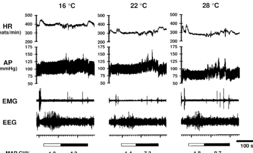
B-3. Effect of ambient temperature (Fig.3)
Changes in ambient temperature (Ta) affect the cardiovascular system during both wake and sleep periods in rats (26, 27). AP and HR increase with decreasing Ta. The increment of AP from non-REM to REM sleep decreases with decreasing Ta. At 28 degrees, HR increases during REM sleep in com-parison to non-REM sleep, while at 16 degrees a decrease is observed. The AP CV% during REM sleep decreases as the Ta is lowered.
C. A possible mechanisms for control of AP
during REM sleep
C-1. The role of the sympathetic nerve and baroreflex Subcutaneously injected guanethidine causes pe-ripheral damage to the sympathetic nervous system.
Fig. 3. Representative recordings showing changes in heart rate (HR), arterial pressure (AP), electromyogram (EMG) and electroencephalogram (EEG) during successive non-REM (indicated by horizontal open columns at bottom) and REM (indicated by horizontal closed columns at bottom) sleep at three ambient temperatures.
MAP CV% : coefficient of variation of mean arterial pressure.
13 The Journal of Medical Investigation Vol.46 1999 13 The Journal of Medical Investigation Vol.46 1999
In rats which have been sympathectomized by treat-ment with guanethidine, increases in REM sleep-associated AP are abolished (28). In humans, changes in muscle sympathetic nerve activity during sleep have been recorded by means of microneurography (29-31). During REM sleep, the muscle sympathetic nerves show an increase in activity compared to waking and non-REM sleep. These observations suggest that the sympathetic nervous system is involved in increases in AP during REM sleep.
It has been suggested by some researchers that the baroreflex may play a crucial role in the increase of AP observed during REM sleep. In fact, there are several reports which show that sino-aortic denervation (SAD) causes a decrease in AP during REM sleep in the rat and cat (31, 32). Recently, however, we have retested the SAD in the rat using a telemetry system for recording AP, and found that in the case of the SAD rats the increases in AP and HR during REM sleep are much larger than those in sham-operated rats (33). We noted a de-crease in AP during REM sleep in the SAD rats in the earlier acute period after surgery. It is possible that the recovery period in the previous experi-ments was too short and, as a result, the SAD rats showed a decrease in AP during REM sleep. The baroreflex buffers the increase in AP and HR during REM sleep in the rat.
C-2. The role of forebrain structure
In order to investigate the role of the forebrain structure, observations relative to the AP change during sleep were carried out in decerebrated cats (34, 35). Although decerebrated cats lose neural in-put from the forebrain structure, they show a REM-sleep like phase. During this REM-REM-sleep like phase, AP decreases tonically with no phasic surge, indicat-ing that the neural connection between the forebrain structure and brain stem is necessary for the in-crease in AP which is observed during REM sleep. The hippocampus is one of the important limbic structures, and shows a rhythmical sinusoidal EEG (theta wave) during REM sleep, especially in rodents. This appearance of a theta wave is indicative of the pronounced activity of the limbic system during REM sleep. During the waking period, the frequency of the theta wave increases when the animal is physically active. The acceleration of theta wave activity pre-cedes the AP surge during REM sleep in the rat (36). Benzodiazepine, which binds to the GABA recep-tor and hyperpolarizes the membrane potential, is known to reduce both hippocampal activity and the
theta frequency. The injection of benzodiazepine reduces the increases in both theta frequency and AP during REM sleep. This also indicates the im-portant role of the forebrain including the limbic system on AP changes during REM sleep.
C-3. Midbrain dopaminergic neurons and AP change during REM sleep
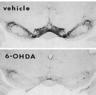
Aminergic neurons are considered to be largely REM-off neurons. It has been reported, however, that dopaminergic neurons do not decrease their firing rate during REM sleep (37). As stimulation of the midbrain dopaminergic system increases AP (38, 39), the possibility exists that the dopaminergic system may play a role in the increase in REM sleep-associated AP. Two dopaminergic systems which have been particularly implicated in cardio-vascular control are the mesolimbic/mesocortical (A10) and the nigrostriatal (A 9) systems. The mesolimbic/mesocortical dopaminergic system origi-nates in the midbrain ventral tegmental area (VTA) and innervates forebrain regions such as the nucleus accumbens, olfactory tubercle, amygdala and hypo-thalamus. We have injected 6-hydroxydopamine (6-OHDA), a drug which is a cathecholaminergic neurotoxin, into the VTA (40, Fig. 4). The increase of AP and HR during REM sleep seen in vehicle-treated rats was suppressed in 6-OHDA-treated rats (Fig.5).
Fig. 4. Examples of brain sections near the injection site, pro-cessed for tyrosine hydroxylase (TH) immunohistochemistry, in a 6-hydroxydopamine (6-OHDA)- and vehicle-treated rat. TH immunoreactive neurons in ventral tegmental area are almost ab-sent in 6-OHDA-treated rat. Neuron loss in the 6-OHDA-treated rat appears to extend to the antero-medial part of the substantia nigra.
H. Sei et al. Blood pressure during REM sleep 14 H. Sei et al. Blood pressure during REM sleep 14
It is reasonably imagined that REM-off neurons including noradrenergic and serotonergic neurons, which are also damaged by 6-OHDA, are not di-rectly involved in the AP increase dur-ing REM sleep, even if their disinhibi-tory effects play an important role on the AP change. This result, therefore, supports the above mentioned hypoth-esis that the dopaminergic system may be involved in increased REM sleep-associated AP.
Oneiric behavior (41, 42) during REM sleep, observed in the cat with a pontine tegmental lesion, suggests that the central motor system actively functions during REM sleep, although the actual behavioral action is inhib-ited by muscle atonia. It has been
reported that the AP and HR increase during exer-cise or voluntary movement, and that the cardio-vascular sympathetic outflow increases immediately before, or concomitantly with, the onset of exercise and voluntary movement (43-45). These findings indicate that, during such behavior, descending autonomic activation originates from the higher nervous system (central command), in order to adjust the blood supply for exercise or movement. Thus, it can be hypothesized that, during REM sleep, AP is increased via a central command in parallel with the activated motor system without any actual observed action, and that the midbrain dopaminergic system is involved in this central command to the AP.
CONCLUSIONS AND PERSPECTIVES
Very little is currently known concerning the mechanism and role of the large autonomic fluctua-tion during REM sleep. Only hints exist as to the involvement of the midbrain dopaminergic neurons on this mechanism. The midbrain dopaminergic neurons have, however, been reported to show a clockwise and regular firing pattern. This regular firing pattern cannot explain the phasic fluctuation of the autonomic nervous system during REM sleep. Clearly, strong inputs from the other sites of the brain into the midbrain dopaminergic system must induce the phasic excitement of the dopamine-associated autonomic control and subsequent phasic events in AP, HR and/or respiration during REM sleep.
Inves-tigations into the interaction among dopaminergic, cholinergic and/or glutamatergic neurons is now ongoing in our laboratory.
If a relationship exists between imagery or emo-tion in dreams and autonomic changes during REM sleep, the question“why does arterial blood pres-sure rise actively during REM sleep?”might be analogous to the question“what mechanism leads to imagery or emotion in dream?”
REFERENCES
1. Parmeggiani PL : The autonomic nervous sys-tem in sleep. In : Kryger MH, Roth T, Dement WC, eds. Principles and Practice of Sleep Medi-cine. W. B. Saunders Company, Philadelphia, 1994, pp. 194-203
2. Jouvet M : Neurophysiology of the state of sleep. Physiol Rev 47 : 117-177, 1967
3. Jouvet M:The role of monoamines and acetylcholine-containing neurons in the regulation of the sleep-waking cycle. Ergeb. Physiol. 64 : 166-307, 1972
4. Jones BE : Basic mechanisms of sleep-wake states. In : Kryger MH, Roth T, Dement WC, eds. Principles and Practice of Sleep Medicine. W. B. Saunders Company, Philadelphia, 1994, pp. 145-162
5. Hobson JA, Stickgold R, Pace-Schott EF : The neuropsychology of REM sleep dreaming. Neuroreport 9 : R 1-R 14, 1998
6. Sakai K : Executive mechanisms of paradoxical
Fig. 5. Representative examples of polygraphic recordings, showing changes in heart rate (HR), arterial blood pressure (AP), electromyogram (EMG) and electroencephalogram (EEG) during sleep in a 6-hydroxydopamine (6-OHDA, right)- and vehicle (left)- treated rat. The upper bars indicate the period of REM sleep.
15 The Journal of Medical Investigation Vol.46 1999 15 The Journal of Medical Investigation Vol.46 1999
sleep. Arch Ital Biol 479 : 225-240, 1988 7. Vanni-Mercier G, Sakai K, Lin JS, Jouvet M :
Mapping of cholinoceptive brain stem structures responsible for the generation of paradoxical sleep in tha cat. Arch Ital Biol 127 : 133-164, 1989
8. Yamamoto K, Mamelak AN, Quattrochi JJ, Hobson JA : A cholinoceptive desynchronized sleep induction zone in the anterodorsal pontine tegmentum : locus of the sensitive region. Neuroscience 39 : 279-293, 1990
9. Sanford LD, Ross RJ, Seggos AE, Morrison AR, Ball WA, Mann GL : Central administration of two 5-HT receptor agonists : effect on REM sleep initiation and PGO waves. Pharmacol Biochem Behav 49 : 93-100, 1994
10. Singh S, Mallick BN : Mild electrical stimu-lation of pontine tegmentum around locus coeruleus reduces rapid eye movement sleep in rats. Neurosci Res 24 : 227-235, 1996 11. Lai YY, Siegel JM : Medullary regions
media-ting atonia. J Neurosci 8 : 4790-4796, 1988 12. Onoe H, Sakai K : Kainate receptors : a novel
mechanism in paradoxical (REM) sleep gen-eration. Neuroreport 6 : 353-356, 1995
13. Kodama T, Lai YY, Siegel JM : Enhanced glutamate release during REM sleep in the rostromedial medulla as measured by in vivo microdialysis. Brain Res 780 : 176-179, 1998 14. Braun AR, Balkin TJ, Wesensten NJ, Gwadry
F, Carson RE, Varga M, Baldwin P, Belenky G, Herscovitch P : Dissociated pattern of activity in visual cortoces and their projections during human rapid eye movement sleep. Science 279 : 91-5, 1998
15. Maquet P, Peters J, Aerts J, Delfiore G, Degueldre C, Luxen A, Franck G : Functional neuroanatomy of human rapid-eye-movement sleep and dreaming. Nature 383 : 163-6, 1996 16. Nofzinger EA, Mintun MA, Wiseman M, Kupfer
DJ, Moore RY : Forebrain activation in REM sleep : an FDG PET study. Brain Res 770 : 192-201, 1997
17. Sei H, Morita Y, Morita H, Hosomi H : Long-term profiles of sleep-related hemodynamic changes in the postoperative chronic cat. Physiol Behav 46 : 499-502, 1989
18. Sei H, Sakai K, Kanamori N, Salvert D, Vanni-Mercier G, Jouvet M : Long-term variations of arterial blood pressure during sleep in freely moving cats. Physiol Behav 55 : 673-679, 1994 19. Smith TL, Coleman TG, Stanek KA, Murphy
WR : Hemodynamic monitoring for 24 h in unanesthetized rats. Am J Physiol H 1335-H 1341, 1987
20. Takezawa H, Hayashi H, Sano H, Saito H, Ebihara S : Circadian and estrous cycle-dependent variations in blood pressure and heart rate in female rats. Am. J Physiol 267 : R1250 -R1256, 1994
21. Schnell CR, Wood JM : Measurement of blood pressure and heart rate by telemetry in con-scious, unrestrained marmosets. Am J Physiol 264 : H1509-H1516, 1993
22. Mancia G, Ferrari A, Gregprini L, Parati G, Pomiddosi G, Bertinieri G, Grassi G, Rienzo M, Pedotti A, Zanchetti A : Blood pressure and heart rate variabilities in normotensive and hypertensive human beings. Circ Res 53 : 96-104, 1983
23. Coca A : Circadian rhythm and blood pressure control : physiological and pathophysiological factors. J Hypertens 12 : S13-S21, 1994
24. Sei H, Sone M, Kanamori N, Morita Y : Light-dark difference in arterial pressure variability during REM sleep in the rat. Chronobiol Int 12 : 389-397, 1995
25. Sei H, Morita Y : Control of brain temperature and arterial pressure during REM sleep in the rat. Cadioangiology 39 : 385, 1996
26. Sei H, Morita Y : Effect of ambient tempera-ture on arterial pressure variability during sleep in the rat. J Sleep Res 5 : 37-41, 1996
27. Sei H, Yamamoto M, Morita Y : Effect of ambi-ent temperature on power spectral density of arterial pressure during sleep in the rat. Arch Ital Biol 134 : 185-190, 1996
28. Bodosi B, Szilagyi A, Varkonyi K, Obal FJ : Effects of sympathectomy and a nitric oxide blocker on the REM sleep-associated variations in blood pressure in rats. J Sleep Res 5 (suppl. 1) : 18, 1996
29. Hornyak M, Cejnar M, Elam M, Matousek M, Wallin G : Sympathetic muscle nerve activity during sleep in man. Brain 114 : 1281-1295, 1991 30. Somers VK, Phil D, Dyken ME, Mark AL, Abboud FM : Sympathetic-nerve activity during sleep in normal subjects. N Engl J Med 328 : 303-307, 1993
31. Mancia C, Zanchetti A : Cardiovascular regula-tion during sleep. In : Orem J, Barnes CD, eds. Physiology in Sleep. Academic Press, New York, 1980, pp. 1-55
32. Junqueira LF, Krieger EM : Blood pressure
H. Sei et al. Blood pressure during REM sleep 16 H. Sei et al. Blood pressure during REM sleep 16
and sleep in the rat in normotension and in neurogenic hypertension. J Physiol 259 : 725-735, 1976
33. Sei H, Morita Y, Tsunooka K, Morita H : Sino-aortic denervation augments the increase in blood pressure seen during paradoxical sleep in the rat. J Sleep Res (in press)
34. Kanamori N, Sakai K, Sei H, Salvert D, Vanni-Mercier G, Yamamoto M, Jouvet M : Power spectral analysis of blood pressure fluctuations during sleep in normal and decerebrated cats. Arch Ital Biol 132 : 105-115, 1994
35. Kanamori N, Sakai K, Sei H, Bouvard A, Salvert D, Vanni-Mercier G, Jouvet M:Effects of decere-bration on blood pressure during paradoxical sleep in cats. Brain Res Bull 37 : 545-549, 1995 36. Sei H, Morita Y : Acceleration of EEG theta wave
precedes the phasic surge of arterial pressure during REM sleep in the rat. Neuroreport 7 : 3059-3062, 1996
37. Jacobs BL : Overview of the activity of brain monoaminergic neurons across the sleep-wake cycle. In : Wauquier A, Monti JM, Gaillard JM, Radulovacki M, eds. Sleep : Neurotransmitters and Neuromodulators. Raven Press, New York, 1985, pp. 1-14
38. Cornish JL, van den Buuse M : Pressor re-sponses to electrical and chemical stimulation of the rat brain A 10 dopaminergic system.
Neurosci Lett 176 : 142-146, 1994
39. Cornish JL, van den Buuse M : Stimulation of the rat mesolimbic dopaminergic system pro-duces a pressor response which is mediated by dopamine D-1 and D-2 receptor activation and the release of vasopressin. Brain Res 701 : 28-38, 1995
40. Sei H, Ikemoto K, Arai R, Morita Y : Injection of 6-hydroxydopamine into the ventral tegmental area suppresses the increase in arterial pres-sure during REM sleep in the rat. Sleep Res Online (http : //www. sro. org/) 2 : 1- 6, 1999 41. Sastre JP, Jouvet M : Le comportment onirique
du chat. Physiol Behav 22 : 979-989, 1979 42. Soh K, Morita Y, Sei H : Relationship between
eye movements and oneiric behavior in cats. Physiol Behav 52 : 553-558, 1992
43. Matsukawa K, Ninomiya I : Changes in renal sympathetic nerve activity, heart rate and arte-rial blood pressure associated with eating in cats. J Physiol 390 : 229-242, 1987
44. Matsukawa K, Mitchell JH, Wall PT, Wilson LB : The effect of static exercise on renal sym-pathetic nerve activity in conscious cats. J Physiol 434 : 453-467, 1991
45. Vissing SF, Hjortso EM : Central motor com-mand activates sympathetic outflow to the cuta-neous circulation in humans. J Physiol 492 : 931-939, 1996
17 The Journal of Medical Investigation Vol.46 1999 17 The Journal of Medical Investigation Vol.46 1999